WB, IP, IHC-P, IF-IC, FC-FP
H Mk
Endogenous
86
Rabbit
#P13010
7520
Product Information
Product Usage Information
| Application | Dilution |
|---|---|
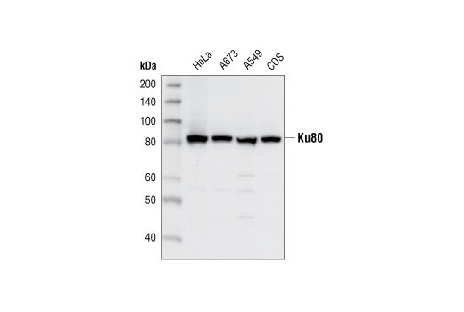
| Western Blotting | 1:1000 |
| Immunoprecipitation | 1:25 |
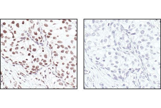
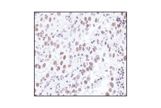
| Immunohistochemistry (Paraffin) | 1:150 - 1:600 |
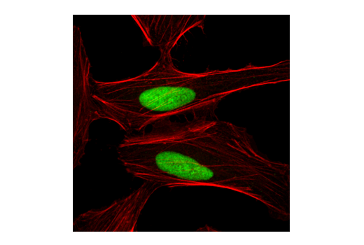
| Immunofluorescence (Immunocytochemistry) | 1:100 - 1:400 |
| Flow Cytometry (Fixed/Permeabilized) | 1:50 - 1:100 |
Storage
Specificity / Sensitivity
Species Reactivity:
Human, Monkey
Species predicted to react based on 100% sequence homology
The antigen sequence used to produce this antibody shares
100% sequence homology with the species listed here, but
reactivity has not been tested or confirmed to work by CST.
Use of this product with these species is not covered under
our
Product Performance Guarantee.
Mouse, Rat
Source / Purification
Polyclonal antibodies are produced by immunizing animals with a synthetic peptide corresponding to the carboxy terminus of mouse Ku80. Antibodies are purified by protein A and peptide affinity chromatography.
Background
Ku is a heterodimeric protein composed of two subunits (Ku70 and Ku80) originally identified by researchers as autoantigens associated with several autoimmune diseases including scleroderma, polymyositis, and systemic lupus erythematosus (1). Ku is an abundant, ubiquitously expressed nuclear protein that binds to and stabilizes the ends of DNA at telomeres or double-stranded DNA breaks (2-5). The Ku70/Ku80 heterodimer has ATP-dependent DNA helicase activity and functions as the DNA-binding regulatory component of DNA-dependent protein kinase (DNA-PK) (6-8). The assembly of the DNA-PK complex at DNA ends is required for nonhomologous end-joining (NHEJ), one mechanism involved in double-stranded DNA break repair and V(D)J recombination (8). DNA-PK has been shown to phosphorylate many proteins, including p53, serum response factor, c-Jun, c-Fos, c-Myc, Oct-1, Sp-1, and RNA polymerase II (1,8). The combined activities of Ku70/Ku80 and DNA-PK implicate Ku in many cellular functions, including cell cycle regulation, DNA replication and repair, telomere maintenance, recombination, and transcriptional activation.
- Tuteja, R. and Tuteja, N. (2000) Crit. Rev. Biochem. Mol. Biol. 35, 1-33.
- Blier, P.R. et al. (1993) J. Biol. Chem. 268, 7594-7601.
- Jin, S. and Weaver, D.T. (1997) EMBO J. 16, 6874-6885.
- Boulton, S.J. and Jackson, S.P. (1998) EMBO J. 17, 1819-1828.
- Gravel, S. et al. (1998) Science 280, 741-744.
- Cao, Q.P. et al. (1994) Biochemistry 33, 8548-8557.
- Lees-Miller, S.P. et al. (1990) Mol. Cell Biol. 10, 6472-6481.
- Collis, S.J. et al. (2005) Oncogene 24, 949-961.
Species Reactivity
Species reactivity is determined by testing in at least one approved application (e.g., western blot).
Western Blot Buffer
IMPORTANT: For western blots, incubate membrane with diluted primary antibody in 5% w/v nonfat dry milk, 1X TBS, 0.1% Tween® 20 at 4°C with gentle shaking, overnight.
Applications Key
WB: Western Blotting IP: Immunoprecipitation IHC-P: Immunohistochemistry (Paraffin) IF-IC: Immunofluorescence (Immunocytochemistry) FC-FP: Flow Cytometry (Fixed/Permeabilized)
Cross-Reactivity Key
H: human M: mouse R: rat Hm: hamster Mk: monkey Vir: virus Mi: mink C: chicken Dm: D. melanogaster X: Xenopus Z: zebrafish B: bovine Dg: dog Pg: pig Sc: S. cerevisiae Ce: C. elegans Hr: horse GP: Guinea Pig Rab: rabbit All: all species expected
Trademarks and Patents
Limited Uses
Except as otherwise expressly agreed in a writing signed by a legally authorized representative of CST, the following terms apply to Products provided by CST, its affiliates or its distributors. Any Customer's terms and conditions that are in addition to, or different from, those contained herein, unless separately accepted in writing by a legally authorized representative of CST, are rejected and are of no force or effect.
Products are labeled with For Research Use Only or a similar labeling statement and have not been approved, cleared, or licensed by the FDA or other regulatory foreign or domestic entity, for any purpose. Customer shall not use any Product for any diagnostic or therapeutic purpose, or otherwise in any manner that conflicts with its labeling statement. Products sold or licensed by CST are provided for Customer as the end-user and solely for research and development uses. Any use of Product for diagnostic, prophylactic or therapeutic purposes, or any purchase of Product for resale (alone or as a component) or other commercial purpose, requires a separate license from CST. Customer shall (a) not sell, license, loan, donate or otherwise transfer or make available any Product to any third party, whether alone or in combination with other materials, or use the Products to manufacture any commercial products, (b) not copy, modify, reverse engineer, decompile, disassemble or otherwise attempt to discover the underlying structure or technology of the Products, or use the Products for the purpose of developing any products or services that would compete with CST products or services, (c) not alter or remove from the Products any trademarks, trade names, logos, patent or copyright notices or markings, (d) use the Products solely in accordance with CST Product Terms of Sale and any applicable documentation, and (e) comply with any license, terms of service or similar agreement with respect to any third party products or services used by Customer in connection with the Products.