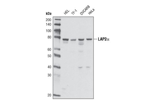
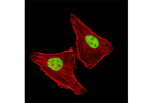
WB, IF-IC
H Mk
Endogenous
76
Mouse IgG1
#P42166
7112
Product Information
Product Usage Information
| Application | Dilution |
|---|---|
| Western Blotting | 1:1000 |
| Immunofluorescence (Immunocytochemistry) | 1:800 |
Storage
Specificity / Sensitivity
Species Reactivity:
Human, Monkey
Source / Purification
Monoclonal antibody is produced by immunizing animals with a synthetic peptide corresponding to residues near the carboxy terminus of human LAP2α protein.
Background
Lamins and lamin associated proteins are the major components of nuclear lamina found between the inner nuclear membrane and the peripheral chromatin. These proteins play important roles in maintaining nuclear structure, chromatin organization, DNA replication, cell cycle regulation, and apoptosis (1-3). Lamins are type V intermediate filaments that are further classified into type A and type B lamin proteins. Type A lamins (including lamin A and the smaller lamin C splice variant) are predominately expressed in terminally differentiated cells, whereas type B lamins (lamin B1, lamin B2) are encoded by distinct genes and are expressed constitutively. Cleavage of lamins by caspases occurs during apoptosis as part of the disassembly of the cell (4-6). A number of lamina-associated proteins contribute to the nuclear lamina and include the lamin B receptor, LAP1, LAP2, emerin, MAN1, otefin, and YA. Several isoforms of lamina-associated polypeptide 2 (LAP2, also known as thymopoietin or TMPO) have been described, with the α, β, and γ isoforms most abundant in humans (7-10). Structurally similar LAP2β and LAP2γ are type II integral membrane proteins. LAP2α has a unique carboxy-terminus that lacks a transmembrane region and results in localization of LAP2α throughout the nucleus where it can associate with lamin A/C (10). LAP2α is also thought to contribute to the nuclear anchorage of retinoblastoma protein (Rb) and control cell cycle progression (11). LAP2α is also targeted for cleavage by caspases, which may contribute to changes in chromatin structure during apoptosis (12).
- Gruenbaum, Y. et al. (2000) J Struct Biol 129, 313-23.
- Goldberg, M. et al. (1999) Crit Rev Eukaryot Gene Expr 9, 285-93.
- Holmer, L. and Worman, H.J. (2001) Cell Mol Life Sci 58, 1741-7.
- Lazebnik, Y.A. et al. (1995) Proc Natl Acad Sci USA 92, 9042-6.
- Oberhammer, F.A. et al. (1994) J Cell Biol 126, 827-37.
- Rao, L. et al. (1996) J Cell Biol 135, 1441-55.
- Furukawa, K. et al. (1995) EMBO J 14, 1626-36.
- Foisner, R. and Gerace, L. (1993) Cell 73, 1267-79.
- Harris, C.A. et al. (1994) Proc Natl Acad Sci USA 91, 6283-7.
- Dechat, T. et al. (2000) J Cell Sci 113 Pt 19, 3473-84.
- Markiewicz, E. et al. (2002) Mol Biol Cell 13, 4401-13.
- Gotzmann, J. et al. (2000) J Cell Sci 113 Pt 21, 3769-80.
Species Reactivity
Species reactivity is determined by testing in at least one approved application (e.g., western blot).
Western Blot Buffer
IMPORTANT: For western blots, incubate membrane with diluted primary antibody in 5% w/v nonfat dry milk, 1X TBS, 0.1% Tween® 20 at 4°C with gentle shaking, overnight.
Applications Key
WB: Western Blotting IF-IC: Immunofluorescence (Immunocytochemistry)
Cross-Reactivity Key
H: human M: mouse R: rat Hm: hamster Mk: monkey Vir: virus Mi: mink C: chicken Dm: D. melanogaster X: Xenopus Z: zebrafish B: bovine Dg: dog Pg: pig Sc: S. cerevisiae Ce: C. elegans Hr: horse GP: Guinea Pig Rab: rabbit All: all species expected
Trademarks and Patents
Limited Uses
Except as otherwise expressly agreed in a writing signed by a legally authorized representative of CST, the following terms apply to Products provided by CST, its affiliates or its distributors. Any Customer's terms and conditions that are in addition to, or different from, those contained herein, unless separately accepted in writing by a legally authorized representative of CST, are rejected and are of no force or effect.
Products are labeled with For Research Use Only or a similar labeling statement and have not been approved, cleared, or licensed by the FDA or other regulatory foreign or domestic entity, for any purpose. Customer shall not use any Product for any diagnostic or therapeutic purpose, or otherwise in any manner that conflicts with its labeling statement. Products sold or licensed by CST are provided for Customer as the end-user and solely for research and development uses. Any use of Product for diagnostic, prophylactic or therapeutic purposes, or any purchase of Product for resale (alone or as a component) or other commercial purpose, requires a separate license from CST. Customer shall (a) not sell, license, loan, donate or otherwise transfer or make available any Product to any third party, whether alone or in combination with other materials, or use the Products to manufacture any commercial products, (b) not copy, modify, reverse engineer, decompile, disassemble or otherwise attempt to discover the underlying structure or technology of the Products, or use the Products for the purpose of developing any products or services that would compete with CST products or services, (c) not alter or remove from the Products any trademarks, trade names, logos, patent or copyright notices or markings, (d) use the Products solely in accordance with CST Product Terms of Sale and any applicable documentation, and (e) comply with any license, terms of service or similar agreement with respect to any third party products or services used by Customer in connection with the Products.