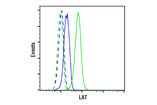
Revision 1
Cell Signaling Technology

Orders: 877-616-CELL (2355) [email protected]

Support: 877-678-TECH (8324)

Web: [email protected] cellsignal.com

3 Trask LaneDanversMassachusetts01923USA
For Research Use Only. Not for Use in Diagnostic Procedures.
Applications:

WB, IHC-P, FC-FP

REACTIVITY:

H M

SENSITIVITY:

Endogenous

MW (kDa):

36, 38

Source/Isotype:

Rabbit IgG

UniProt ID:

#O43561

Entrez-Gene Id:

27040

Product Information

Product Usage Information

This product is the carrier free version of product #45533. All data were generated using the same antibody clone in the standard formulation which contains BSA and glycerol.

This formulation is ideal for use with technologies requiring specialized or custom antibody labeling, including fluorophores, metals, lanthanides, and oligonucleotides. It is not recommended for ChIP, ChIP-seq, CUT&RUN or CUT&Tag assays. If you require a carrier free formulation for chromatin profiling, please contact us. Optimal dilutions/concentrations should be determined by the end user.

Formulation

Supplied in 1X PBS, BSA and Azide Free.

For standard formulation of this product see product #45533

Storage

Store at -20°C. This product will freeze at -20°C so it is recommended to aliquot into single-use vials to avoid multiple freeze/thaw cycles. A slight precipitate may be present and can be dissolved by gently vortexing. This will not interfere with antibody performance.

Specificity / Sensitivity

LAT (E3U6J) XP® Rabbit mAb (BSA and Azide Free) recognizes endogenous levels of total LAT protein.

Species Reactivity:

Human, Mouse

Source / Purification

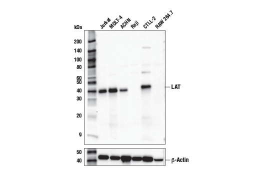
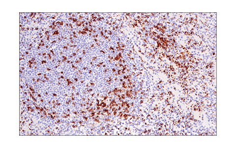
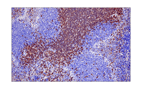
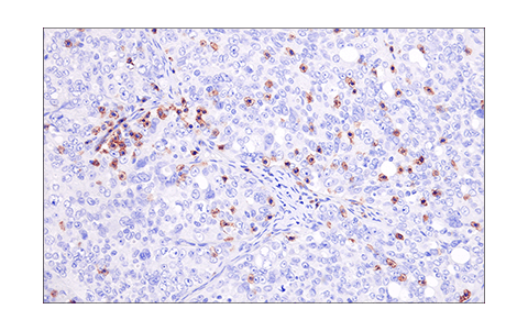
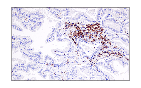
Monoclonal antibody is produced by immunizing animals with a synthetic peptide corresponding to residues surrounding Leu88 of human LAT protein.

Background

LAT, a transmembrane adaptor protein expressed in T, NK, and mast cells, is an important mediator for T cell receptor (TCR) signaling (1). Upon TCR engagement, activated Zap-70 phosphorylates LAT at multiple conserved tyrosine residues within SH2 binding motifs, exposing these motifs as the docking sites for downstream signaling targets (2,3). The phosphorylation of LAT at Tyr171 and Tyr220 enables the binding of Grb2, Gads/SLP-76, PLCγ1, and PI3 kinase through their SH2 domain and translocates them to the membrane. This process eventually leads to activation of the corresponding signaling pathways (1-4).

  1. Wonerow, P. and Watson, S.P. (2001) Oncogene 20, 6273-83.
  2. Zhang, W. et al. (1998) Cell 92, 83-92.
  3. Paz, P. E. et al. (2001) Biochem. J. 356, 461-71.
  4. Zhang, W. et al. (2000) J. Biol. Chem. 275, 23355-61.

Species Reactivity

Species reactivity is determined by testing in at least one approved application (e.g., western blot).

Applications Key

WB: Western Blotting IHC-P: Immunohistochemistry (Paraffin) FC-FP: Flow Cytometry (Fixed/Permeabilized)

Cross-Reactivity Key

H: human M: mouse R: rat Hm: hamster Mk: monkey Vir: virus Mi: mink C: chicken Dm: D. melanogaster X: Xenopus Z: zebrafish B: bovine Dg: dog Pg: pig Sc: S. cerevisiae Ce: C. elegans Hr: horse GP: Guinea Pig Rab: rabbit All: all species expected

Trademarks and Patents

Cell Signaling Technology is a trademark of Cell Signaling Technology, Inc.
SignalStain is a trademark of Cell Signaling Technology, Inc.
XP is a registered trademark of Cell Signaling Technology, Inc.
All other trademarks are the property of their respective owners. Visit cellsignal.com/trademarks for more information.

Limited Uses

Except as otherwise expressly agreed in a writing signed by a legally authorized representative of CST, the following terms apply to Products provided by CST, its affiliates or its distributors. Any Customer's terms and conditions that are in addition to, or different from, those contained herein, unless separately accepted in writing by a legally authorized representative of CST, are rejected and are of no force or effect.

Products are labeled with For Research Use Only or a similar labeling statement and have not been approved, cleared, or licensed by the FDA or other regulatory foreign or domestic entity, for any purpose. Customer shall not use any Product for any diagnostic or therapeutic purpose, or otherwise in any manner that conflicts with its labeling statement. Products sold or licensed by CST are provided for Customer as the end-user and solely for research and development uses. Any use of Product for diagnostic, prophylactic or therapeutic purposes, or any purchase of Product for resale (alone or as a component) or other commercial purpose, requires a separate license from CST. Customer shall (a) not sell, license, loan, donate or otherwise transfer or make available any Product to any third party, whether alone or in combination with other materials, or use the Products to manufacture any commercial products, (b) not copy, modify, reverse engineer, decompile, disassemble or otherwise attempt to discover the underlying structure or technology of the Products, or use the Products for the purpose of developing any products or services that would compete with CST products or services, (c) not alter or remove from the Products any trademarks, trade names, logos, patent or copyright notices or markings, (d) use the Products solely in accordance with CST Product Terms of Sale and any applicable documentation, and (e) comply with any license, terms of service or similar agreement with respect to any third party products or services used by Customer in connection with the Products.

Revision 1
#41708

LAT (E3U6J) XP® Rabbit mAb (BSA and Azide Free)

Western Blotting Image 1: LAT (E3U6J) XP® Rabbit mAb (BSA and Azide Free) Expand Image
Western blot analysis of extracts from various cell lines using LAT (E3U6J) XP® Rabbit mAb (upper) or β-Actin (D6A8) Rabbit mAb #8457 (lower). Data were generated using the standard formulation of this product.
Immunohistochemistry Image 1: LAT (E3U6J) XP® Rabbit mAb (BSA and Azide Free) Expand Image
Immunohistochemical analysis of paraffin-embedded Jurkat cell pellet (left, positive) or Raji cell pellet (right, negative) using LAT (E3U6J) XP® Rabbit mAb. Data were generated using the standard formulation of this product.
Immunohistochemistry Image 2: LAT (E3U6J) XP® Rabbit mAb (BSA and Azide Free) Expand Image
Immunohistochemical analysis of paraffin-embedded human colon carcinoma using LAT (E3U6J) XP® Rabbit mAb. Data were generated using the standard formulation of this product.
Immunohistochemistry Image 3: LAT (E3U6J) XP® Rabbit mAb (BSA and Azide Free) Expand Image
Immunohistochemical analysis of paraffin-embedded human lung adenocarcinoma using LAT (E3U6J) XP® Rabbit mAb (left) compared to concentration-matched Rabbit (DA1E) mAb IgG XP® Isotype Control #3900 (right). Data were generated using the standard formulation of this product.
Immunohistochemistry Image 4: LAT (E3U6J) XP® Rabbit mAb (BSA and Azide Free) Expand Image
Immunohistochemical analysis of paraffin-embedded human Non-Hodgkin's lymphoma using LAT (E3U6J) XP® Rabbit mAb. Data were generated using the standard formulation of this product.
Immunohistochemistry Image 5: LAT (E3U6J) XP® Rabbit mAb (BSA and Azide Free) Expand Image
Immunohistochemical analysis of paraffin-embedded mouse spleen using LAT (E3U6J) XP® Rabbit mAb. Data were generated using the standard formulation of this product.
Immunohistochemistry Image 6: LAT (E3U6J) XP® Rabbit mAb (BSA and Azide Free) Expand Image
Immunohistochemical analysis of paraffin-embedded human serous papillary carcinoma of the ovary using LAT (E3U6J) XP® Rabbit mAb. Data were generated using the standard formulation of this product.
Immunohistochemistry Image 7: LAT (E3U6J) XP® Rabbit mAb (BSA and Azide Free) Expand Image
Immunohistochemical analysis of paraffin-embedded human prostate carcinoma using LAT (E3U6J) XP® Rabbit mAb. Data were generated using the standard formulation of this product.
Flow Cytometry Image 1: LAT (E3U6J) XP® Rabbit mAb (BSA and Azide Free) Expand Image
Flow cytometric analysis of Daudi cells (blue) and Jurkat cells (green) using LAT (E3U6J) XP® Rabbit mAb (solid lines) or a concentration-matched Rabbit (DA1E) mAb IgG XP® Isotype Control #3900 (dashed lines). Anti-rabbit IgG (H+L), F(ab')2 Fragment (Alexa Fluor® 488 Conjugate) #4412 was used as a secondary antibody. Data were generated using the standard formulation of this product.