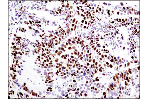
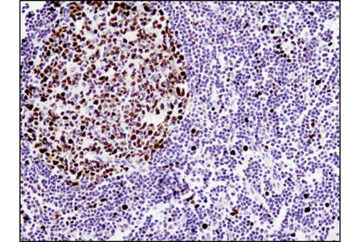
WB, IP, IHC-P, IF-IC
H Mk
Endogenous
125
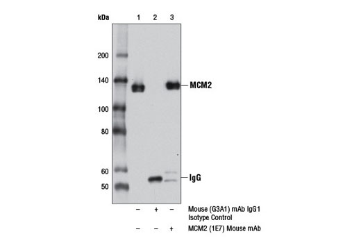
Mouse IgG1
#P49736
4171
Product Information
Product Usage Information
| Application | Dilution |
|---|---|
| Western Blotting | 1:1000 |
| Immunoprecipitation | 1:200 |
| Immunohistochemistry (Paraffin) | 1:400 |
| Immunofluorescence (Immunocytochemistry) | 1:200 |
Storage
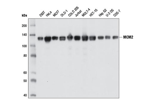
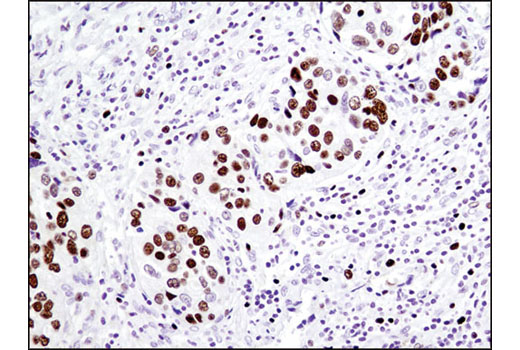
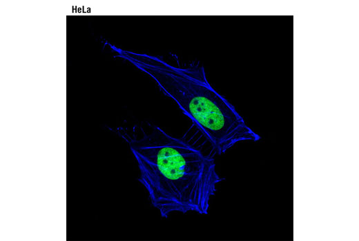
Specificity / Sensitivity
Species Reactivity:
Human, Monkey
Source / Purification
Monoclonal antibody is produced by immunizing animals with recombinant protein specific to the amino terminus of human MCM2 protein.
Background
The minichromosome maintenance (MCM) 2-7 proteins are a family of six related proteins required for initiation and elongation of DNA replication. MCM2-7 bind together to form the heterohexameric MCM complex that is thought to act as a replicative helicase at the DNA replication fork (1-5). This complex is a key component of the pre-replication complex (pre-RC) (reviewed in 1). Cdc6 and CDT1 recruit the MCM complex to the origin recognition complex (ORC) during late mitosis/early G1 phase forming the pre-RC and licensing the DNA for replication (reviewed in 2). Licensing of the chromatin permits the DNA to replicate only once per cell cycle, thereby helping to ensure that genetic alterations and malignant cell growth do not occur (reviewed in 3). Phosphorylation of the MCM2, MCM3, MCM4, and MCM6 subunits appears to regulate MCM complex activity and the initiation of DNA synthesis (6-8). CDK1 phosphorylation of MCM3 at Ser112 during late mitosis/early G1 phase has been shown to initiate complex formation and chromatin loading in vitro (8). Phosphorylation of MCM2 at serine 139 by cdc7/dbf4 coincides with the initiation of DNA replication (9). MCM proteins are removed during DNA replication, causing chromatin to become unlicensed through inhibition of pre-RC reformation. Studies have shown that the MCM complex is involved in checkpoint control by protecting the structure of the replication fork and assisting in restarting replication by recruiting checkpoint proteins after arrest (reviewed in 3,10).
- Lei, M. and Tye, B.K. (2001) J Cell Sci 114, 1447-54.
- Lygerou, Z. and Nurse, P. (2000) Science 290, 2271-3.
- Forsburg, S.L. (2004) Microbiol Mol Biol Rev 68, 109-31.
- Tye, B.K. and Sawyer, S. (2000) J Biol Chem 275, 34833-6.
- Labib, K. et al. (2000) Science 288, 1643-7.
- Charych, D.H. et al. (2008) J Cell Biochem 104, 1075-86.
- Masai, H. et al. (2006) J Biol Chem 281, 39249-61.
- Lin, D.I. et al. (2008) Proc Natl Acad Sci USA 105, 8079-84.
- Tsuji, T. et al. (2006) Mol Biol Cell 17, 4459-72.
- Bailis, J.M. et al. (2008) Mol Cell Biol 28, 1724-38.
Species Reactivity
Species reactivity is determined by testing in at least one approved application (e.g., western blot).
Western Blot Buffer
IMPORTANT: For western blots, incubate membrane with diluted primary antibody in 5% w/v nonfat dry milk, 1X TBS, 0.1% Tween® 20 at 4°C with gentle shaking, overnight.
Applications Key
WB: Western Blotting IP: Immunoprecipitation IHC-P: Immunohistochemistry (Paraffin) IF-IC: Immunofluorescence (Immunocytochemistry)
Cross-Reactivity Key
H: human M: mouse R: rat Hm: hamster Mk: monkey Vir: virus Mi: mink C: chicken Dm: D. melanogaster X: Xenopus Z: zebrafish B: bovine Dg: dog Pg: pig Sc: S. cerevisiae Ce: C. elegans Hr: horse GP: Guinea Pig Rab: rabbit All: all species expected
Trademarks and Patents
Limited Uses
Except as otherwise expressly agreed in a writing signed by a legally authorized representative of CST, the following terms apply to Products provided by CST, its affiliates or its distributors. Any Customer's terms and conditions that are in addition to, or different from, those contained herein, unless separately accepted in writing by a legally authorized representative of CST, are rejected and are of no force or effect.
Products are labeled with For Research Use Only or a similar labeling statement and have not been approved, cleared, or licensed by the FDA or other regulatory foreign or domestic entity, for any purpose. Customer shall not use any Product for any diagnostic or therapeutic purpose, or otherwise in any manner that conflicts with its labeling statement. Products sold or licensed by CST are provided for Customer as the end-user and solely for research and development uses. Any use of Product for diagnostic, prophylactic or therapeutic purposes, or any purchase of Product for resale (alone or as a component) or other commercial purpose, requires a separate license from CST. Customer shall (a) not sell, license, loan, donate or otherwise transfer or make available any Product to any third party, whether alone or in combination with other materials, or use the Products to manufacture any commercial products, (b) not copy, modify, reverse engineer, decompile, disassemble or otherwise attempt to discover the underlying structure or technology of the Products, or use the Products for the purpose of developing any products or services that would compete with CST products or services, (c) not alter or remove from the Products any trademarks, trade names, logos, patent or copyright notices or markings, (d) use the Products solely in accordance with CST Product Terms of Sale and any applicable documentation, and (e) comply with any license, terms of service or similar agreement with respect to any third party products or services used by Customer in connection with the Products.