| Product Includes | Product # | Quantity | Mol. Wt | Isotype/Source |
|---|---|---|---|---|
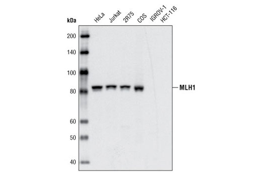
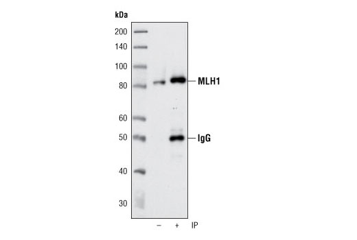
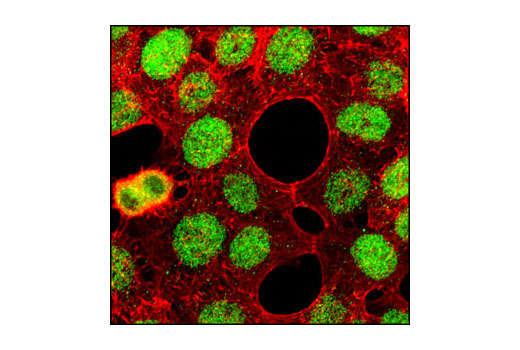
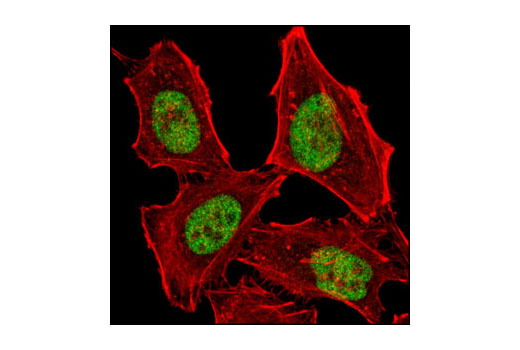
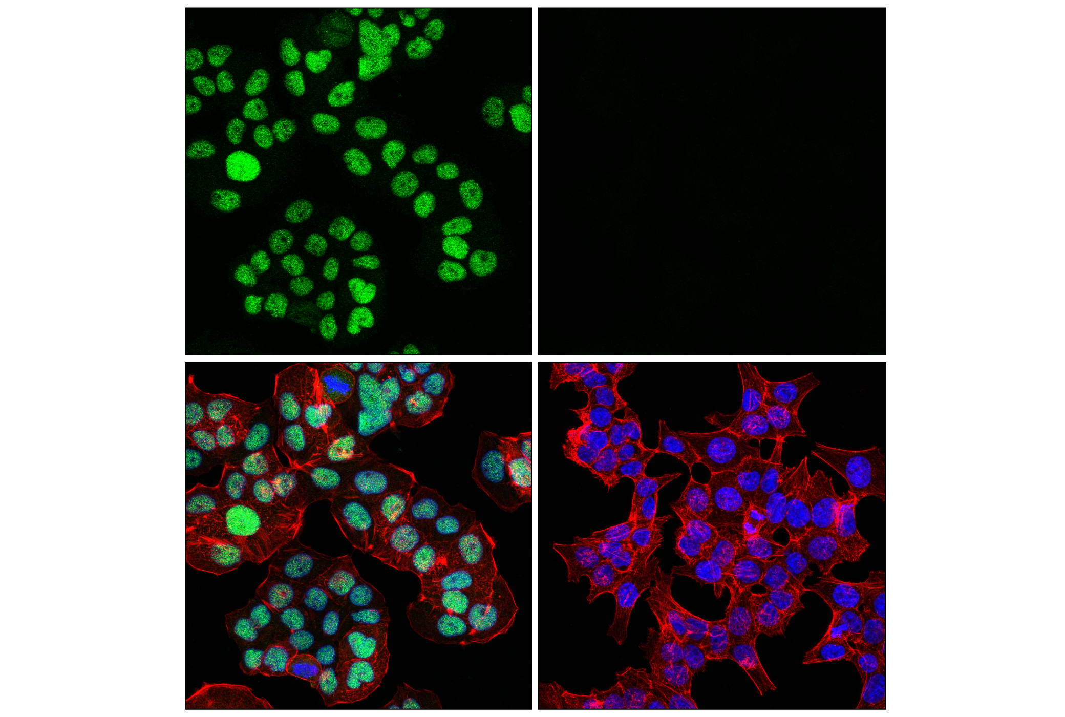
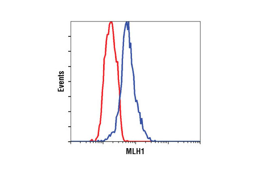
| MLH1 (4C9C7) Mouse mAb | 3515 | 40 µl | 85 kDa | Mouse IgG1 |
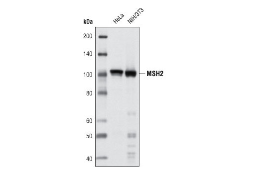
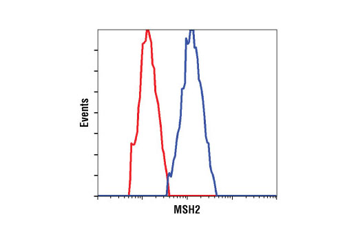
| MSH2 (D24B5) XP® Rabbit mAb | 2017 | 40 µl | 100 kDa | Rabbit IgG |
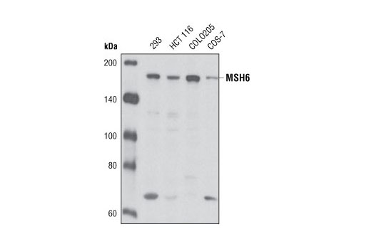
| MSH6 (L990) Antibody | 3996 | 40 µl | 160 kDa | Rabbit |
| Anti-rabbit IgG, HRP-linked Antibody | 7074 | 100 µl | Goat | |
| Anti-mouse IgG, HRP-linked Antibody | 7076 | 100 µl | Horse |
Please visit cellsignal.com for individual component applications, species cross-reactivity, dilutions, protocols, and additional product information.
Description
Mismatch Repair Antibody Sampler Kit provides an economical means to investigate the DNA mismatch repair system within the cell. The kit contains primary and secondary antibodies to perform four western blots with each antibody.
Storage
Background
The DNA mismatch repair system (MMR) repairs post-replication DNA, inhibits recombination between nonidentical DNA sequences, and induces both checkpoint and apoptotic responses following certain types of DNA damage (1). MSH2 (MutS homologue 2) forms the hMutS-α dimer with MSH6 and is an essential component of the mismatch repair process. hMutS-α is part of the BRCA1-associated surveillance complex (BASC), a complex that also contains BRCA1, MLH1, ATM, BLM, PMS2 proteins, and the Rad50-Mre11-NBS1 complex (2). Mutations in MSH6 and other MMR proteins have been found in a large proportion of hereditary nonpolyposis colorectal cancer (Lynch Syndrome), the most common form of inherited colorectal cancer in the Western world (3). Mutations in MSH6 have been shown to occur in glioblastoma in response to temozolomide therapy and to promote temozolomide resistance (4).
Background References
Trademarks and Patents
Limited Uses
Except as otherwise expressly agreed in a writing signed by a legally authorized representative of CST, the following terms apply to Products provided by CST, its affiliates or its distributors. Any Customer's terms and conditions that are in addition to, or different from, those contained herein, unless separately accepted in writing by a legally authorized representative of CST, are rejected and are of no force or effect.
Products are labeled with For Research Use Only or a similar labeling statement and have not been approved, cleared, or licensed by the FDA or other regulatory foreign or domestic entity, for any purpose. Customer shall not use any Product for any diagnostic or therapeutic purpose, or otherwise in any manner that conflicts with its labeling statement. Products sold or licensed by CST are provided for Customer as the end-user and solely for research and development uses. Any use of Product for diagnostic, prophylactic or therapeutic purposes, or any purchase of Product for resale (alone or as a component) or other commercial purpose, requires a separate license from CST. Customer shall (a) not sell, license, loan, donate or otherwise transfer or make available any Product to any third party, whether alone or in combination with other materials, or use the Products to manufacture any commercial products, (b) not copy, modify, reverse engineer, decompile, disassemble or otherwise attempt to discover the underlying structure or technology of the Products, or use the Products for the purpose of developing any products or services that would compete with CST products or services, (c) not alter or remove from the Products any trademarks, trade names, logos, patent or copyright notices or markings, (d) use the Products solely in accordance with CST Product Terms of Sale and any applicable documentation, and (e) comply with any license, terms of service or similar agreement with respect to any third party products or services used by Customer in connection with the Products.