| Product Includes | Product # | Quantity | Mol. Wt | Isotype/Source |
|---|---|---|---|---|
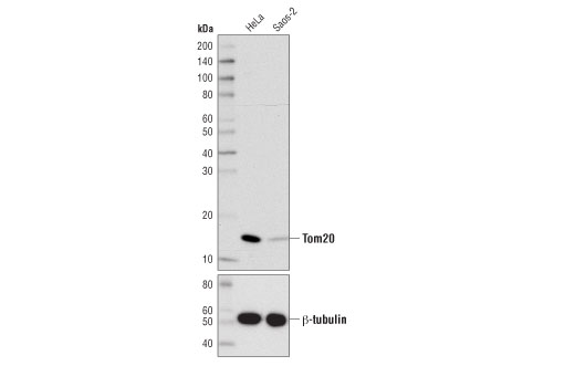
| Tom20 (D8T4N) Rabbit mAb | 42406 | 20 µl | 16 kDa | Rabbit IgG |
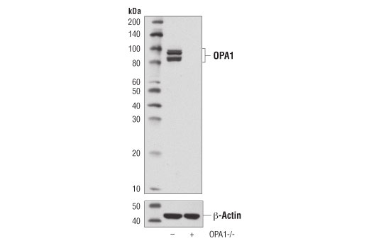
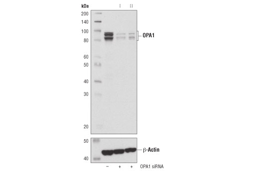
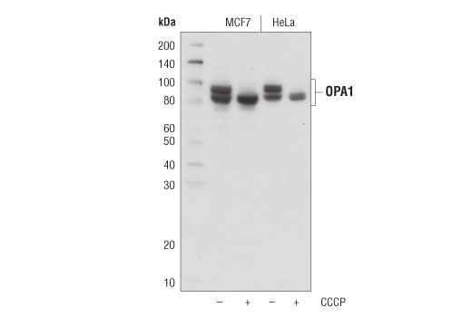
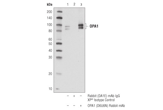
| OPA1 (D6U6N) Rabbit mAb | 80471 | 20 µl | 80-100 kDa | Rabbit IgG |
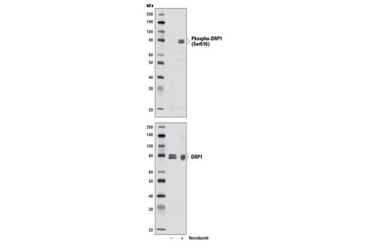
| Phospho-DRP1 (Ser616) (D9A1) Rabbit mAb | 4494 | 20 µl | 78-82 kDa | Rabbit IgG |
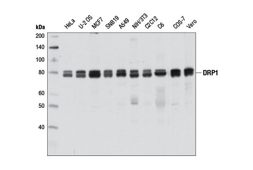
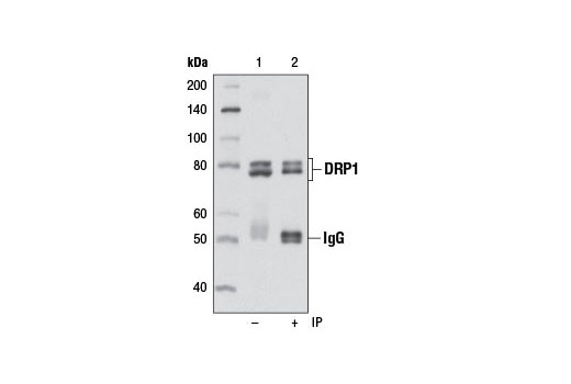
| DRP1 (D8H5) Rabbit mAb | 5391 | 20 µl | 78-82 kDa | Rabbit IgG |
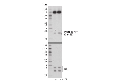
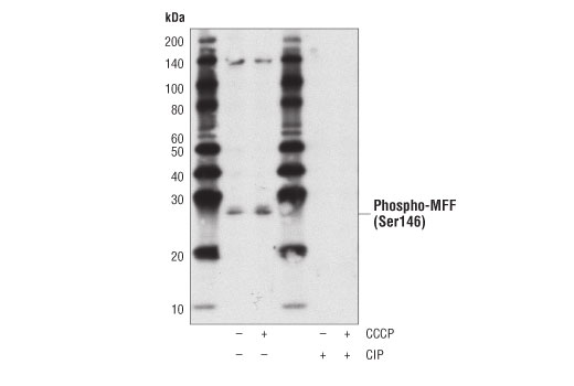
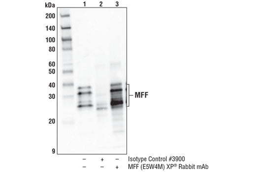
| Phospho-MFF (Ser146) Antibody | 49281 | 20 µl | 25, 27 kDa | Rabbit |
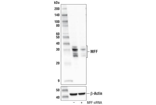
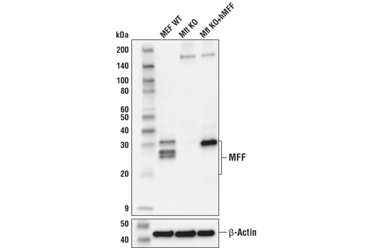
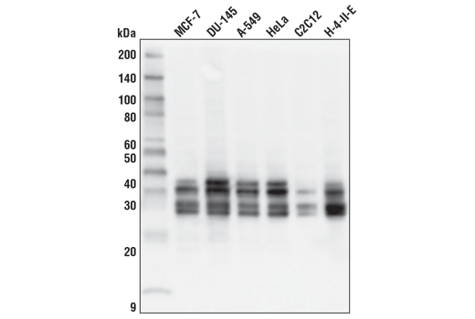
| MFF (E5W4M) XP® Rabbit mAb | 84580 | 20 µl | 25, 27, 30, 35 kDa | Rabbit IgG |
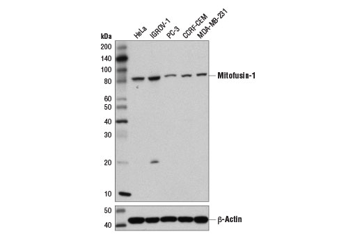
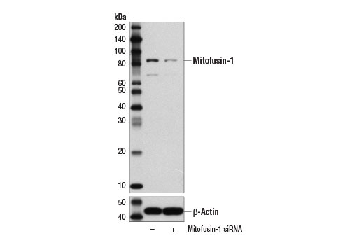
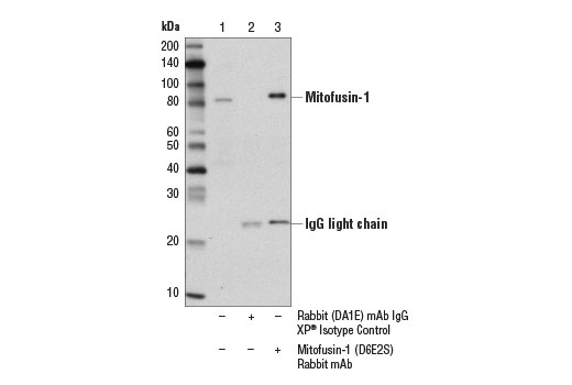
| Mitofusin-1 (D6E2S) Rabbit mAb | 14739 | 20 µl | 82 kDa | Rabbit IgG |
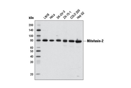
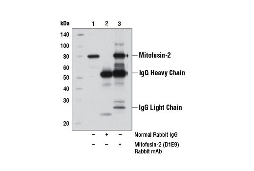
| Mitofusin-2 (D1E9) Rabbit mAb | 11925 | 20 µl | 80 kDa | Rabbit IgG |
| Anti-rabbit IgG, HRP-linked Antibody | 7074 | 100 µl | Goat |
Please visit cellsignal.com for individual component applications, species cross-reactivity, dilutions, protocols, and additional product information.
Description
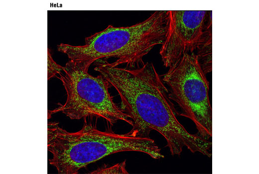
The Mitochondrial Dynamics Antibody Sampler Kit provides an economical means to examine signaling involved in mitochondrial dynamics. The kit contains enough primary antibody to perform two western blot experiments.
Storage
Background
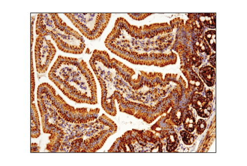
Import of proteins into the mitochondria is regulated by the translocase of the outer mitochondrial membrane (TOM) complex, which facilitates transport through the outer mitochondrial membrane, and a complementary translocase of the inner membrane (TIM) complex, responsible for protein transport to the mitochondrial matrix. The TOM complex consists of the receptors Tom20, Tom22, and Tom70, and the channel-forming protein Tom40 (1). Tom20 is localized in the outer mitochondrial membrane and initially recognizes precursors with a presequence to facilitate protein import across the outer mitochondrial membrane (2).
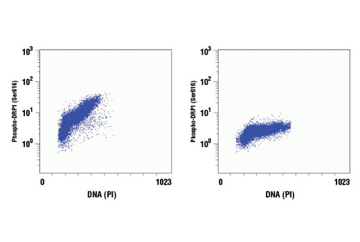
Changes in mitochondrial dynamics regulated by environmental cues affect mitochondrial size and shape and have been shown to dramatically impact mitochondrial metabolism, apoptosis, and autophagy (3). These processes are largely controlled by mitochondrial dynamin-related GTPases, including mitofusin-1, mitofusin-2, OPA1, and DRP1. DRP1 regulates mitochondrial fission, while the mitofusins and OPA1 control fusion at the outer and inner mitochondrial membrane, respectively. These proteins are tightly regulated. OPA1 activity is regulated through alternative splicing and post-translational modifications, including complex proteolytic processing by multiple proteases (4-9). In addition, OPA1 expression can be induced under conditions of metabolic demand through a pathway involving Parkin induced NF-κB activation (10). DRP1 is regulated in part through multiple phosphorylation sites (11). Phosphorylation of DRP1 at Ser616 by MAPK or during mitosis by CDKs stimulates mitochondrial fission (12-14). In contrast, PKA dependent phosphorylation of DRP1 at Ser637 inhibits its GTPase activity and mitochondrial fission (15,16). Mitochondrial fission factor (MFF) is a tail-anchored protein that resides within the outer mitochondrial membrane and is part of the mitochondrial fission complex. MFF participates in mitochondrial fission by serving as one of multiple receptors for the GTPase dynamin-related protein 1 (Drp1) (17-20). AMPK directly phosphorylates MFF at two sites to allow for enhanced recruitment of Drp1 to the mitochondria (21).
- Chacinska, A. et al. (2009) Cell 138, 628-44.
- Saitoh, T. et al. (2007) EMBO J 26, 4777-87.
- Kasahara, A. and Scorrano, L. (2014) Trends Cell Biol 24, 761-70.
- Delettre, C. et al. (2001) Hum Genet 109, 584-91.
- Olichon, A. et al. (2007) Cell Death Differ 14, 682-92.
- Ishihara, N. et al. (2006) EMBO J 25, 2966-77.
- Cipolat, S. et al. (2006) Cell 126, 163-75.
- Griparic, L. et al. (2007) J Cell Biol 178, 757-64.
- Merkwirth, C. et al. (2008) Genes Dev 22, 476-88.
- Müller-Rischart, A.K. et al. (2013) Mol Cell 49, 908-21.
- Knott, A.B. et al. (2008) Nat Rev Neurosci 9, 505-18.
- Kashatus, J.A. et al. (2015) Mol Cell 57, 537-51.
- Kashatus, D.F. et al. (2011) Nat Cell Biol 13, 1108-15.
- Taguchi, N. et al. (2007) J Biol Chem 282, 11521-9.
- Chang, C.R. and Blackstone, C. (2007) J Biol Chem 282, 21583-7.
- Cribbs, J.T. and Strack, S. (2007) EMBO Rep 8, 939-44.
- Liu, R. and Chan, D.C. (2015) Mol Biol Cell 26, 4466-77.
- Shen, Q. et al. (2014) Mol Biol Cell 25, 145-59.
- Losón, O.C. et al. (2013) Mol Biol Cell 24, 659-67.
- Otera, H. et al. (2010) J Cell Biol 191, 1141-58.
- Toyama, E.Q. et al. (2016) Science 351, 275-281.
Background References
Trademarks and Patents
Limited Uses
Except as otherwise expressly agreed in a writing signed by a legally authorized representative of CST, the following terms apply to Products provided by CST, its affiliates or its distributors. Any Customer's terms and conditions that are in addition to, or different from, those contained herein, unless separately accepted in writing by a legally authorized representative of CST, are rejected and are of no force or effect.
Products are labeled with For Research Use Only or a similar labeling statement and have not been approved, cleared, or licensed by the FDA or other regulatory foreign or domestic entity, for any purpose. Customer shall not use any Product for any diagnostic or therapeutic purpose, or otherwise in any manner that conflicts with its labeling statement. Products sold or licensed by CST are provided for Customer as the end-user and solely for research and development uses. Any use of Product for diagnostic, prophylactic or therapeutic purposes, or any purchase of Product for resale (alone or as a component) or other commercial purpose, requires a separate license from CST. Customer shall (a) not sell, license, loan, donate or otherwise transfer or make available any Product to any third party, whether alone or in combination with other materials, or use the Products to manufacture any commercial products, (b) not copy, modify, reverse engineer, decompile, disassemble or otherwise attempt to discover the underlying structure or technology of the Products, or use the Products for the purpose of developing any products or services that would compete with CST products or services, (c) not alter or remove from the Products any trademarks, trade names, logos, patent or copyright notices or markings, (d) use the Products solely in accordance with CST Product Terms of Sale and any applicable documentation, and (e) comply with any license, terms of service or similar agreement with respect to any third party products or services used by Customer in connection with the Products.