| Product Includes | Product # | Quantity | Mol. Wt | Isotype/Source |
|---|---|---|---|---|
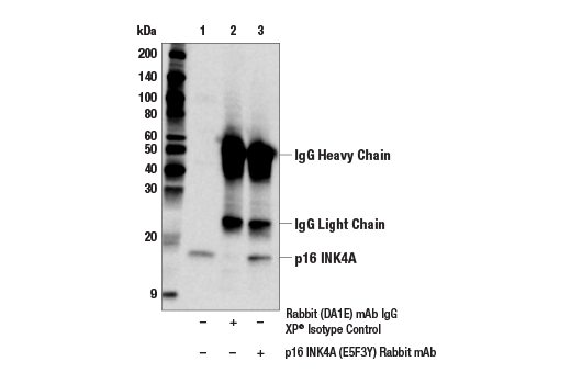
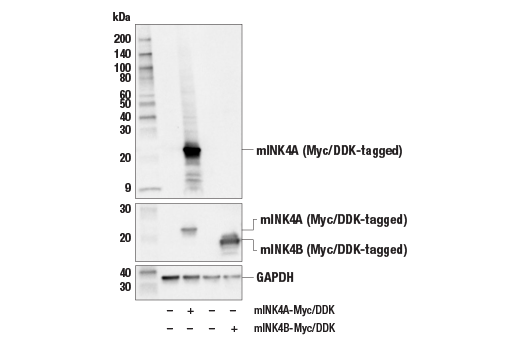
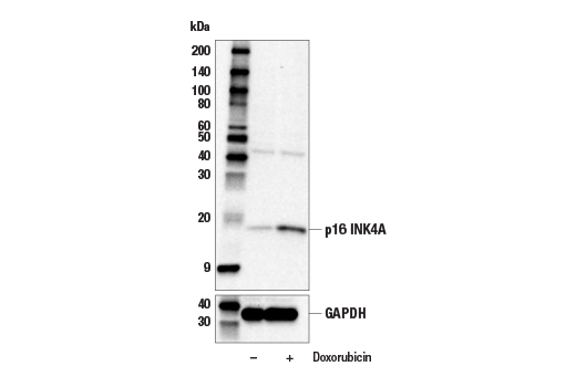
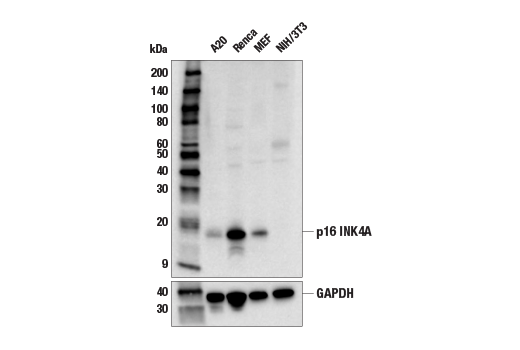
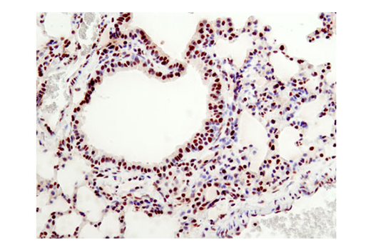
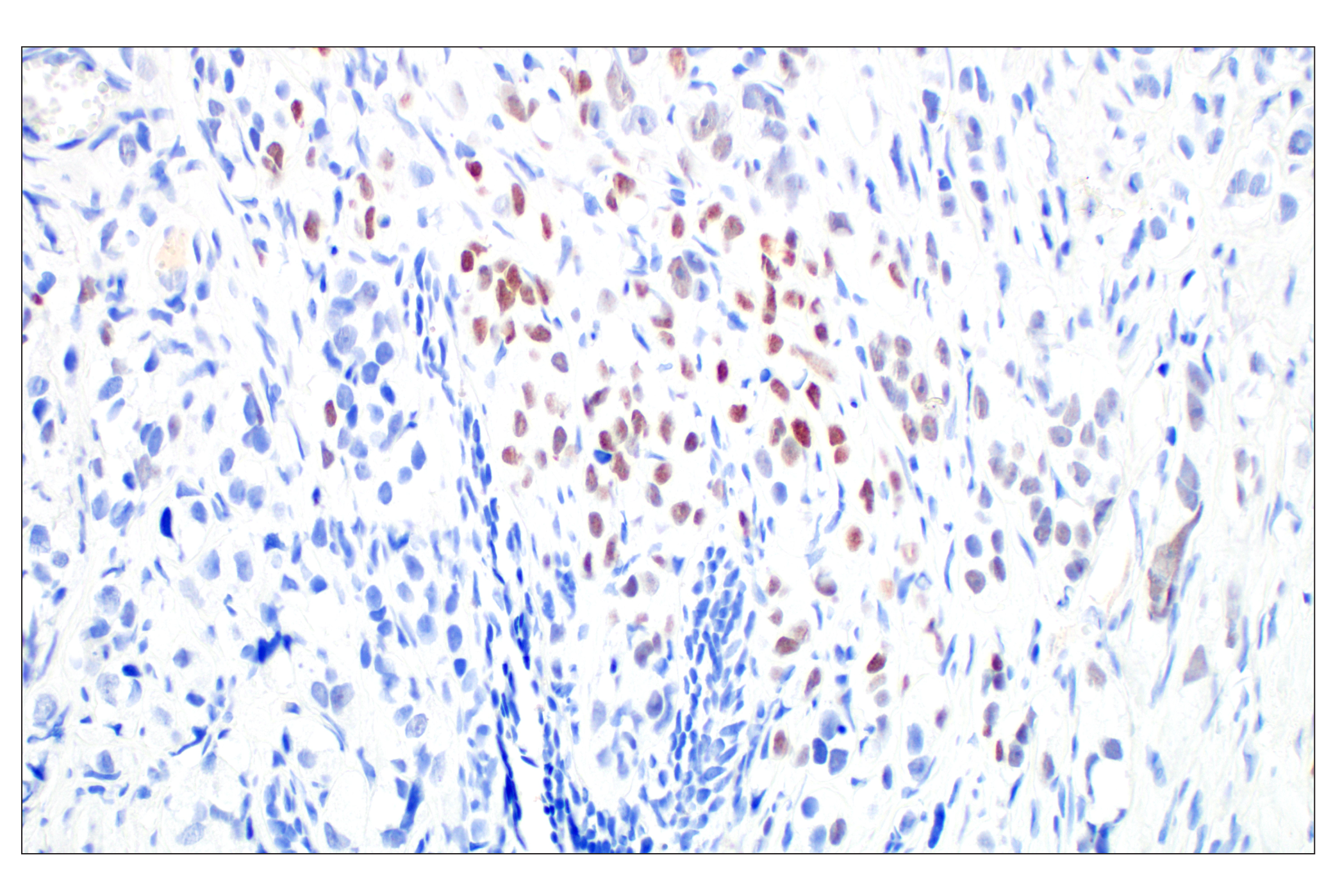
| p16 INK4A (E5F3Y) Rabbit mAb | 29271 | 20 µl | 16 kDa | Rabbit IgG |
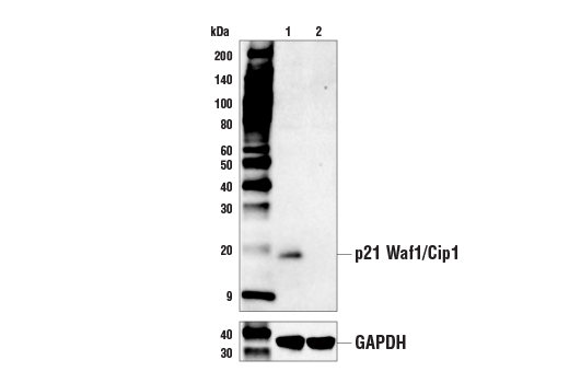
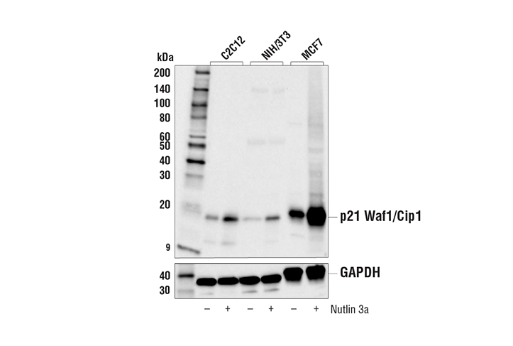
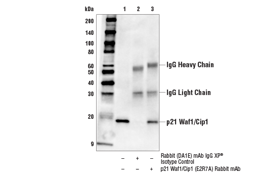
| p21 Waf1/Cip1 (E2R7A) Rabbit mAb | 37543 | 20 µl | 21 kDa | Rabbit IgG |
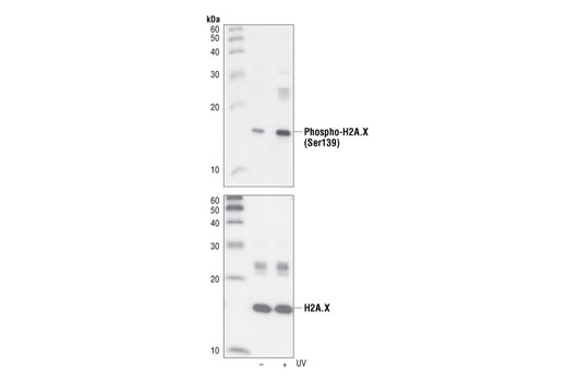
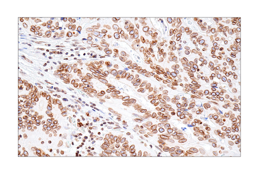
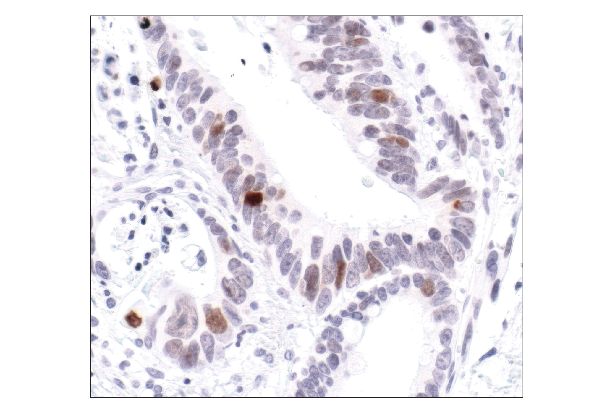
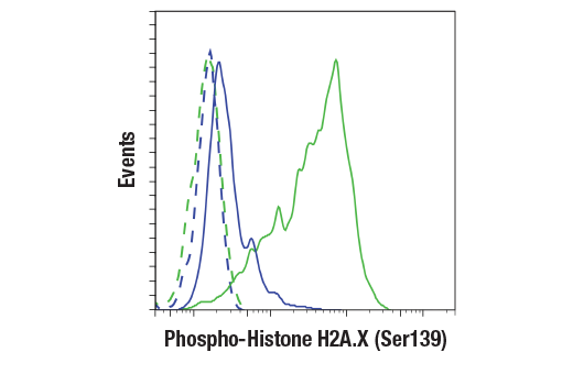
| Phospho-Histone H2A.X (Ser139) (20E3) Rabbit mAb | 9718 | 20 µl | 15 kDa | Rabbit IgG |
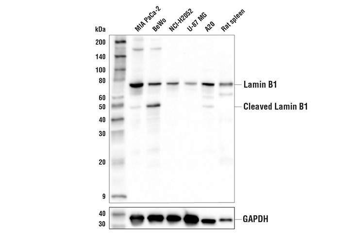
| Lamin B1 (E6M5T) Rabbit mAb | 17416 | 20 µl | 68, 45 kDa | Rabbit IgG |
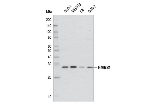
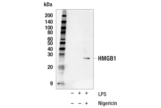
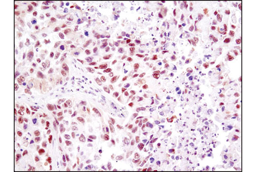
| HMGB1 (D3E5) Rabbit mAb | 6893 | 20 µl | 29 kDa | Rabbit IgG |
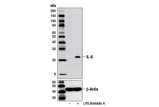
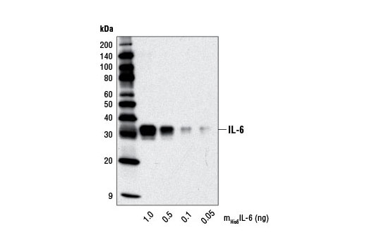
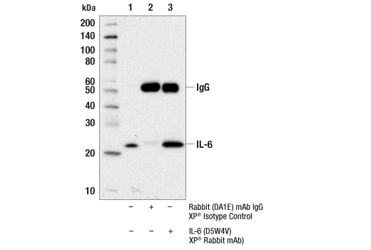
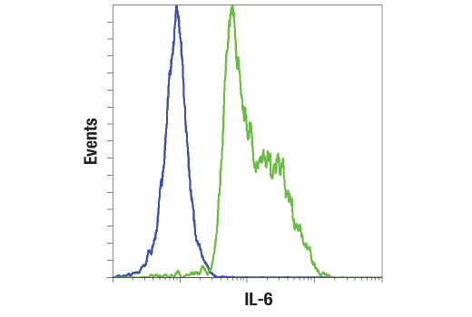
| IL-6 (D5W4V) XP® Rabbit mAb | 12912 | 20 µl | 24 kDa | Rabbit IgG |
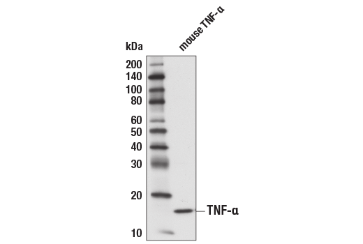
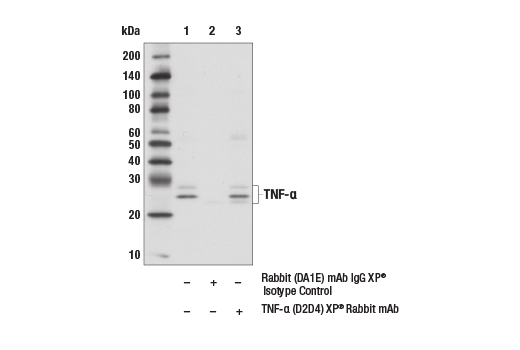
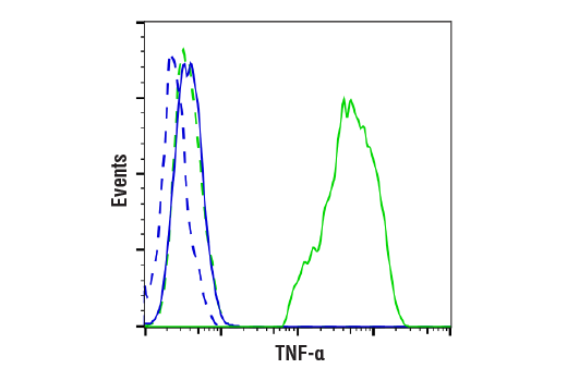
| TNF-α (D2D4) XP® Rabbit mAb | 11948 | 20 µl | 17,25,28 kDa | Rabbit IgG |
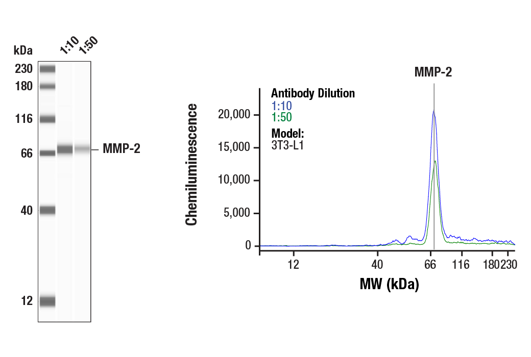
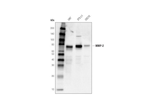
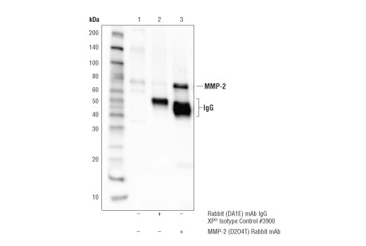
| MMP-2 (D2O4T) Rabbit mAb | 87809 | 20 µl | 64,72 kDa | Rabbit IgG |
| Anti-rabbit IgG, HRP-linked Antibody | 7074 | 100 µl | Goat |
Please visit cellsignal.com for individual component applications, species cross-reactivity, dilutions, protocols, and additional product information.
Description
The Mouse Reactive Senescence Marker Antibody Sampler Kit provides an economical means of detecting multiple markers of cellular senescence in mouse samples. The kit includes enough antibodies to perform two western blot experiments with each primary antibody.
Storage
Background
Senescence is characterized by stable stress-induced proliferative arrest and resistance to mitogenic stimuli, as well as the secretion of proteins such as cytokines, growth factors, and proteases. These secreted proteins comprise the senescence-associated secretory phenotype (SASP). Senescent cells are thought to accumulate as an organism ages, and contribute to age-related diseases, including cancer, through promotion of inflammation and disruption of normal cellular function (1,2).
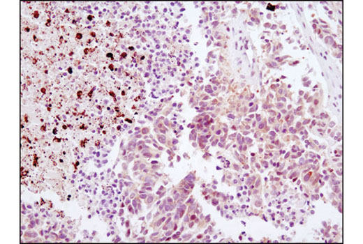
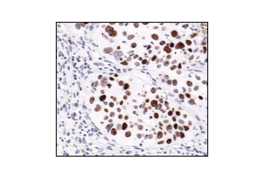
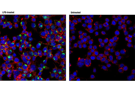
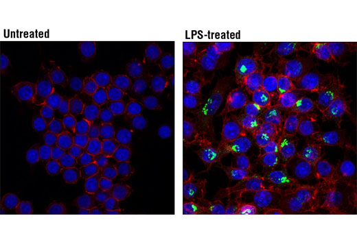
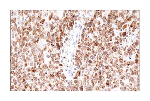
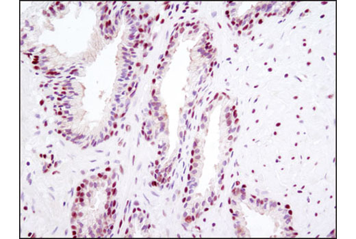
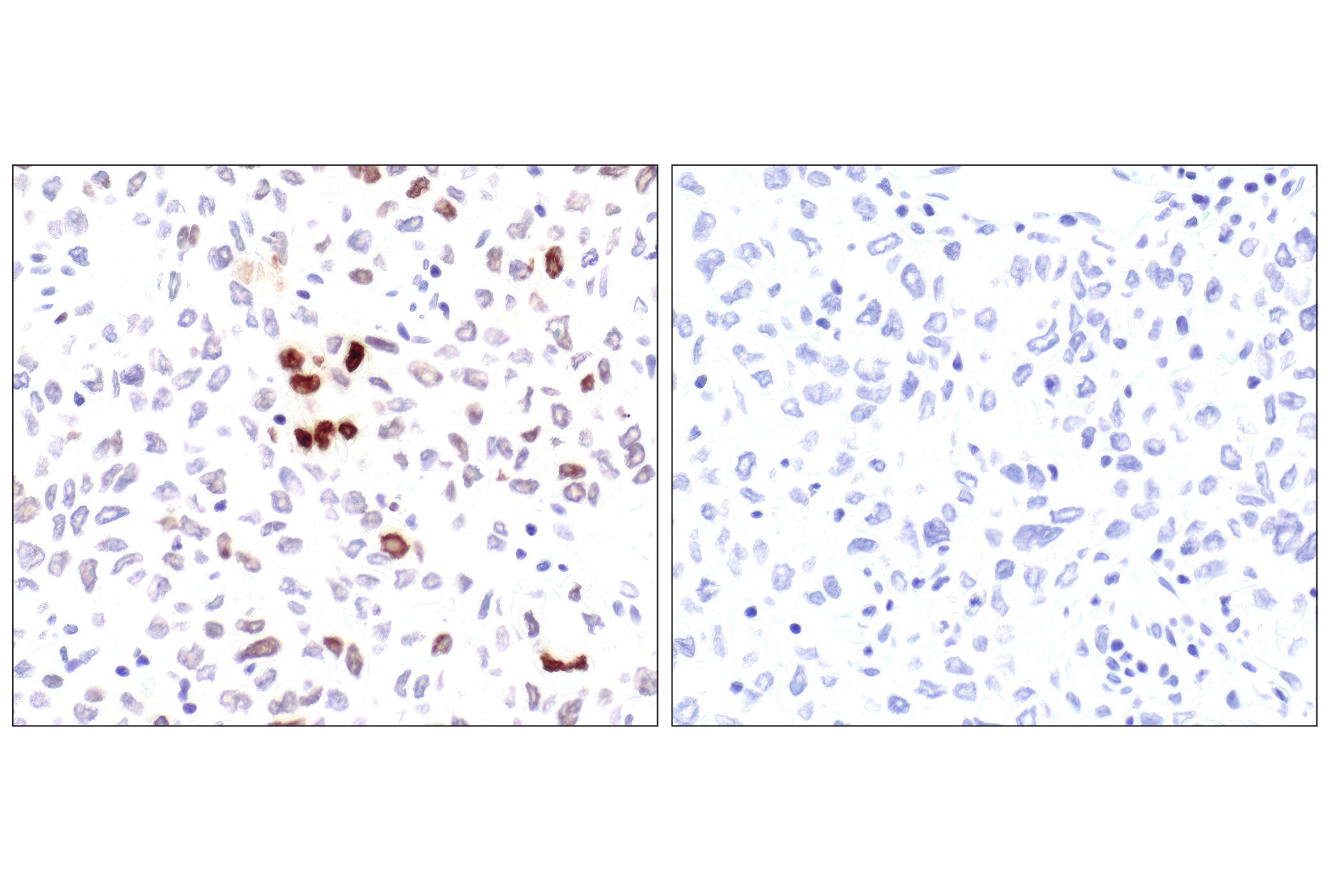
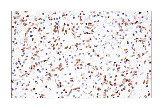
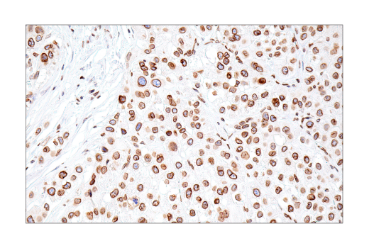
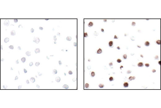
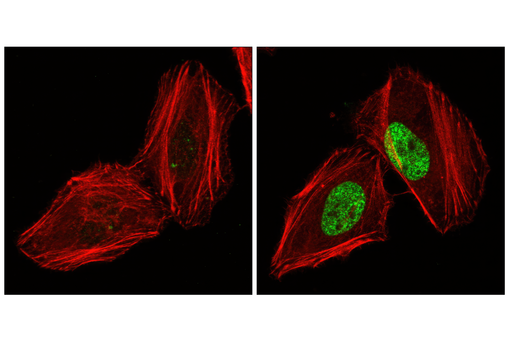
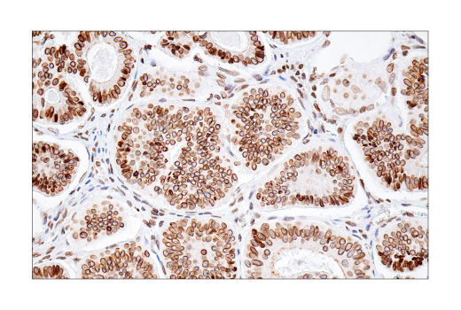
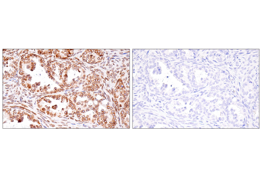
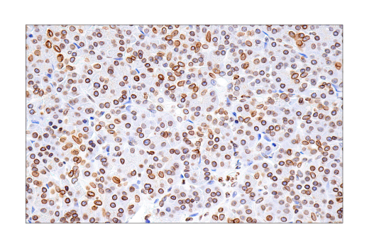
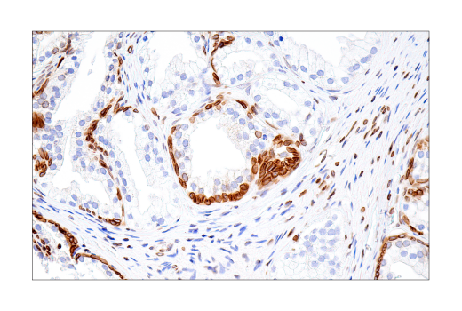
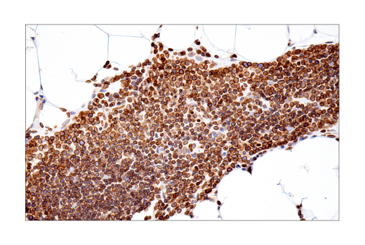
Because there is no single biomarker that can be used to definitively identify senescent cells, researchers must rely on a collection of biomarkers commonly associated with senescence (3). The Mouse Reactive Senescence Marker Antibody Sampler Kit provides a collection of antibodies to commonly used biomarkers of senescence-associated cell cycle arrest (p16 INK4A, p21 Waf1/Cip1), senescence-associated DNA damage (gamma-Histone H2A.X), and the SASP (HMGB1, IL-6, TNF-α, MMP2). The kit also includes an antibody to lamin B1, which is frequently reduced in senescent cells.
Background References
Trademarks and Patents
Limited Uses
Except as otherwise expressly agreed in a writing signed by a legally authorized representative of CST, the following terms apply to Products provided by CST, its affiliates or its distributors. Any Customer's terms and conditions that are in addition to, or different from, those contained herein, unless separately accepted in writing by a legally authorized representative of CST, are rejected and are of no force or effect.
Products are labeled with For Research Use Only or a similar labeling statement and have not been approved, cleared, or licensed by the FDA or other regulatory foreign or domestic entity, for any purpose. Customer shall not use any Product for any diagnostic or therapeutic purpose, or otherwise in any manner that conflicts with its labeling statement. Products sold or licensed by CST are provided for Customer as the end-user and solely for research and development uses. Any use of Product for diagnostic, prophylactic or therapeutic purposes, or any purchase of Product for resale (alone or as a component) or other commercial purpose, requires a separate license from CST. Customer shall (a) not sell, license, loan, donate or otherwise transfer or make available any Product to any third party, whether alone or in combination with other materials, or use the Products to manufacture any commercial products, (b) not copy, modify, reverse engineer, decompile, disassemble or otherwise attempt to discover the underlying structure or technology of the Products, or use the Products for the purpose of developing any products or services that would compete with CST products or services, (c) not alter or remove from the Products any trademarks, trade names, logos, patent or copyright notices or markings, (d) use the Products solely in accordance with CST Product Terms of Sale and any applicable documentation, and (e) comply with any license, terms of service or similar agreement with respect to any third party products or services used by Customer in connection with the Products.