WB, IP, IF-IC
H
Endogenous
85, 90
Rabbit IgG
#O75676
8986
Product Information
Product Usage Information
| Application | Dilution |
|---|---|
| Western Blotting | 1:1000 |
| Immunoprecipitation | 1:50 |
| Immunofluorescence (Immunocytochemistry) | 1:200 |
Storage
Specificity / Sensitivity
Species Reactivity:
Human
Source / Purification
Monoclonal antibody is produced by immunizing animals with a synthetic peptide corresponding to the region surrounding Pro751 of human MSK2.
Background
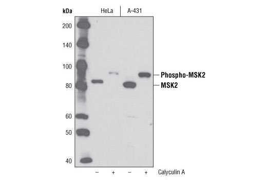
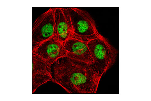
Mitogen- and stress-activated protein kinase 1 (MSK1) and MSK2 are serine/threonine kinases that promote immediate early gene transcription in stress- or mitogen-induced cells (1-4,7, 8) and LPS-stimulated macrophages (9). MSK2, also known as RSKB, contains two catalytic domains and has been shown to interact directly with p38 MAP kinase (10). MSK2 is phosphorylated and activated in response to tumor necrosis factor, epidermal growth factor or phorbol ester in HeLa cells or murine embryonic fibroblasts (MEFs) in a p38- and ERK-dependent manner (8,11). Phosphorylation on residues Ser196 and Thr568 within the activation loop of both catalytic domains is required for full kinase activation (11). Both MSK1 and MSK2 contain a functional nuclear localization sequence that is sufficient and required for nuclear targeting (10). Consistent with their nuclear localization, these kinases play an important role in regulating transcriptional responses to stress and mitogens. Activation of MSK2 in HeLa cells or MEFs results in rapid phosphorylation of histone H3, HMG-14, CREB and ATF1 and acetylation of histone H3 associated with immediate early gene transcription (3,4,6,7).
- Ananieva, O. et al. (2008) Nat Immunol 9, 1028-36.
- Sury, M.D. et al. (2006) Free Radic Biol Med 41, 1372-83.
- Duncan, E.A. et al. (2006) J Biol Chem 281, 12521-5.
- Darragh, J. et al. (2005) Biochem J 390, 749-59.
- Doehn, U. et al. (2004) Biochem J 382, 425-31.
- Davie, J.R. (2003) Sci STKE 2003, PE33.
- Soloaga, A. et al. (2003) EMBO J 22, 2788-97.
- Wiggin, G.R. et al. (2002) Mol Cell Biol 22, 2871-81.
- Caivano, M. and Cohen, P. (2000) J Immunol 164, 3018-25.
- Tomás-Zuber, M. et al. (2001) J Biol Chem 276, 5892-9.
- Tomás-Zuber, M. et al. (2000) J Biol Chem 275, 23549-58.
Species Reactivity
Species reactivity is determined by testing in at least one approved application (e.g., western blot).
Western Blot Buffer
IMPORTANT: For western blots, incubate membrane with diluted primary antibody in 5% w/v BSA, 1X TBS, 0.1% Tween® 20 at 4°C with gentle shaking, overnight.
Applications Key
WB: Western Blotting IP: Immunoprecipitation IF-IC: Immunofluorescence (Immunocytochemistry)
Cross-Reactivity Key
H: human M: mouse R: rat Hm: hamster Mk: monkey Vir: virus Mi: mink C: chicken Dm: D. melanogaster X: Xenopus Z: zebrafish B: bovine Dg: dog Pg: pig Sc: S. cerevisiae Ce: C. elegans Hr: horse GP: Guinea Pig Rab: rabbit All: all species expected
Trademarks and Patents
Limited Uses
Except as otherwise expressly agreed in a writing signed by a legally authorized representative of CST, the following terms apply to Products provided by CST, its affiliates or its distributors. Any Customer's terms and conditions that are in addition to, or different from, those contained herein, unless separately accepted in writing by a legally authorized representative of CST, are rejected and are of no force or effect.
Products are labeled with For Research Use Only or a similar labeling statement and have not been approved, cleared, or licensed by the FDA or other regulatory foreign or domestic entity, for any purpose. Customer shall not use any Product for any diagnostic or therapeutic purpose, or otherwise in any manner that conflicts with its labeling statement. Products sold or licensed by CST are provided for Customer as the end-user and solely for research and development uses. Any use of Product for diagnostic, prophylactic or therapeutic purposes, or any purchase of Product for resale (alone or as a component) or other commercial purpose, requires a separate license from CST. Customer shall (a) not sell, license, loan, donate or otherwise transfer or make available any Product to any third party, whether alone or in combination with other materials, or use the Products to manufacture any commercial products, (b) not copy, modify, reverse engineer, decompile, disassemble or otherwise attempt to discover the underlying structure or technology of the Products, or use the Products for the purpose of developing any products or services that would compete with CST products or services, (c) not alter or remove from the Products any trademarks, trade names, logos, patent or copyright notices or markings, (d) use the Products solely in accordance with CST Product Terms of Sale and any applicable documentation, and (e) comply with any license, terms of service or similar agreement with respect to any third party products or services used by Customer in connection with the Products.