| Product Includes | Product # | Quantity | Mol. Wt | Isotype/Source |
|---|---|---|---|---|
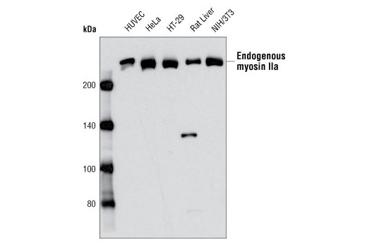
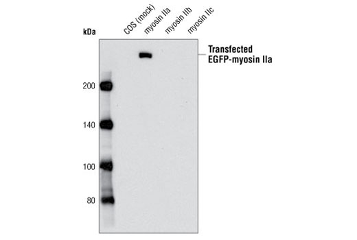
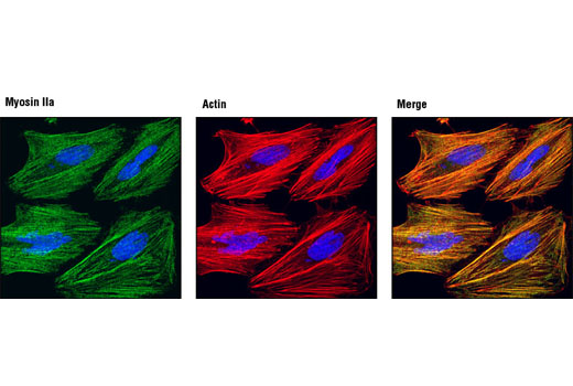
| Myosin IIa Antibody | 3403 | 40 µl | 230 kDa | Rabbit |
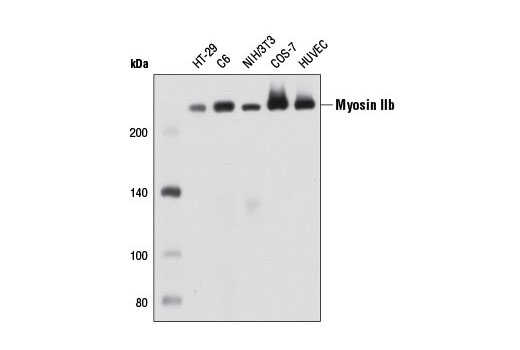
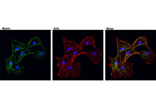
| Myosin IIb (D8H8) XP® Rabbit mAb | 8824 | 40 µl | 230 kDa | Rabbit IgG |
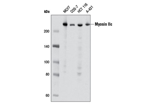
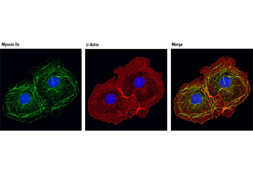
| Myosin IIc (D4A7) Rabbit mAb | 8189 | 40 µl | 230 kDa | Rabbit IgG |
| Anti-rabbit IgG, HRP-linked Antibody | 7074 | 100 µl | Goat |
Please visit cellsignal.com for individual component applications, species cross-reactivity, dilutions, protocols, and additional product information.
Description
The Myosin II Isoform Antibody Sampler Kit is an economical way to examine the total protein levels of myosin II isoforms a, b, and c. The kit includes enough primary and secondary antibodies to perform four Western blot experiments.
Storage
Background
Nonmuscle myosin is an actin-based motor protein essential to cell motility, cell division, migration, adhesion, and polarity. The holoenzyme consists of two identical heavy chains and two sets of light chains. The light chains (MLCs) regulate myosin II activity and stability. The heavy chains (NMHCs) are encoded by three genes, MYH9, MYH10, and MYH14, which generate three different nonmuscle myosin II isoforms, IIa, IIb, and IIc, respectively (reviewed in 1). While all three isoforms perform the same enzymatic tasks, binding to and contracting actin filaments coupled to ATP hydrolysis, their cellular functions do not appear to be redundant and they have different subcellular distributions (2-5). The carboxy-terminal tail domain of myosin II is important in isoform-specific subcellular localization (6). Research studies have shown that phosphorylation of myosin IIa at Ser1943 contributes to the regulation of breast cancer cell migration (7).
- Conti, M.A. and Adelstein, R.S. (2008) J Cell Sci 121, 11-18.
- Sandquist, J.C. et al. (2006) J Biol Chem 281, 35873-83.
- Even-Ram, S. et al. (2007) Nat Cell Biol 9, 299-309.
- Vicente-Manzanares, M. et al. (2007) J Cell Biol 176, 573-80.
- Wylie, S.R. and Chantler, P.D. (2008) Mol Biol Cell 19, 3956-68.
- Sandquist, J.C. and Means, A.R. (2008) Mol Biol Cell 19, 5156-67.
- Dulyaninova, N.G. et al. (2007) Mol Biol Cell 18, 3144-55.
Background References
Trademarks and Patents
Limited Uses
Except as otherwise expressly agreed in a writing signed by a legally authorized representative of CST, the following terms apply to Products provided by CST, its affiliates or its distributors. Any Customer's terms and conditions that are in addition to, or different from, those contained herein, unless separately accepted in writing by a legally authorized representative of CST, are rejected and are of no force or effect.
Products are labeled with For Research Use Only or a similar labeling statement and have not been approved, cleared, or licensed by the FDA or other regulatory foreign or domestic entity, for any purpose. Customer shall not use any Product for any diagnostic or therapeutic purpose, or otherwise in any manner that conflicts with its labeling statement. Products sold or licensed by CST are provided for Customer as the end-user and solely for research and development uses. Any use of Product for diagnostic, prophylactic or therapeutic purposes, or any purchase of Product for resale (alone or as a component) or other commercial purpose, requires a separate license from CST. Customer shall (a) not sell, license, loan, donate or otherwise transfer or make available any Product to any third party, whether alone or in combination with other materials, or use the Products to manufacture any commercial products, (b) not copy, modify, reverse engineer, decompile, disassemble or otherwise attempt to discover the underlying structure or technology of the Products, or use the Products for the purpose of developing any products or services that would compete with CST products or services, (c) not alter or remove from the Products any trademarks, trade names, logos, patent or copyright notices or markings, (d) use the Products solely in accordance with CST Product Terms of Sale and any applicable documentation, and (e) comply with any license, terms of service or similar agreement with respect to any third party products or services used by Customer in connection with the Products.