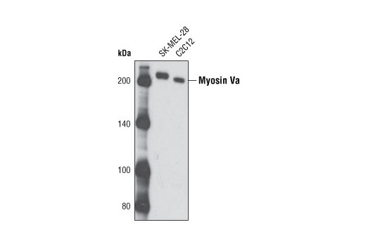
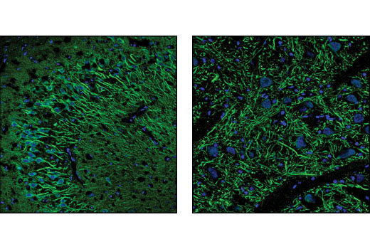
WB, IP, IF-F
H M R
Endogenous
207
Rabbit
#Q99104
17918
Product Information
Product Usage Information
| Application | Dilution |
|---|---|
| Western Blotting | 1:1000 |
| Immunoprecipitation | 1:100 |
| Immunofluorescence (Frozen) | 1:50 |
Storage
Specificity / Sensitivity
Species Reactivity:
Human, Mouse, Rat
Species predicted to react based on 100% sequence homology
The antigen sequence used to produce this antibody shares
100% sequence homology with the species listed here, but
reactivity has not been tested or confirmed to work by CST.
Use of this product with these species is not covered under
our
Product Performance Guarantee.
Monkey, Chicken, Pig
Source / Purification
Polyclonal antibodies are produced by immunizing animals with a synthetic peptide corresponding to residues near the carboxy terminus of human myosin Va.
Background
Myosin Va is a molecular motor protein involved in the transport of organelles, vesicles and other cellular cargo along actin filaments (reviewed in 1). The molecule consists of two identical heavy chains, which dimerize via helical domains in a coiled coil structure. The amino-terminal motor domains of the heavy chains contain both the ATPase and the actin-binding activities of myosin Va. The globular tail domains act in a regulatory capacity, binding the myosin Va cargo (2) or inhibiting motor activity by binding the head domains and preventing ATP consumption (3). Mutation of the murine dilute gene, which encodes myosin Va, causes defects in coat pigmentation as well as severe neurological defects (4). In melanocytes, the coiled coil structure of myosin Va is important in regulating the trafficking of melanosomes in conjunction with melanophilin and Rab27a (5). Myosin Va regulates trafficking and exocytosis of secretory granules in neuroendocrine cells (reviewed in 6) as well as RNA transport and distribution (7).
- Desnos, C. et al. (2007) Biol Cell 99, 411-23.
- Wu, X. et al. (1997) J Cell Sci 110 ( Pt 7), 847-59.
- Li, X.D. et al. (2006) J Biol Chem 281, 21789-98.
- Mercer, J.A. et al. (1991) Nature 349, 709-13.
- Hume, A.N. et al. (2006) Mol Biol Cell 17, 4720-35.
- Eichler, T.W. et al. (2006) Biochem Soc Trans 34, 671-4.
- Salerno, V.P. et al. (2008) Cell Motil Cytoskeleton 65, 422-33.
Species Reactivity
Species reactivity is determined by testing in at least one approved application (e.g., western blot).
Western Blot Buffer
IMPORTANT: For western blots, incubate membrane with diluted primary antibody in 5% w/v BSA, 1X TBS, 0.1% Tween® 20 at 4°C with gentle shaking, overnight.
Applications Key
WB: Western Blotting IP: Immunoprecipitation IF-F: Immunofluorescence (Frozen)
Cross-Reactivity Key
H: human M: mouse R: rat Hm: hamster Mk: monkey Vir: virus Mi: mink C: chicken Dm: D. melanogaster X: Xenopus Z: zebrafish B: bovine Dg: dog Pg: pig Sc: S. cerevisiae Ce: C. elegans Hr: horse GP: Guinea Pig Rab: rabbit All: all species expected
Trademarks and Patents
Limited Uses
Except as otherwise expressly agreed in a writing signed by a legally authorized representative of CST, the following terms apply to Products provided by CST, its affiliates or its distributors. Any Customer's terms and conditions that are in addition to, or different from, those contained herein, unless separately accepted in writing by a legally authorized representative of CST, are rejected and are of no force or effect.
Products are labeled with For Research Use Only or a similar labeling statement and have not been approved, cleared, or licensed by the FDA or other regulatory foreign or domestic entity, for any purpose. Customer shall not use any Product for any diagnostic or therapeutic purpose, or otherwise in any manner that conflicts with its labeling statement. Products sold or licensed by CST are provided for Customer as the end-user and solely for research and development uses. Any use of Product for diagnostic, prophylactic or therapeutic purposes, or any purchase of Product for resale (alone or as a component) or other commercial purpose, requires a separate license from CST. Customer shall (a) not sell, license, loan, donate or otherwise transfer or make available any Product to any third party, whether alone or in combination with other materials, or use the Products to manufacture any commercial products, (b) not copy, modify, reverse engineer, decompile, disassemble or otherwise attempt to discover the underlying structure or technology of the Products, or use the Products for the purpose of developing any products or services that would compete with CST products or services, (c) not alter or remove from the Products any trademarks, trade names, logos, patent or copyright notices or markings, (d) use the Products solely in accordance with CST Product Terms of Sale and any applicable documentation, and (e) comply with any license, terms of service or similar agreement with respect to any third party products or services used by Customer in connection with the Products.