WB, IP, IHC-P, IF-IC, ChIP
H
Endogenous
270
Rabbit IgG
#O75376
9611
Product Information
Product Usage Information
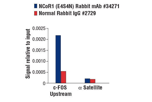
For optimal ChIP and ChIP-seq results, use 10 μl of antibody and 10 μg of chromatin (approximately 4 × 10^6 cells) per IP. This antibody has been validated using SimpleChIP® Enzymatic Chromatin IP Kits.
| Application | Dilution |
|---|---|
| Western Blotting | 1:1000 |
| Immunoprecipitation | 1:200 |
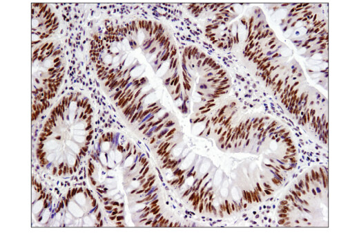
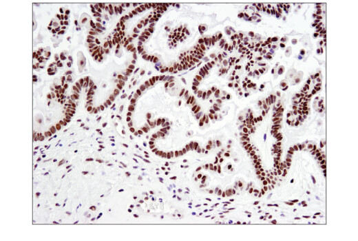
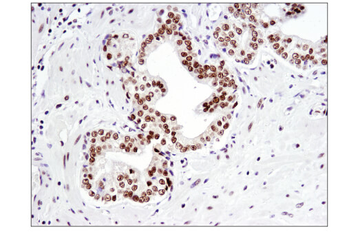
| Immunohistochemistry (Paraffin) | 1:100 |
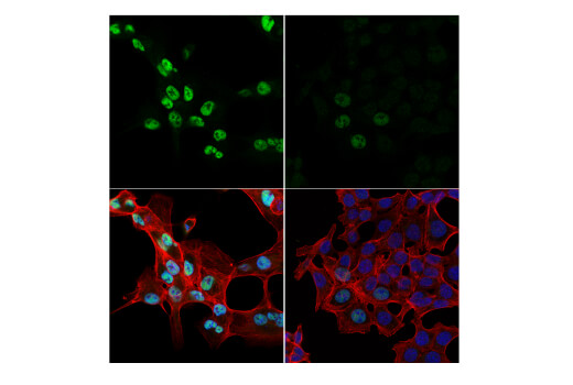
| Immunofluorescence (Immunocytochemistry) | 1:800 |
| Chromatin IP | 1:50 |
Storage
Specificity / Sensitivity
Species Reactivity:
Human
Source / Purification
Monoclonal antibody is produced by immunizing animals with recombinant protein specific to the carboxy terminus of human NCoR1 protein.
Background
The most well characterized nuclear receptor corepressors are SMRT (silencing mediator for retinoic acid and thyroid hormone receptors) and its close paralog NCoR1 (nuclear receptor corepressor) (1,2). NCoR1 functions to transcriptionally silence various unliganded, DNA bound non-steroidal nuclear receptors by serving as a large molecular scaffold that bridges the receptors with multiple chromatin remodeling factors that repress nuclear receptor-mediated gene transcription, in part, through deacetylation of core histones surrounding target promoters. Indeed, the N-terminal portion of NCoR1 possesses multiple distinct transcriptional repression domains (RDs) reponsible for the recruitment of additional components of the corepressor complex such as HDACs, mSin3, GPS2, and TBL1/TBLR1. In between the RDs lies a pair of potent repressor motifs known as SANT motifs (SWI3, ADA2, N-CoR, and TFIIIB), which recruit HDAC3 and histones to the repressor complex in order to enhance HDAC3 activity (3). The C-terminal portion of NCoR1 contains multiple nuclear receptor interaction domains (NDs), each of which contains a conserved CoRNR box (or L/I-X-X-I/V-I) motif that allow for binding to various unliganded nuclear hormone receptors such as thyroid hormone (THR) and retinoic acid (RAR) receptors (4,5).
Recent genetic studies in mice have not only corroborated the wealth of biochemical studies involving NCoR1 but have also provided significant insight regarding the function of NCoR1 in mammalian development and physiology. Although it has been observed that loss of Ncor1 does not affect early embyonic development, likely due to compensation by Smrt, embryonic lethality ultimately results during mid-gestation, largely due to defects in erythropoesis and thymopoesis (6). Another study demonstrated that the NDs of NCoR1 are critical for its ability to function in a physiological setting as a transcriptional repressor of hepatic THR and Liver X Receptor (LXR) (7).
- Chen, J.D. and Evans, R.M. (1995) Nature 377, 454-7.
- Hörlein, A.J. et al. (1995) Nature 377, 397-404.
- Jones, P.L. and Shi, Y.B. (2003) Curr Top Microbiol Immunol 274, 237-68.
- Downes, M. et al. (1996) Nucleic Acids Res 24, 4379-86.
- Wong, C.W. and Privalsky, M.L. (1998) Mol Cell Biol 18, 5724-33.
- Jepsen, K. et al. (2000) Cell 102, 753-63.
- Astapova, I. et al. (2008) Proc Natl Acad Sci U S A 105, 19544-9.
Species Reactivity
Species reactivity is determined by testing in at least one approved application (e.g., western blot).
Western Blot Buffer
IMPORTANT: For western blots, incubate membrane with diluted primary antibody in 5% w/v BSA, 1X TBS, 0.1% Tween® 20 at 4°C with gentle shaking, overnight.
Applications Key
WB: Western Blotting IP: Immunoprecipitation IHC-P: Immunohistochemistry (Paraffin) IF-IC: Immunofluorescence (Immunocytochemistry) ChIP: Chromatin IP
Cross-Reactivity Key
H: human M: mouse R: rat Hm: hamster Mk: monkey Vir: virus Mi: mink C: chicken Dm: D. melanogaster X: Xenopus Z: zebrafish B: bovine Dg: dog Pg: pig Sc: S. cerevisiae Ce: C. elegans Hr: horse GP: Guinea Pig Rab: rabbit All: all species expected
Trademarks and Patents
Limited Uses
Except as otherwise expressly agreed in a writing signed by a legally authorized representative of CST, the following terms apply to Products provided by CST, its affiliates or its distributors. Any Customer's terms and conditions that are in addition to, or different from, those contained herein, unless separately accepted in writing by a legally authorized representative of CST, are rejected and are of no force or effect.
Products are labeled with For Research Use Only or a similar labeling statement and have not been approved, cleared, or licensed by the FDA or other regulatory foreign or domestic entity, for any purpose. Customer shall not use any Product for any diagnostic or therapeutic purpose, or otherwise in any manner that conflicts with its labeling statement. Products sold or licensed by CST are provided for Customer as the end-user and solely for research and development uses. Any use of Product for diagnostic, prophylactic or therapeutic purposes, or any purchase of Product for resale (alone or as a component) or other commercial purpose, requires a separate license from CST. Customer shall (a) not sell, license, loan, donate or otherwise transfer or make available any Product to any third party, whether alone or in combination with other materials, or use the Products to manufacture any commercial products, (b) not copy, modify, reverse engineer, decompile, disassemble or otherwise attempt to discover the underlying structure or technology of the Products, or use the Products for the purpose of developing any products or services that would compete with CST products or services, (c) not alter or remove from the Products any trademarks, trade names, logos, patent or copyright notices or markings, (d) use the Products solely in accordance with CST Product Terms of Sale and any applicable documentation, and (e) comply with any license, terms of service or similar agreement with respect to any third party products or services used by Customer in connection with the Products.