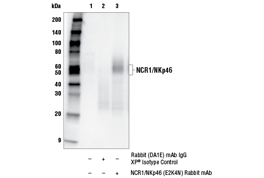
WB, IP
M
Endogenous
50-60
Rabbit IgG
#Q8C567
17086
Product Information
Product Usage Information
| Application | Dilution |
|---|---|
| Western Blotting | 1:1000 |
| Immunoprecipitation | 1:100 |
Storage
Specificity / Sensitivity
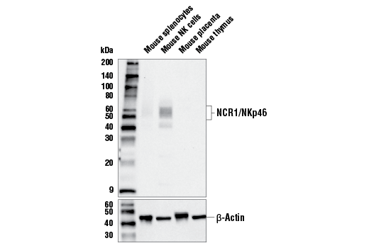
Species Reactivity:
Mouse
Source / Purification
Monoclonal antibody is produced by immunizing animals with recombinant protein specific to the amino terminus of mouse NCR1/NKp46 protein.
Background
The family of natural cytotoxicity receptors (NCRs) contains NCR1 (NKp46/CD335), NCR2 (NKp44/CD336), and NCR3 (NKp30/CD337). They are single-pass type I transmembrane proteins belonging to the immunoglobin (Ig) superfamily. Various pathogen-derived as well as host-encoded molecules have been identified as ligands for the NCRs. They were originally identified by their ability to evoke the cytotoxicity of natural killer (NK) cells towards tumor cells in vitro and animal models have demonstrated that NCRs play a role in vivo in tumor surveillance, viral infection, and pregnancy (1-5).
NCR1/NKp46 is considered a universal marker for NK cells, and recent studies found that it is also expressed by other cells, such as group 1 innate lymphoid cells (ILC1), a subset of group 3 ILCs (NCR+ ILC3), and γδ T cells (2-4). NCR1/NKp46 is also expressed on and considered as a diagnostic marker and therapeutic target for some malignant NK, NKT, and T cell lymphomas (6,7). Cross-linking NCR1/NKp46 with antibodies can activate NK cells and this has been pursued as a promising therapeutic avenue (8).
- Sivori, S. et al. (1997) J Exp Med 186, 1129-36.
- Kruse, P.H. et al. (2014) Immunol Cell Biol 92, 221-9.
- Pazina, T. et al. (2017) Front Immunol 8, 369.
- Barrow, A.D. et al. (2019) Front Immunol 10, 909.
- Meza Guzman, L.G. et al. (2020) Cancers (Basel) 12, 952.
- Laribi, K. et al. (2018) Recent Pat Anticancer Drug Discov 13, 308-40.
- Cheminant, M. et al. (2019) Gut 68, 1396-405.
- Gauthier, L. et al. (2019) Cell 177, 1701-1713.e16.
Species Reactivity
Species reactivity is determined by testing in at least one approved application (e.g., western blot).
Western Blot Buffer
IMPORTANT: For western blots, incubate membrane with diluted primary antibody in 5% w/v nonfat dry milk, 1X TBS, 0.1% Tween® 20 at 4°C with gentle shaking, overnight.
Applications Key
WB: Western Blotting IP: Immunoprecipitation
Cross-Reactivity Key
H: human M: mouse R: rat Hm: hamster Mk: monkey Vir: virus Mi: mink C: chicken Dm: D. melanogaster X: Xenopus Z: zebrafish B: bovine Dg: dog Pg: pig Sc: S. cerevisiae Ce: C. elegans Hr: horse GP: Guinea Pig Rab: rabbit All: all species expected
Trademarks and Patents
Limited Uses
Except as otherwise expressly agreed in a writing signed by a legally authorized representative of CST, the following terms apply to Products provided by CST, its affiliates or its distributors. Any Customer's terms and conditions that are in addition to, or different from, those contained herein, unless separately accepted in writing by a legally authorized representative of CST, are rejected and are of no force or effect.
Products are labeled with For Research Use Only or a similar labeling statement and have not been approved, cleared, or licensed by the FDA or other regulatory foreign or domestic entity, for any purpose. Customer shall not use any Product for any diagnostic or therapeutic purpose, or otherwise in any manner that conflicts with its labeling statement. Products sold or licensed by CST are provided for Customer as the end-user and solely for research and development uses. Any use of Product for diagnostic, prophylactic or therapeutic purposes, or any purchase of Product for resale (alone or as a component) or other commercial purpose, requires a separate license from CST. Customer shall (a) not sell, license, loan, donate or otherwise transfer or make available any Product to any third party, whether alone or in combination with other materials, or use the Products to manufacture any commercial products, (b) not copy, modify, reverse engineer, decompile, disassemble or otherwise attempt to discover the underlying structure or technology of the Products, or use the Products for the purpose of developing any products or services that would compete with CST products or services, (c) not alter or remove from the Products any trademarks, trade names, logos, patent or copyright notices or markings, (d) use the Products solely in accordance with CST Product Terms of Sale and any applicable documentation, and (e) comply with any license, terms of service or similar agreement with respect to any third party products or services used by Customer in connection with the Products.