| Product Includes | Product # | Quantity | Mol. Wt | Isotype/Source |
|---|---|---|---|---|
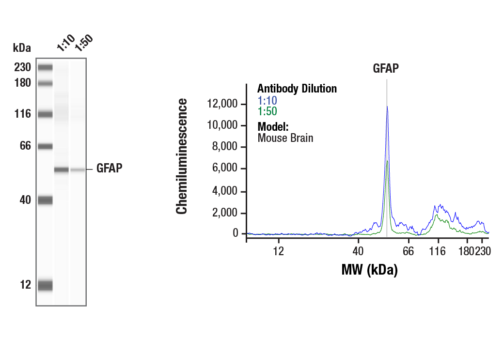
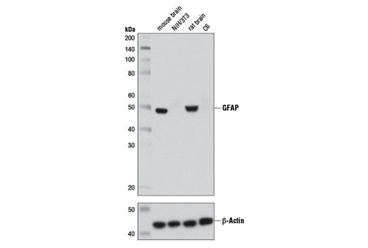
| GFAP (D1F4Q) XP® Rabbit mAb | 12389 | 20 µl | 50 kDa | Rabbit IgG |
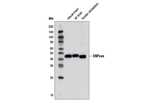
| CNPase (D83E10) XP® Rabbit mAb | 5664 | 20 µl | 47 kDa | Rabbit IgG |
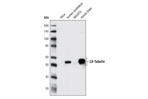
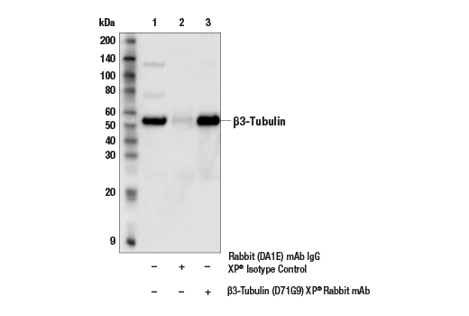
| β3-Tubulin (D71G9) XP® Rabbit mAb | 5568 | 20 µl | 55 kDa | Rabbit IgG |
| Nestin (Rat-401) Mouse mAb | 4760 | 20 µl | Mouse IgG1 | |
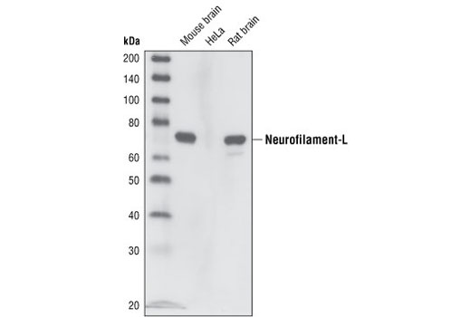
| Neurofilament-L (C28E10) Rabbit mAb | 2837 | 20 µl | 70 kDa | Rabbit IgG |
Please visit cellsignal.com for individual component applications, species cross-reactivity, dilutions, protocols, and additional product information.
Description
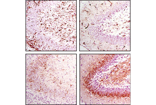
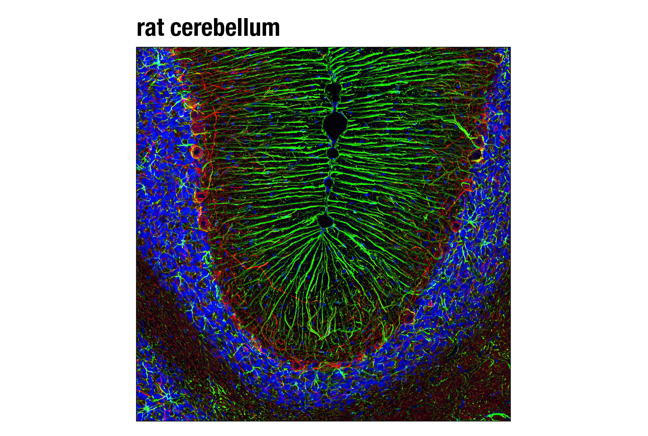
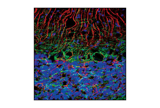
The Neuronal Marker IF Antibody Sampler Kit provides an economical means for labeling neuronal structures by immunofluorescence (IF-F). This kit includes enough primary antibody to perform at least forty IF-F tests or two western blot experiments per primary antibody.
Storage
Background
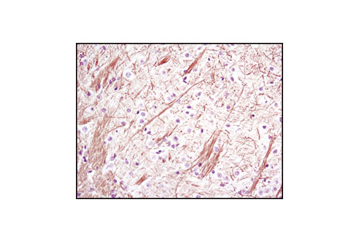
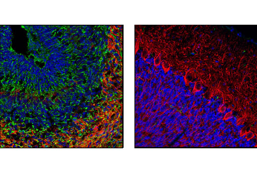
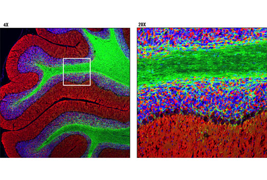
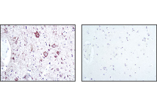
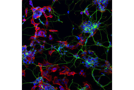
The antibodies in this kit serve as neuronal markers to determine protein localization in neurons. The cytoskeleton consists of three types of cytosolic fibers: microfilaments (actin filaments), intermediate filaments, and microtubules. Neurofilaments are the major intermediate filaments found in neurons and consist of light (NFL), medium (NFM), and heavy (NFH) subunits (1). Nestin is an intermediate filament family member protein that is structurally related to the neurofilament proteins (2). Globular tubulin subunits comprise the microtubule building block, with α/β-tubulin heterodimers forming the tubulin subunit common to all eukaryotic cells (3). High CNPase expression is seen in oligodendrocytes and Schwann cells as CNPase accounts for roughly 4% of the total myelin protein in the central nervous system (4). CNPase binds to tubulin heterodimers and plays a role in tubulin polymerization and oligodendrocyte process outgrowth (5). GFAP filaments are characteristic of differentiated and mature brain astrocytes. Thus, GFAP is commonly used by investigators as a marker for intracranial and intraspinal tumors arising from astrocytes (6).
- Al-Chalabi, A. and Miller, C.C. (2003) Bioessays 25, 346-55.
- Michalczyk, K. and Ziman, M. (2005) Histol Histopathol 20, 665-71.
- Westermann, S. and Weber, K. (2003) Nat Rev Mol Cell Biol 4, 938-47.
- Kozlov, G. et al. (2003) J Biol Chem 278, 46021-8.
- Lee, J. et al. (2005) J Cell Biol 170, 661-73.
- Goebel, H.H. et al. (1987) Acta Histochem Suppl 34, 81-93.
Background References
Trademarks and Patents
Limited Uses
Except as otherwise expressly agreed in a writing signed by a legally authorized representative of CST, the following terms apply to Products provided by CST, its affiliates or its distributors. Any Customer's terms and conditions that are in addition to, or different from, those contained herein, unless separately accepted in writing by a legally authorized representative of CST, are rejected and are of no force or effect.
Products are labeled with For Research Use Only or a similar labeling statement and have not been approved, cleared, or licensed by the FDA or other regulatory foreign or domestic entity, for any purpose. Customer shall not use any Product for any diagnostic or therapeutic purpose, or otherwise in any manner that conflicts with its labeling statement. Products sold or licensed by CST are provided for Customer as the end-user and solely for research and development uses. Any use of Product for diagnostic, prophylactic or therapeutic purposes, or any purchase of Product for resale (alone or as a component) or other commercial purpose, requires a separate license from CST. Customer shall (a) not sell, license, loan, donate or otherwise transfer or make available any Product to any third party, whether alone or in combination with other materials, or use the Products to manufacture any commercial products, (b) not copy, modify, reverse engineer, decompile, disassemble or otherwise attempt to discover the underlying structure or technology of the Products, or use the Products for the purpose of developing any products or services that would compete with CST products or services, (c) not alter or remove from the Products any trademarks, trade names, logos, patent or copyright notices or markings, (d) use the Products solely in accordance with CST Product Terms of Sale and any applicable documentation, and (e) comply with any license, terms of service or similar agreement with respect to any third party products or services used by Customer in connection with the Products.