| Product Includes | Product # | Quantity | Mol. Wt | Isotype/Source |
|---|---|---|---|---|
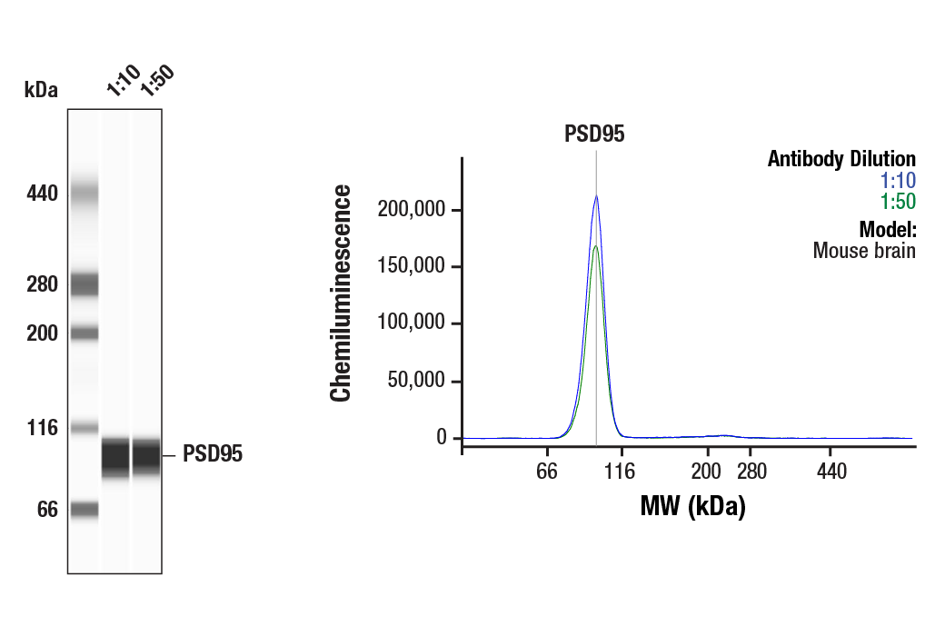
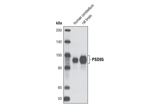
| PSD95 (D27E11) XP® Rabbit mAb | 3450 | 40 µl | 95 kDa | Rabbit IgG |
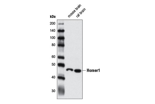
| Homer1 Antibody | 8231 | 40 µl | 46 kDa | Rabbit |
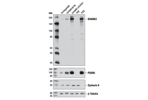
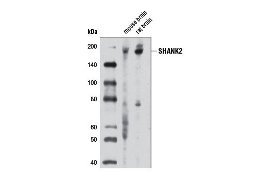
| SHANK2 Antibody | 12218 | 40 µl | 165 kDa | Rabbit |
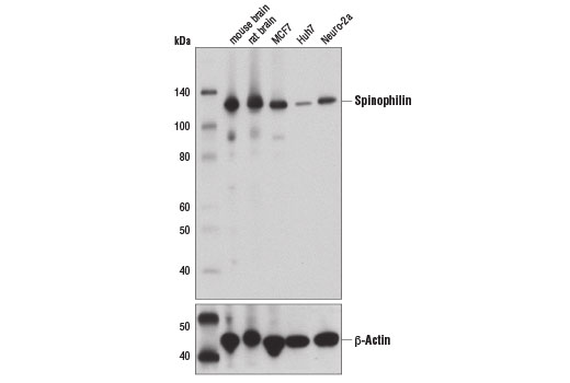
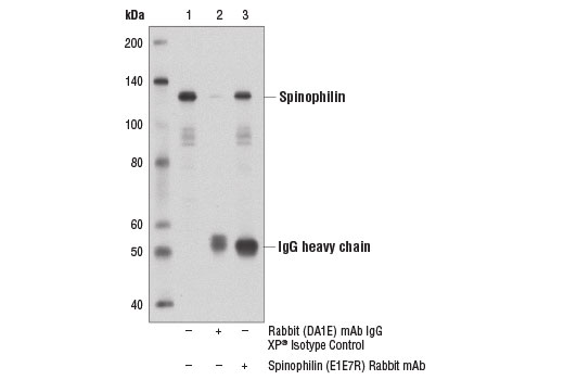
| Spinophilin (E1E7R) Rabbit mAb | 14136 | 40 µl | 130 kDa | Rabbit IgG |
| Anti-rabbit IgG, HRP-linked Antibody | 7074 | 100 µl | Goat |
Please visit cellsignal.com for individual component applications, species cross-reactivity, dilutions, protocols, and additional product information.
Description
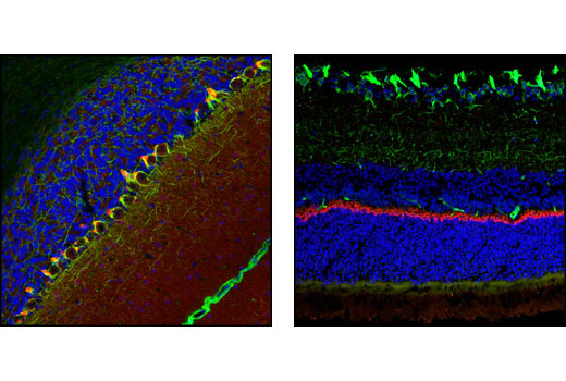
The Neuronal Scaffold Proteins Antibody Sampler Kit provides an economical means of evaluating four major scaffolding proteins. The kit includes enough primary antibody to perform four western blot experiments.
Storage
Background
Scaffold proteins are composed of protein-interaction domains that tether multiple components of a signaling pathway to form signal transduction complexes. This organization of signaling molecules can help enhance signaling specificity and speed. Scaffold proteins are central components in neuronal synapses, where dynamic trafficking of synaptic proteins occurs. Mutations in scaffold proteins could have significant impact on synaptic structure and function. Postsynaptic density protein 95 (PSD95) is a member of the membrane-associated guanylate kinase (MAGUK) family of proteins and a scaffolding protein involved in the assembly and function of the postsynaptic density complex (1,2). SHANK proteins act as scaffolds at the neuronal post-synaptic density (PSD), where they play a critical role in PSD assembly of excitatory synapses during development (3,4). While recruitment of SHANK proteins to the synapse is independent of their interaction with Homer (5), proper synaptic targeting of SHANK1 is mediated by interactions between its PDZ domain and PSD proteins (6). Homer proteins (1-3) are scaffolds, composed of an EVH protein–binding domain, a coiled-coil domain and a leucine zipper domain. The EVH domain is a protein-protein binding module that binds to the proline-rich motifs of G-protein–coupled receptors (GPCRs), inositol 1,4,5-triphosphate (IP3) receptors (IP3Rs), ryanodine receptors, and TRP channels (7,8). The coiled-coil and the leucine zipper domains cause multimerization of Homers and assemble signaling proteins complexes. Spinophilin is a protein phosphatase 1 regulatory protein that interacts with a large number of proteins, including ion channel components and G-protein-coupled receptors (GPCRs). Spinophilin also interacts with actin filaments; phosphorylation of spinophilin at Ser94 and Ser177 disrupts this interaction (9,10).
- Cao, J. et al. (2005) J Cell Biol 168, 117-26.
- Chetkovich, D.M. et al. (2002) J Neurosci 22, 6415-25.
- Grabrucker, A.M. et al. (2011) Trends Cell Biol 21, 594-603.
- Boeckers, T.M. et al. (1999) J Neurosci 19, 6506-18.
- Boeckers, T.M. et al. (2005) J Neurochem 92, 519-24.
- Sala, C. et al. (2001) Neuron 31, 115-30.
- Fagni, L. et al. (2002) Sci STKE 2002, re8.
- Yuan, J.P. et al. (2003) Cell 114, 777-89.
- Sarrouilhe, D. et al. (2006) Biochimie 88, 1099-113.
- Hsieh-Wilson, L.C. et al. (2003) J Biol Chem 278, 1186-94.
Background References
Trademarks and Patents
Limited Uses
Except as otherwise expressly agreed in a writing signed by a legally authorized representative of CST, the following terms apply to Products provided by CST, its affiliates or its distributors. Any Customer's terms and conditions that are in addition to, or different from, those contained herein, unless separately accepted in writing by a legally authorized representative of CST, are rejected and are of no force or effect.
Products are labeled with For Research Use Only or a similar labeling statement and have not been approved, cleared, or licensed by the FDA or other regulatory foreign or domestic entity, for any purpose. Customer shall not use any Product for any diagnostic or therapeutic purpose, or otherwise in any manner that conflicts with its labeling statement. Products sold or licensed by CST are provided for Customer as the end-user and solely for research and development uses. Any use of Product for diagnostic, prophylactic or therapeutic purposes, or any purchase of Product for resale (alone or as a component) or other commercial purpose, requires a separate license from CST. Customer shall (a) not sell, license, loan, donate or otherwise transfer or make available any Product to any third party, whether alone or in combination with other materials, or use the Products to manufacture any commercial products, (b) not copy, modify, reverse engineer, decompile, disassemble or otherwise attempt to discover the underlying structure or technology of the Products, or use the Products for the purpose of developing any products or services that would compete with CST products or services, (c) not alter or remove from the Products any trademarks, trade names, logos, patent or copyright notices or markings, (d) use the Products solely in accordance with CST Product Terms of Sale and any applicable documentation, and (e) comply with any license, terms of service or similar agreement with respect to any third party products or services used by Customer in connection with the Products.