WB
H M R Mk
Endogenous
78
Rabbit
#Q9Y5A7
51667
Product Information
Product Usage Information
| Application | Dilution |
|---|---|
| Western Blotting | 1:1000 |
Storage
Specificity / Sensitivity
Species Reactivity:
Human, Mouse, Rat, Monkey
Source / Purification
Polyclonal antibodies are produced by immunizing animals with a synthetic peptide corresponding to residues near the carboxy terminus of human NUB1 protein. Antibodies are purified by protein A and peptide affinity chromatography.
Background
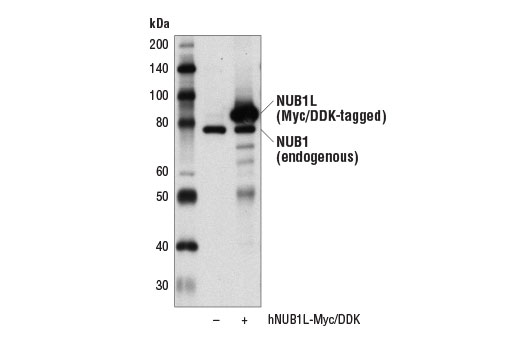
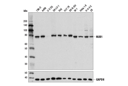
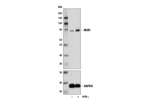
Both the NEDD8 ultimate buster 1 (NUB1) and the related NUB1L isoform are interferon-inducible adaptor proteins that negatively regulate ubiquitin-like protein NEDD8 (1,2). NUB1 protein contains an amino terminal ubiquitin-like (UBL) domain and multiple carboxy terminal ubiquitin-associated (UBA) domains. The NUB1L isoform is generated by alternative splicing and contains an extra UBA domain relative to NUB1 (2). Research studies indicate that NUB1 and NUB1L non-covalently bind NEDD8 and facilitate delivery of both NEDD8 monomers and NEDD8 conjugates to the proteasome for degradation (2-5). In addition, NUB1L binds and enhances the proteasomal degradation of the FAT10 ubiquitin-like protein (6). Additional research shows that NUB1 negatively regulates cell proliferation, likely due to inhibition of NEDD8 conjugation to SCF ubiquitin ligases, which leads to inhibition of p27 and cyclin E ubiquitination (3,7). NUB1 has been identified as a putative therapeutic target in Huntington's disease as NUB1 promotes a decrease in levels of mutant HTT protein (8).
- Kamitani, T. et al. (2001) J Biol Chem 276, 46655-60.
- Tanaka, T. et al. (2003) J Biol Chem 278, 32905-13.
- Kito, K. et al. (2001) J Biol Chem 276, 20603-9.
- Tanji, K. et al. (2005) Biochem Biophys Res Commun 337, 116-20.
- Liu, S. et al. (2013) J Biol Chem 288, 31339-49.
- Hipp, M.S. et al. (2004) J Biol Chem 279, 16503-10.
- Hosono, T. et al. (2010) Br J Cancer 102, 873-82.
- Lu, B. et al. (2013) Nat Neurosci 16, 562-70.
Species Reactivity
Species reactivity is determined by testing in at least one approved application (e.g., western blot).
Western Blot Buffer
IMPORTANT: For western blots, incubate membrane with diluted primary antibody in 5% w/v BSA, 1X TBS, 0.1% Tween® 20 at 4°C with gentle shaking, overnight.
Applications Key
WB: Western Blotting
Cross-Reactivity Key
H: human M: mouse R: rat Hm: hamster Mk: monkey Vir: virus Mi: mink C: chicken Dm: D. melanogaster X: Xenopus Z: zebrafish B: bovine Dg: dog Pg: pig Sc: S. cerevisiae Ce: C. elegans Hr: horse GP: Guinea Pig Rab: rabbit All: all species expected
Trademarks and Patents
Limited Uses
Except as otherwise expressly agreed in a writing signed by a legally authorized representative of CST, the following terms apply to Products provided by CST, its affiliates or its distributors. Any Customer's terms and conditions that are in addition to, or different from, those contained herein, unless separately accepted in writing by a legally authorized representative of CST, are rejected and are of no force or effect.
Products are labeled with For Research Use Only or a similar labeling statement and have not been approved, cleared, or licensed by the FDA or other regulatory foreign or domestic entity, for any purpose. Customer shall not use any Product for any diagnostic or therapeutic purpose, or otherwise in any manner that conflicts with its labeling statement. Products sold or licensed by CST are provided for Customer as the end-user and solely for research and development uses. Any use of Product for diagnostic, prophylactic or therapeutic purposes, or any purchase of Product for resale (alone or as a component) or other commercial purpose, requires a separate license from CST. Customer shall (a) not sell, license, loan, donate or otherwise transfer or make available any Product to any third party, whether alone or in combination with other materials, or use the Products to manufacture any commercial products, (b) not copy, modify, reverse engineer, decompile, disassemble or otherwise attempt to discover the underlying structure or technology of the Products, or use the Products for the purpose of developing any products or services that would compete with CST products or services, (c) not alter or remove from the Products any trademarks, trade names, logos, patent or copyright notices or markings, (d) use the Products solely in accordance with CST Product Terms of Sale and any applicable documentation, and (e) comply with any license, terms of service or similar agreement with respect to any third party products or services used by Customer in connection with the Products.