| Product Includes | Product # | Quantity | Mol. Wt | Isotype/Source |
|---|---|---|---|---|
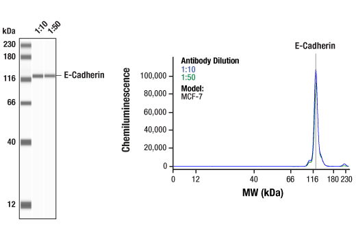
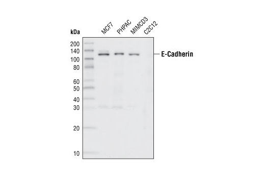
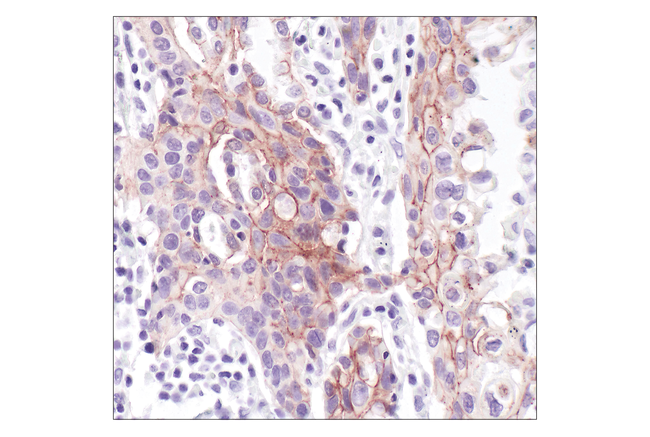
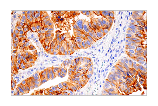
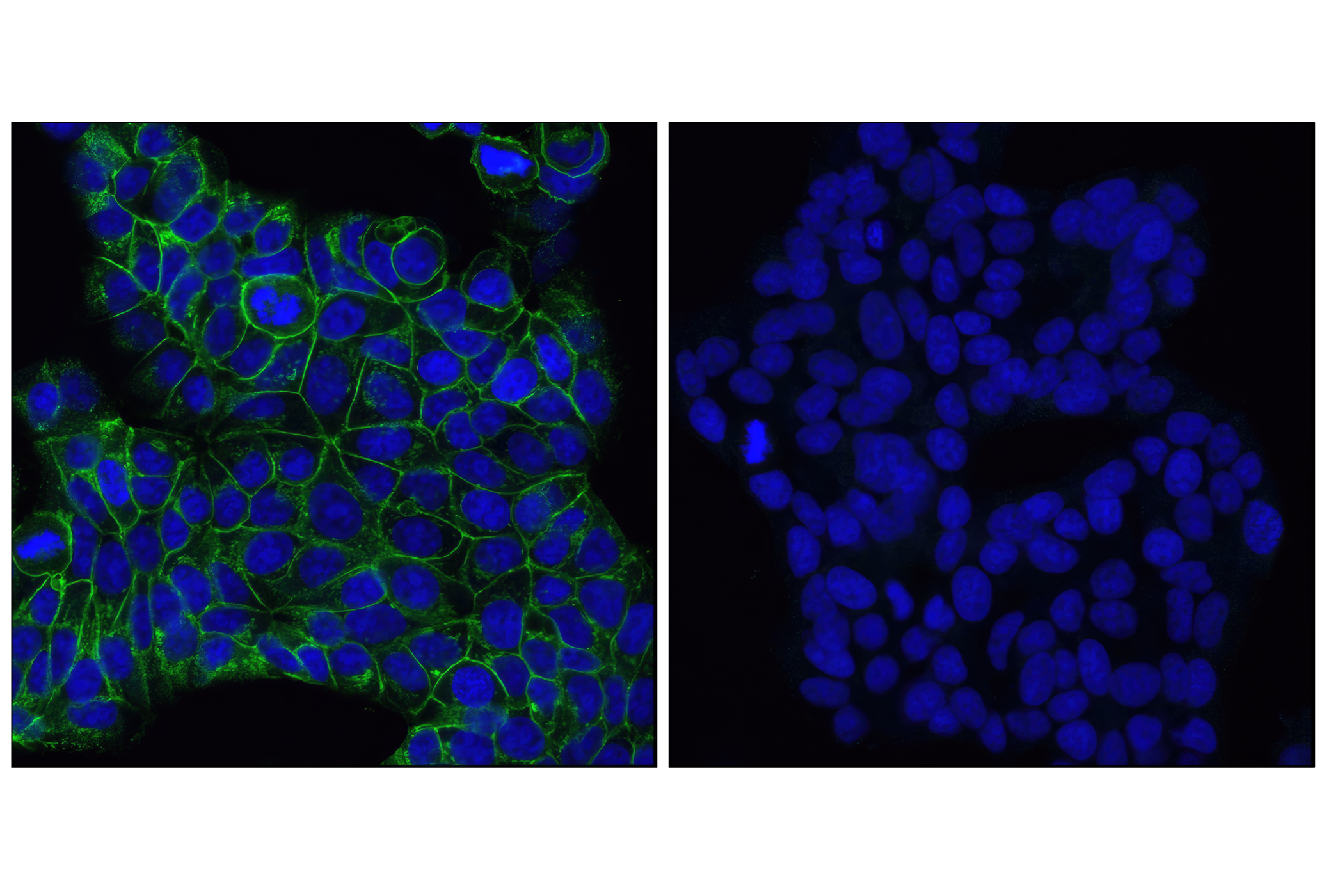
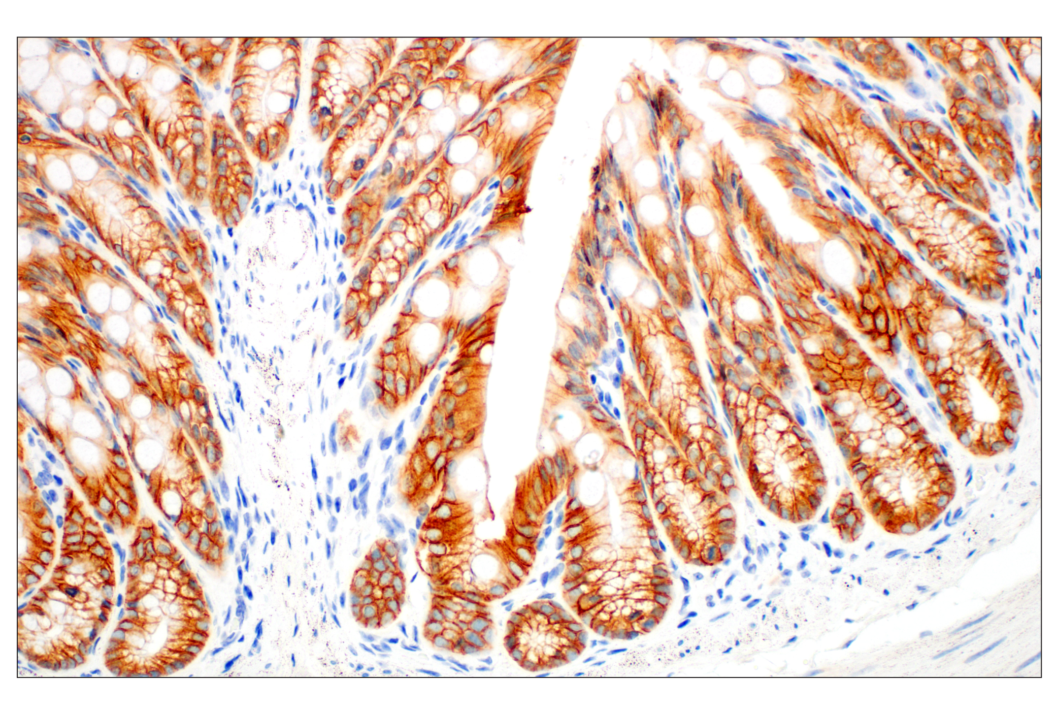
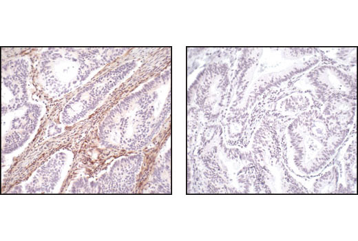
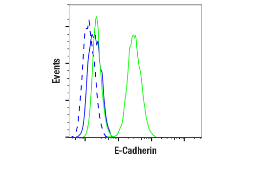
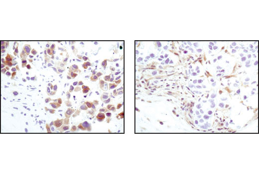
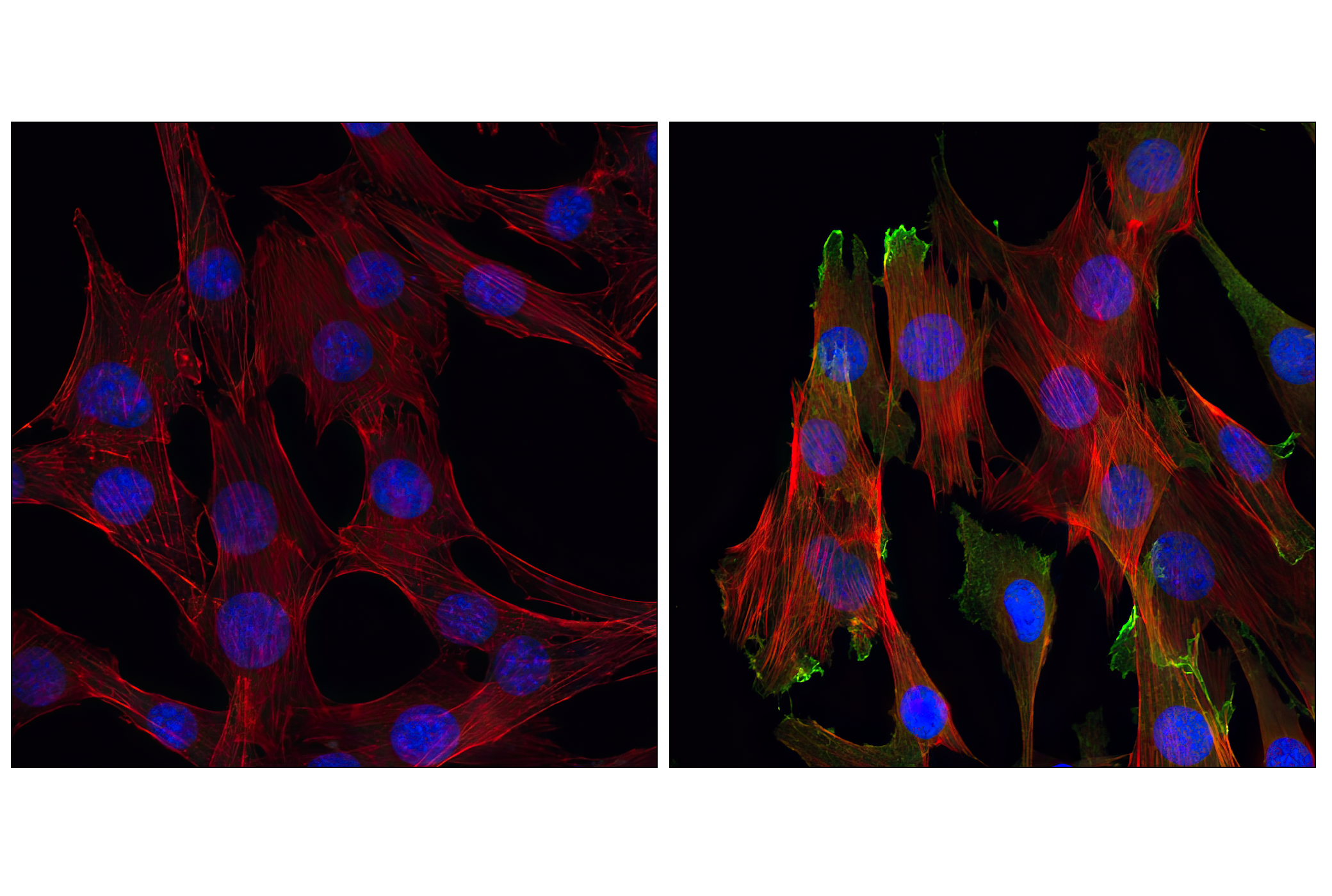
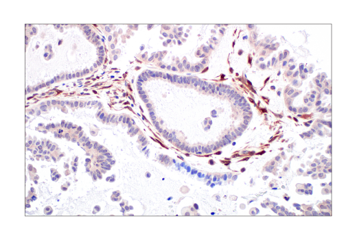
| E-Cadherin (24E10) Rabbit mAb | 3195 | 20 µl | 135 kDa | Rabbit IgG |
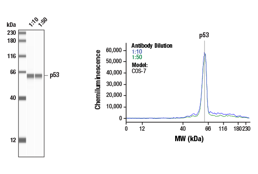
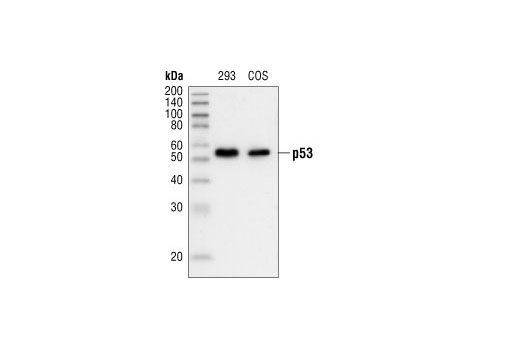
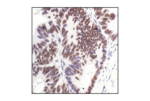
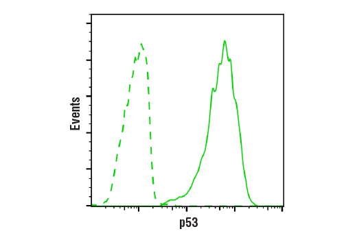
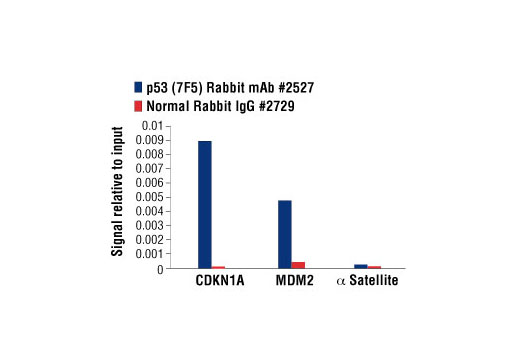
| p53 (7F5) Rabbit mAb | 2527 | 20 µl | 53 kDa | Rabbit IgG |
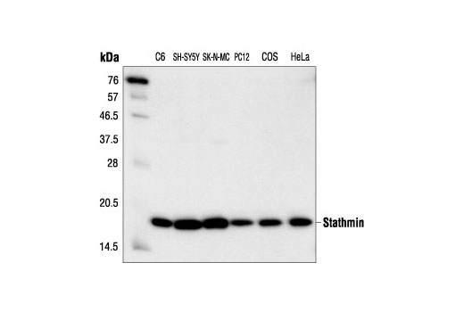
| Stathmin Antibody | 3352 | 20 µl | 19 kDa | Rabbit |
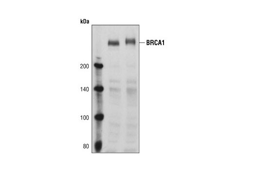
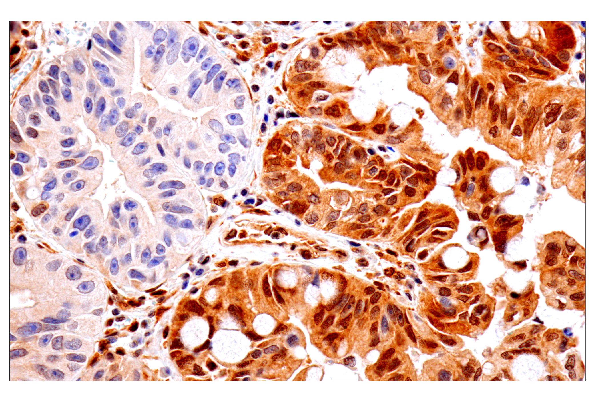
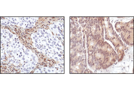
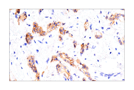
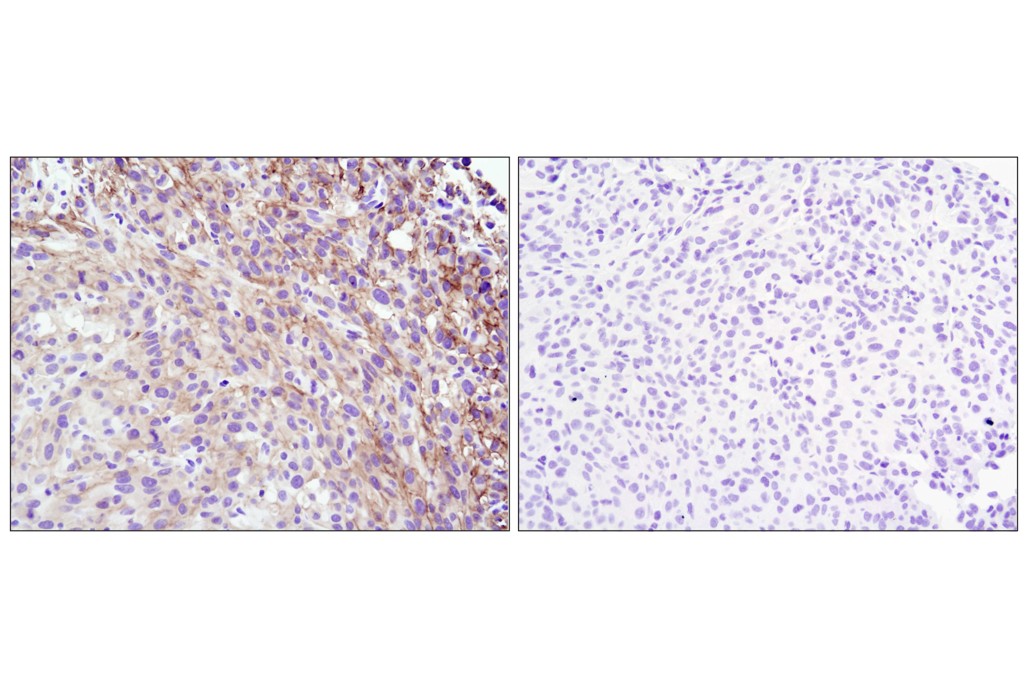
| BRCA1 Antibody | 9010 | 20 µl | 220 kDa | Rabbit |
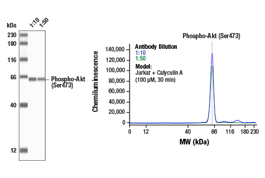
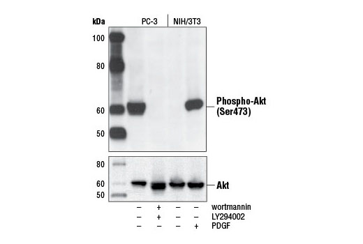
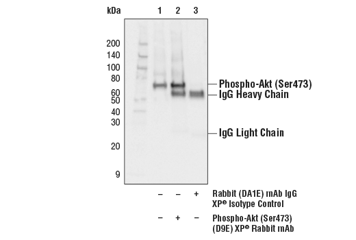
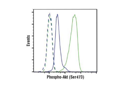
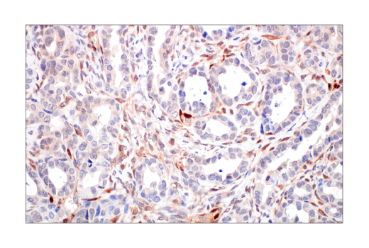
| Phospho-Akt (Ser473) (D9E) XP® Rabbit mAb | 4060 | 20 µl | 60 kDa | Rabbit IgG |
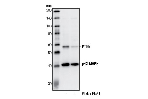
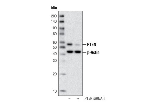
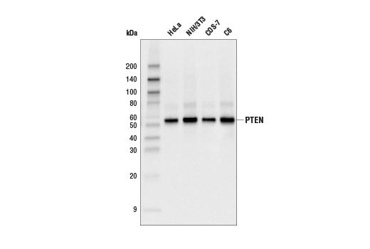
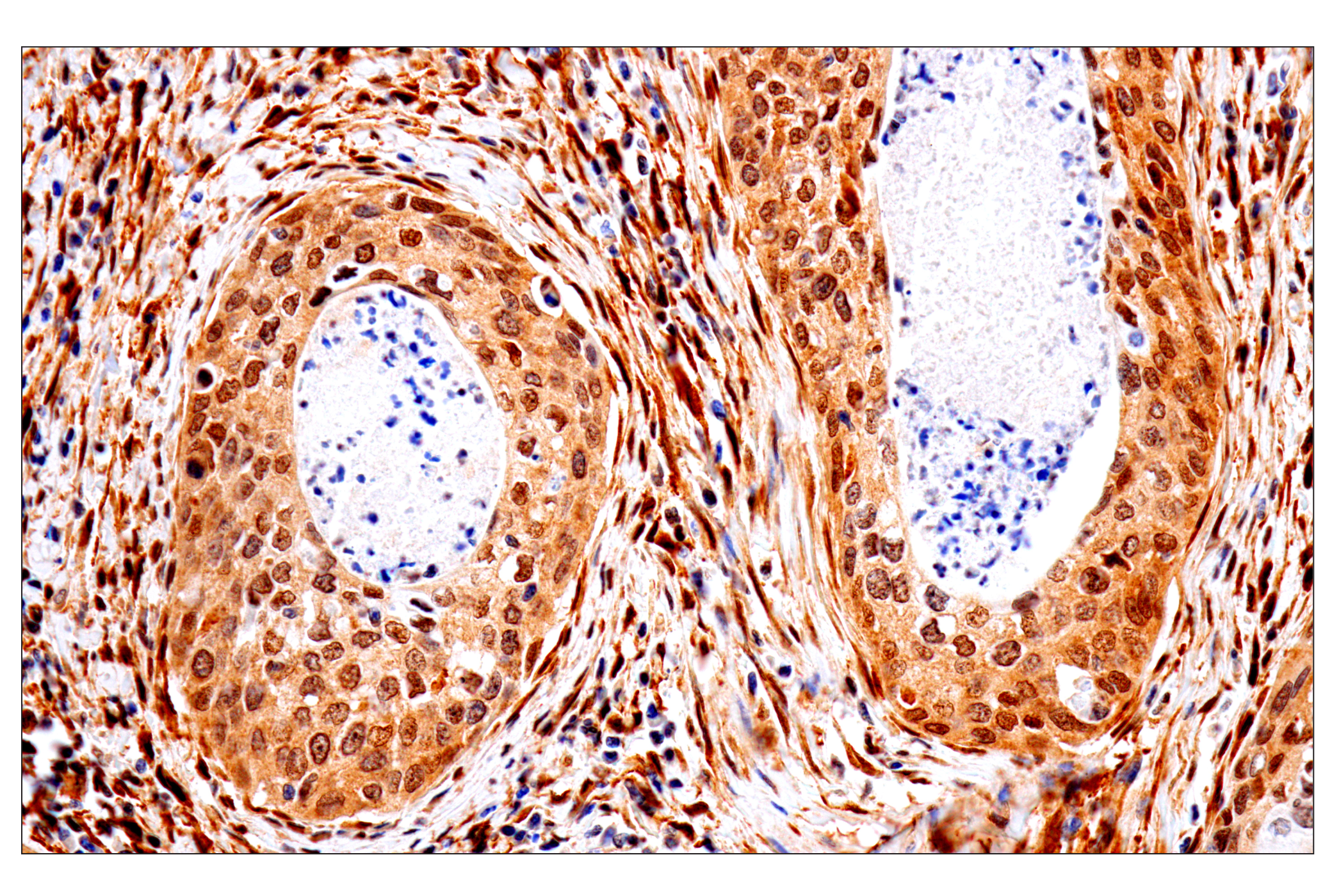
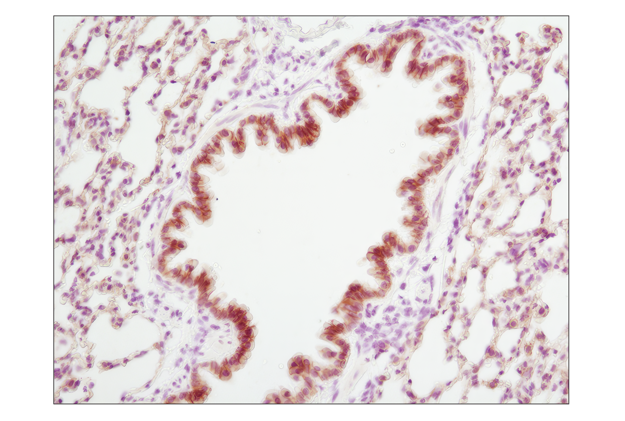
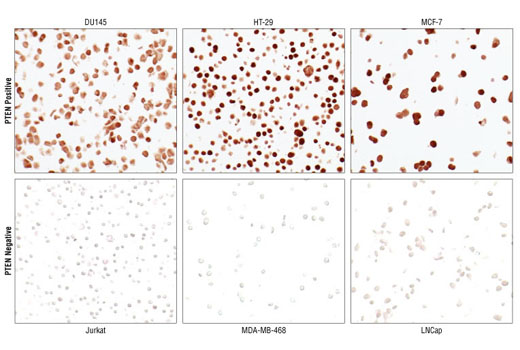
| PTEN (138G6) Rabbit mAb | 9559 | 20 µl | 54 kDa | Rabbit IgG |
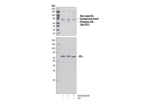
| Phospho-Estrogen Receptor α (Ser167) (D1A3) Rabbit mAb | 5587 | 20 µl | 66 kDa | Rabbit IgG |
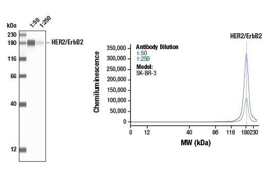
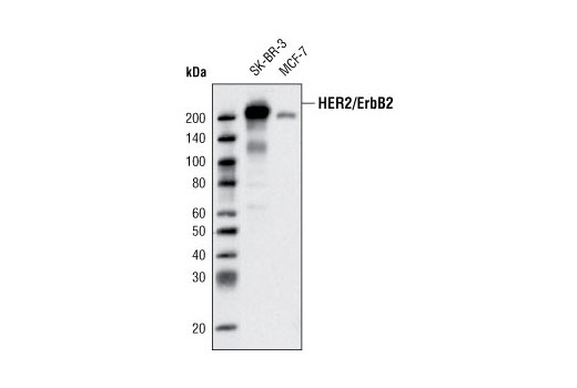
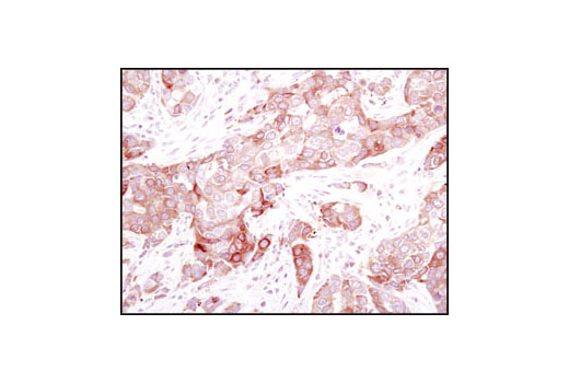
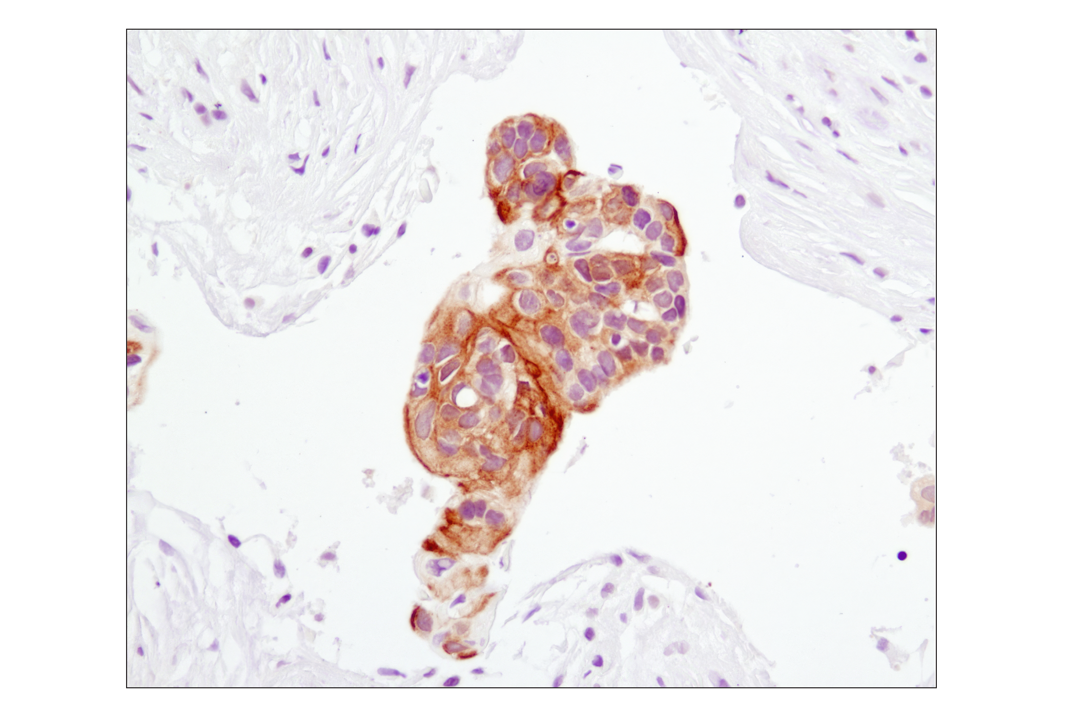
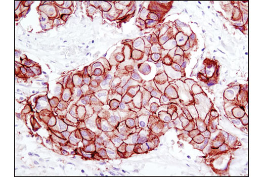
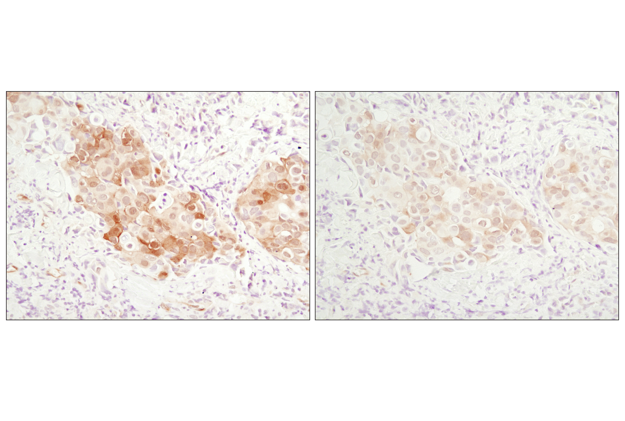
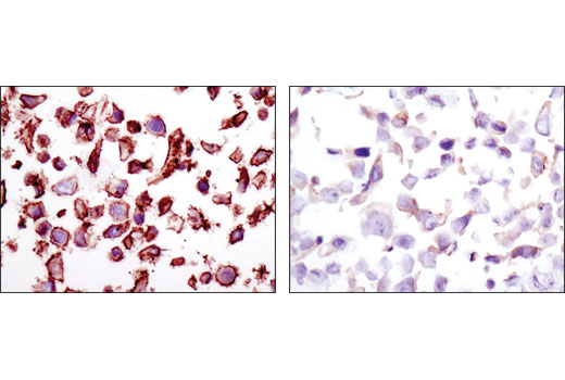
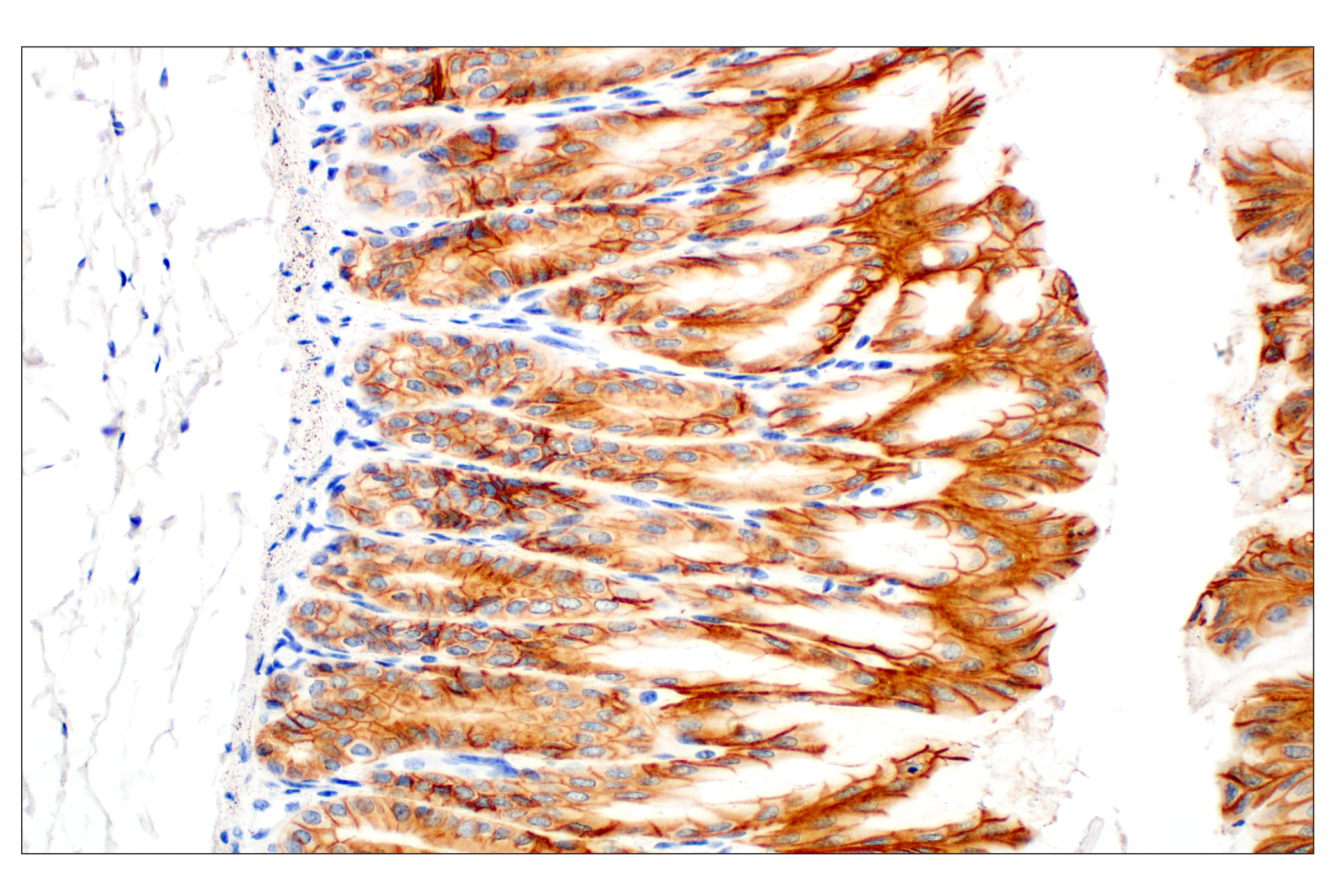
| HER2/ErbB2 (D8F12) XP® Rabbit mAb | 4290 | 20 µl | 185 kDa | Rabbit IgG |
| Anti-rabbit IgG, HRP-linked Antibody | 7074 | 100 µl | Goat |
Please visit cellsignal.com for individual component applications, species cross-reactivity, dilutions, protocols, and additional product information.
Description
The Oncogenes and Tumor Suppressor Antibody Sampler Kit offers an economical means of investigating proteins commonly involved in the biological pathways behind oncogenesis, tumor metastasis, and cancer pathology. The kit contains enough primary and secondary antibody to perform four western blot experiments with each antibody.
Storage
Background
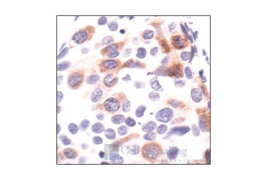
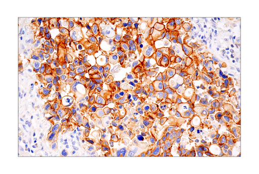
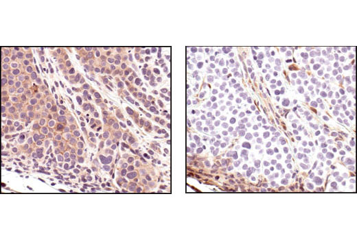
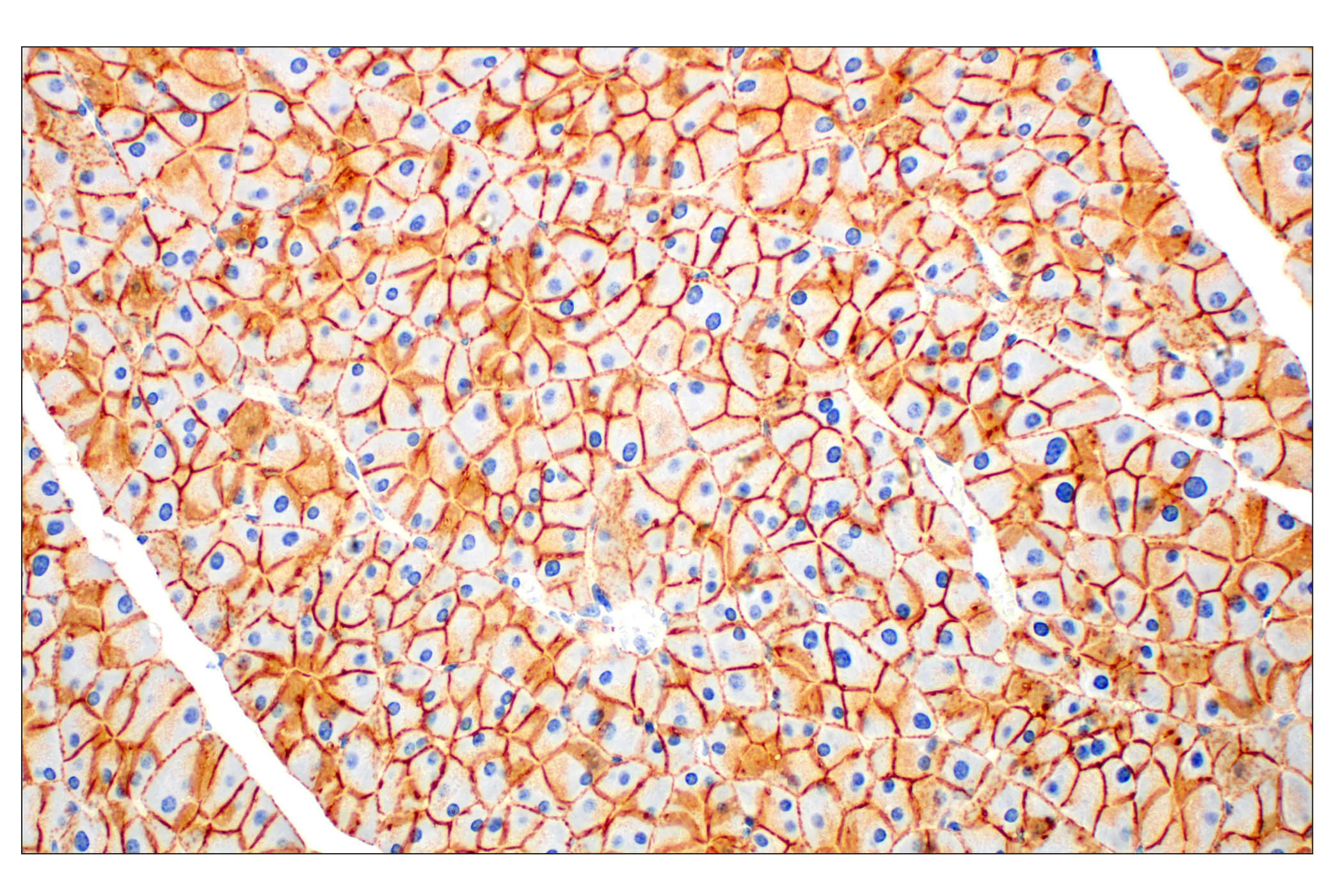
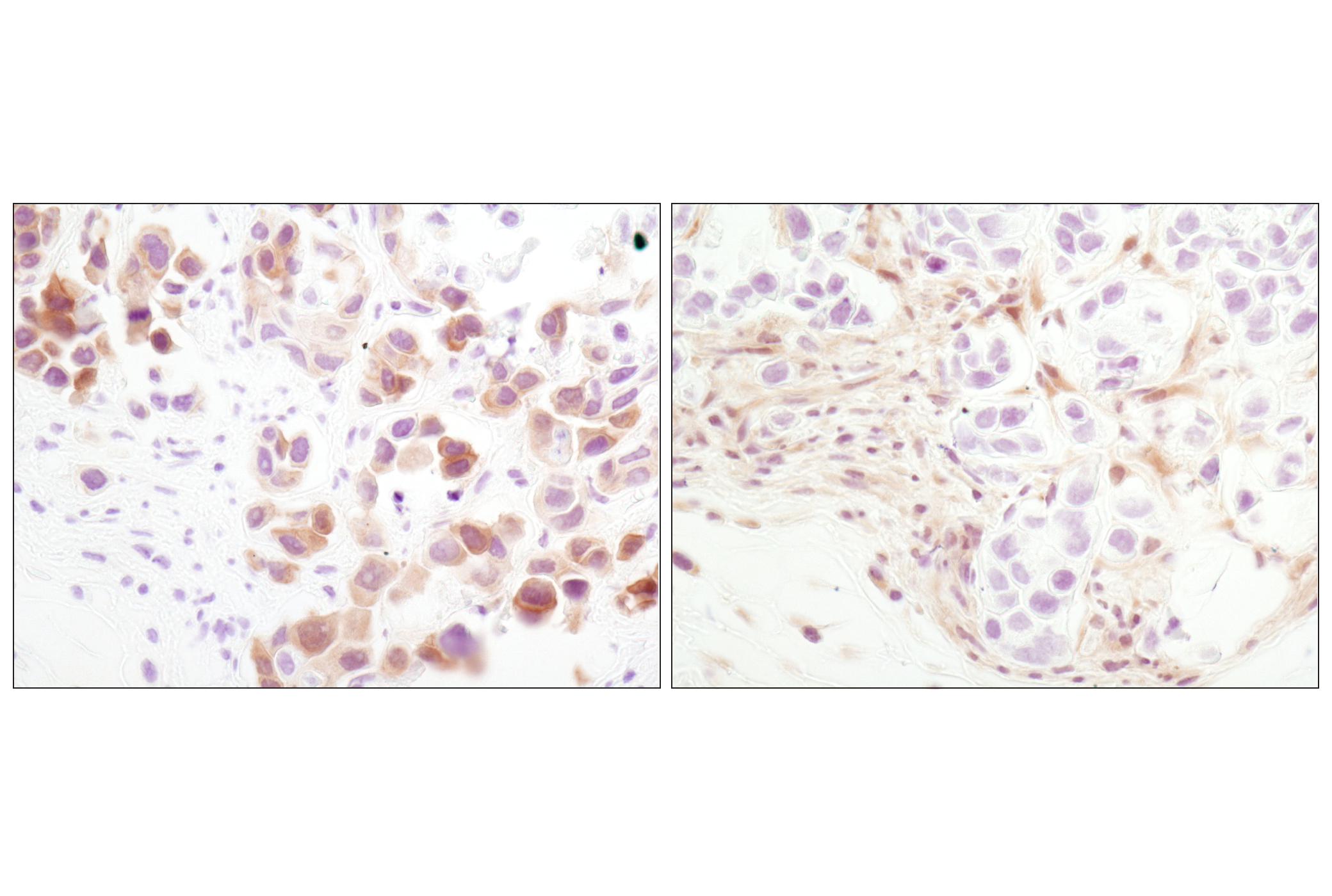
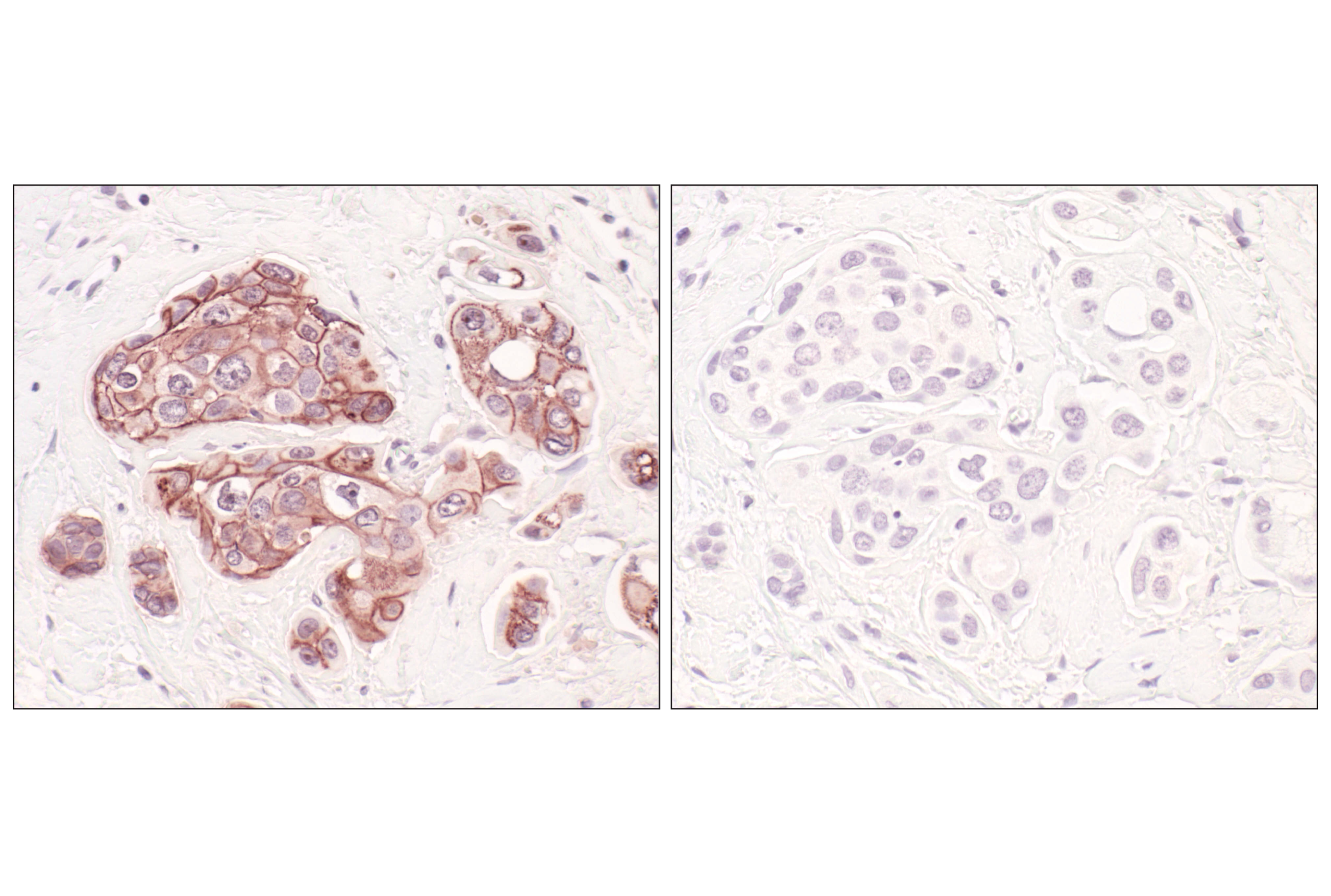
Oncogenesis is a multistep process leading to sequential alterations in several oncogenes, tumor-suppressor genes, and microRNA genes (1,2). These alterations often disrupt the expression, function, and/or activity of proteins regulating cell growth and programmed cell death. Many of the molecular mechanisms and biological pathways driving oncogenesis and cancer pathology have been identified. The signal transduction pathways regulating apoptosis, cell-cycle progression, cell adhesion, cell migration, and DNA damage responses are often disrupted. HER2/ErbB2 (3), E-Cadherin (4), p53 (5,6), Stathmin (7), BRCA1 (8,9), Akt (10), PTEN (11), and Estrogen Receptor α (12) function in many of these pathways.
- Berger, A.H. et al. (2011) Nature 476, 163-9.
- Peltomäki, P. (2012) Exp Cell Res 318, 299-310.
- Dittadi, R. and Gion, M. (2000) J Natl Cancer Inst 92, 1443-4.
- Hazan, R.B. et al. (2004) Ann N Y Acad Sci 1014, 155-63.
- Rahman, N. and Stratton, M.R. (1998) Annu Rev Genet 32, 95-121.
- Freed-Pastor, W.A. and Prives, C. (2012) Genes Dev 26, 1268-86.
- Belletti, B. and Baldassarre, G. (2011) Expert Opin Ther Targets 15, 1249-66.
- Gayther, S.A. et al. (1999) Am J Hum Genet 65, 1021-9.
- Scully, R. and Livingston, D.M. (2000) Nature 408, 429-32.
- Jazirehi, A.R. et al. (2012) Am J Cancer Res 2, 178-91.
- Saal, L.H. et al. (2008) Nat Genet 40, 102-7.
- Pópulo, H. et al. (2012) Int J Mol Sci 13, 1886-918.
Background References
Trademarks and Patents
Limited Uses
Except as otherwise expressly agreed in a writing signed by a legally authorized representative of CST, the following terms apply to Products provided by CST, its affiliates or its distributors. Any Customer's terms and conditions that are in addition to, or different from, those contained herein, unless separately accepted in writing by a legally authorized representative of CST, are rejected and are of no force or effect.
Products are labeled with For Research Use Only or a similar labeling statement and have not been approved, cleared, or licensed by the FDA or other regulatory foreign or domestic entity, for any purpose. Customer shall not use any Product for any diagnostic or therapeutic purpose, or otherwise in any manner that conflicts with its labeling statement. Products sold or licensed by CST are provided for Customer as the end-user and solely for research and development uses. Any use of Product for diagnostic, prophylactic or therapeutic purposes, or any purchase of Product for resale (alone or as a component) or other commercial purpose, requires a separate license from CST. Customer shall (a) not sell, license, loan, donate or otherwise transfer or make available any Product to any third party, whether alone or in combination with other materials, or use the Products to manufacture any commercial products, (b) not copy, modify, reverse engineer, decompile, disassemble or otherwise attempt to discover the underlying structure or technology of the Products, or use the Products for the purpose of developing any products or services that would compete with CST products or services, (c) not alter or remove from the Products any trademarks, trade names, logos, patent or copyright notices or markings, (d) use the Products solely in accordance with CST Product Terms of Sale and any applicable documentation, and (e) comply with any license, terms of service or similar agreement with respect to any third party products or services used by Customer in connection with the Products.