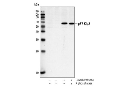
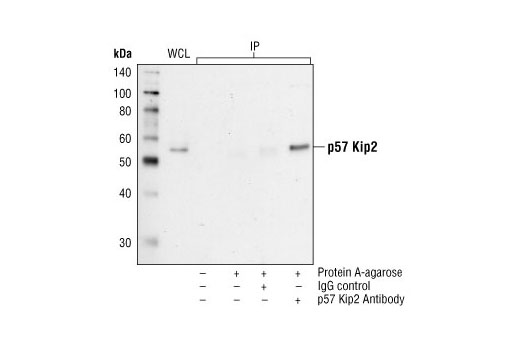
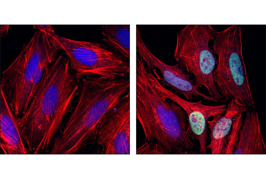
WB, IP, IF-IC
H
Endogenous
57
Rabbit
#P49918
1028
Product Information
Product Usage Information
| Application | Dilution |
|---|---|
| Western Blotting | 1:1000 |
| Immunoprecipitation | 1:50 |
| Immunofluorescence (Immunocytochemistry) | 1:100 |
Storage
Specificity / Sensitivity
Species Reactivity:
Human
Source / Purification
Polyclonal antibodies are produced by immunizing animals with a synthetic peptide corresponding to amino acids near the carboxy terminus of human p57 Kip2. Antibodies are purified by protein A and peptide affinity chromatography.
Background
p27 Kip1 is a member of the Cip/Kip family of cyclin-dependent kinase inhibitors. Like its relatives, p57 Kip2 and p21 Waf1/Cip1, the ability to enforce the G1 restriction point is derived from its inhibitory binding to CDK2/cyclin E and other CDK/cyclin complexes. Expression levels of p27 are upregulated in quiescent cells and in cells treated with cAMP or other negative cell cycle regulators. Downregulation of p27 can be induced by treatment with interleukin-2 or other mitogens; this involves phosphorylation of p27 and its degradation by the ubiquitin-proteasome pathway (1-4).
p57 Kip2 (Cyclin-dependent kinase inhibitor 1C) functions as a tumor suppressor. Mutations of p57 Kip2 have been associated with numerous human malignancies as well as Beckwith–Wiedemann syndrome (BWS), characterized by an increased risk of childhood cancer. The amino-terminal CDK inhibitory domain, common to the family, binds to and inhibits CDK/cyclin complexes and restricts cell cycle progression (5). The unique central region of p57 Kip2 interactes with LIMK-1, a downstream effector of the Rho family of GTPases. By sequestering LIMK-1 in the nucleus, p57 Kip2 disrupts actin dynamics within cells and may be linked to its essential role in embryonic development (6). In addition, the carboxyl-terminal QT domain of p57KIP2 binds to and inhibits JNK/SAPK activity regulating cellular apoptosis and differentiation (7). Expression levels of human p57 Kip2 are more restricted then other CDK inhibitors (8) and are controlled by ubiquitination/degradation via the Skp1/Cul1/F-box-type E3 ubiquitin ligase complex SCF-Skp2. This effect is dependent on Thr310 (9). A similar threonine phosphorylation site in p27 Kip1, Thr187, has also been shown to regulate protein stability (10).
- Lloyd, R.V. et al. (1999) Am J Pathol 154, 313-23.
- Polyak, K. et al. (1994) Genes Dev 8, 9-22.
- Kato, J.Y. et al. (1994) Cell 79, 487-96.
- Vlach, J. et al. (1997) EMBO J 16, 5334-44.
- Pateras, I.S. et al. (2009) Mol Cancer Res 7, 1902-19.
- Yokoo, T. et al. (2003) J Biol Chem 278, 52919-23.
- Chang, T.S. et al. (2003) J Biol Chem 278, 48092-8.
- Lee, M.H. et al. (1995) Genes Dev 9, 639-49.
- Kamura, T. et al. (2003) Proc Natl Acad Sci U S A 100, 10231-6.
- Ishida, N. et al. (2000) J Biol Chem 275, 25146-54.
Species Reactivity
Species reactivity is determined by testing in at least one approved application (e.g., western blot).
Western Blot Buffer
IMPORTANT: For western blots, incubate membrane with diluted primary antibody in 5% w/v BSA, 1X TBS, 0.1% Tween® 20 at 4°C with gentle shaking, overnight.
Applications Key
WB: Western Blotting IP: Immunoprecipitation IF-IC: Immunofluorescence (Immunocytochemistry)
Cross-Reactivity Key
H: human M: mouse R: rat Hm: hamster Mk: monkey Vir: virus Mi: mink C: chicken Dm: D. melanogaster X: Xenopus Z: zebrafish B: bovine Dg: dog Pg: pig Sc: S. cerevisiae Ce: C. elegans Hr: horse GP: Guinea Pig Rab: rabbit All: all species expected
Trademarks and Patents
Limited Uses
Except as otherwise expressly agreed in a writing signed by a legally authorized representative of CST, the following terms apply to Products provided by CST, its affiliates or its distributors. Any Customer's terms and conditions that are in addition to, or different from, those contained herein, unless separately accepted in writing by a legally authorized representative of CST, are rejected and are of no force or effect.
Products are labeled with For Research Use Only or a similar labeling statement and have not been approved, cleared, or licensed by the FDA or other regulatory foreign or domestic entity, for any purpose. Customer shall not use any Product for any diagnostic or therapeutic purpose, or otherwise in any manner that conflicts with its labeling statement. Products sold or licensed by CST are provided for Customer as the end-user and solely for research and development uses. Any use of Product for diagnostic, prophylactic or therapeutic purposes, or any purchase of Product for resale (alone or as a component) or other commercial purpose, requires a separate license from CST. Customer shall (a) not sell, license, loan, donate or otherwise transfer or make available any Product to any third party, whether alone or in combination with other materials, or use the Products to manufacture any commercial products, (b) not copy, modify, reverse engineer, decompile, disassemble or otherwise attempt to discover the underlying structure or technology of the Products, or use the Products for the purpose of developing any products or services that would compete with CST products or services, (c) not alter or remove from the Products any trademarks, trade names, logos, patent or copyright notices or markings, (d) use the Products solely in accordance with CST Product Terms of Sale and any applicable documentation, and (e) comply with any license, terms of service or similar agreement with respect to any third party products or services used by Customer in connection with the Products.