WB, IP, IF-IC, FC-FP
H
Endogenous
68
Rabbit IgG
#Q9UM07
23569
Product Information
Product Usage Information
| Application | Dilution |
|---|---|
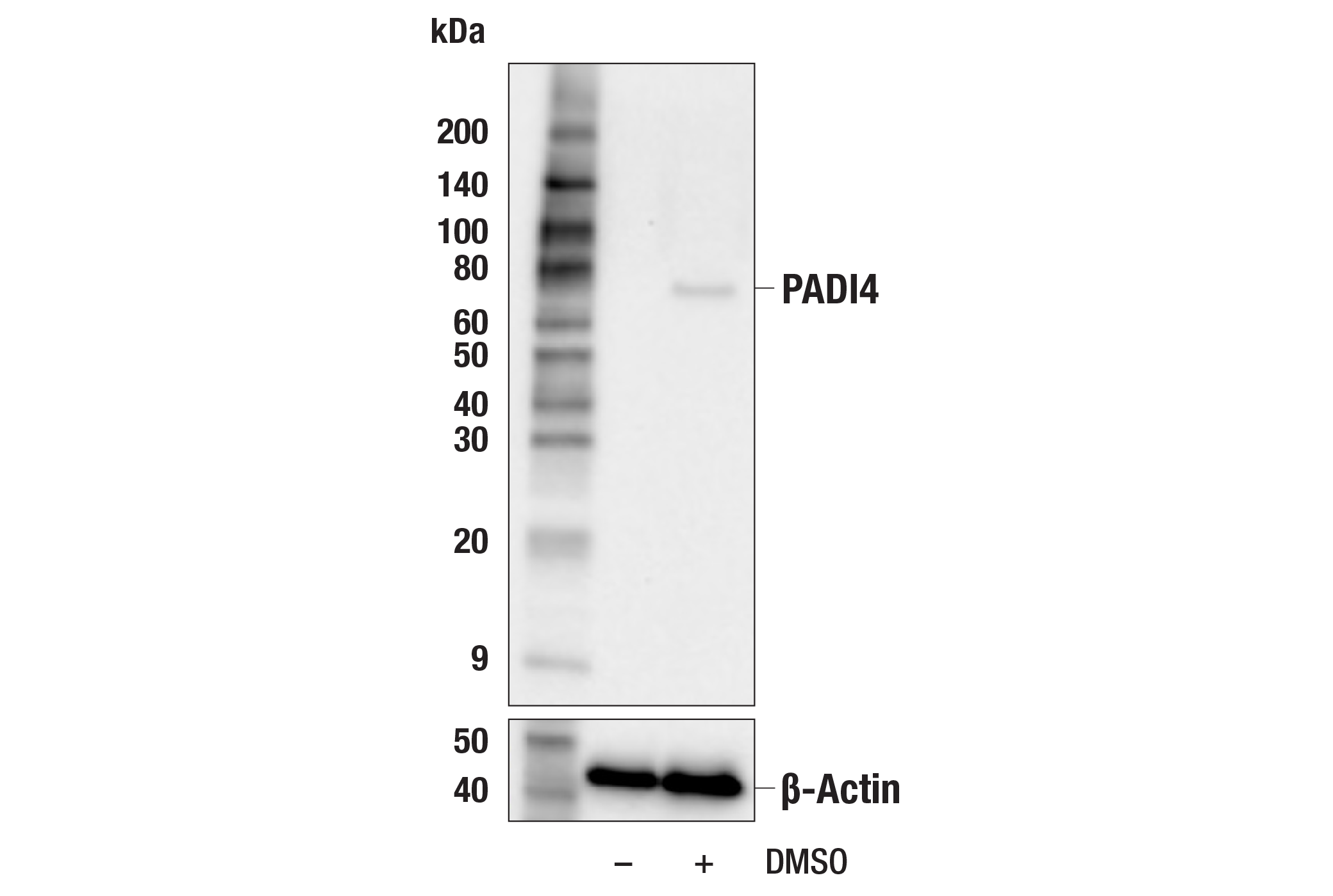
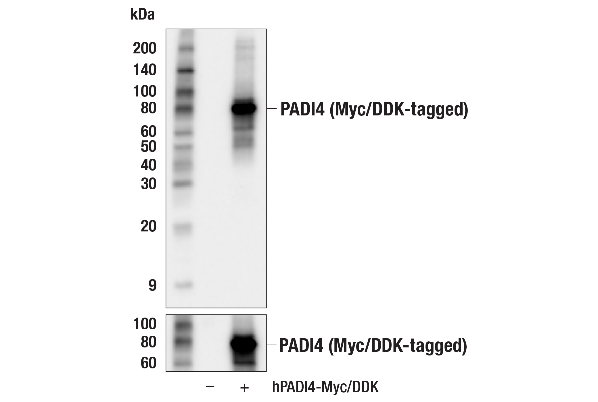
| Western Blotting | 1:1000 |
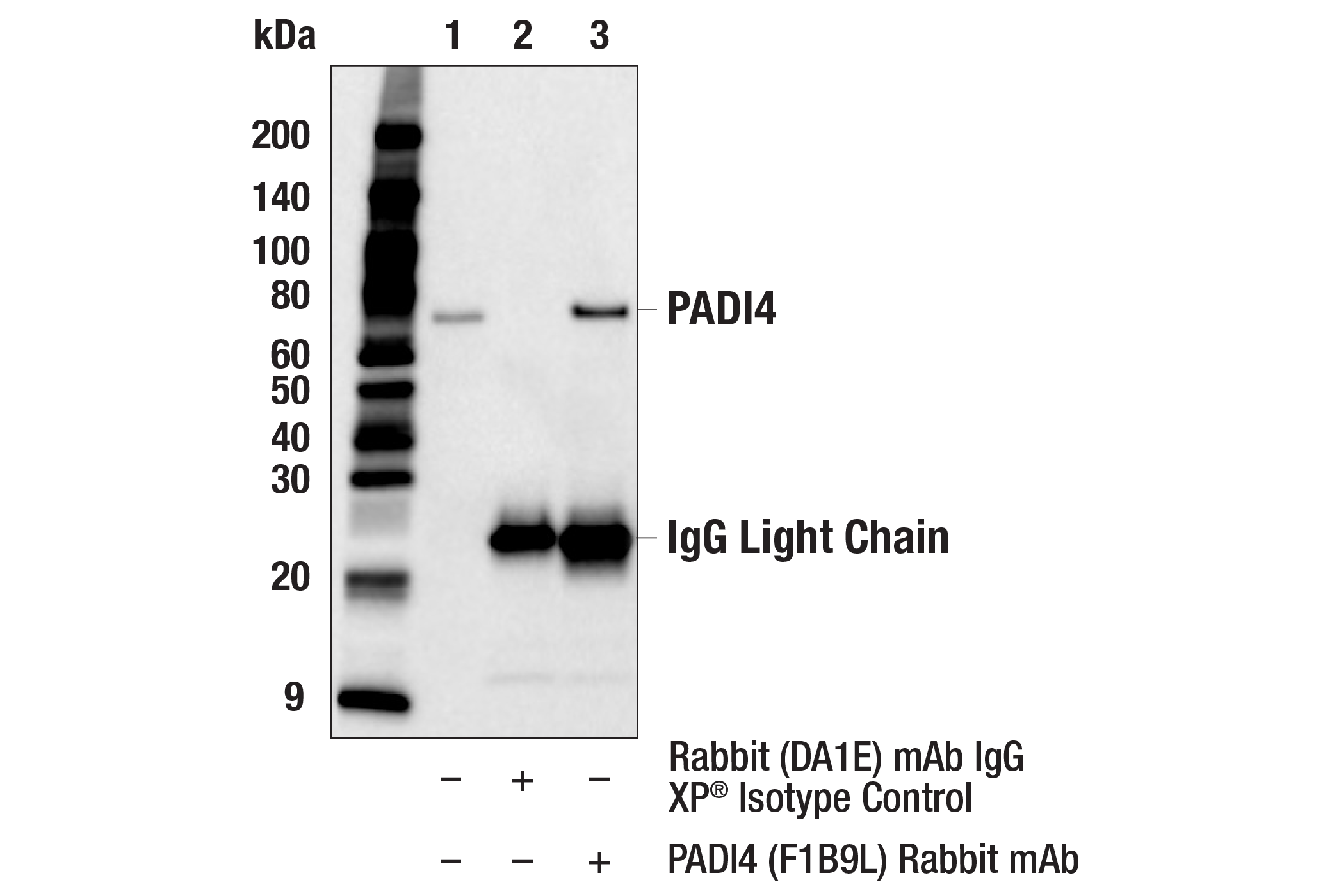
| Immunoprecipitation | 1:50 |
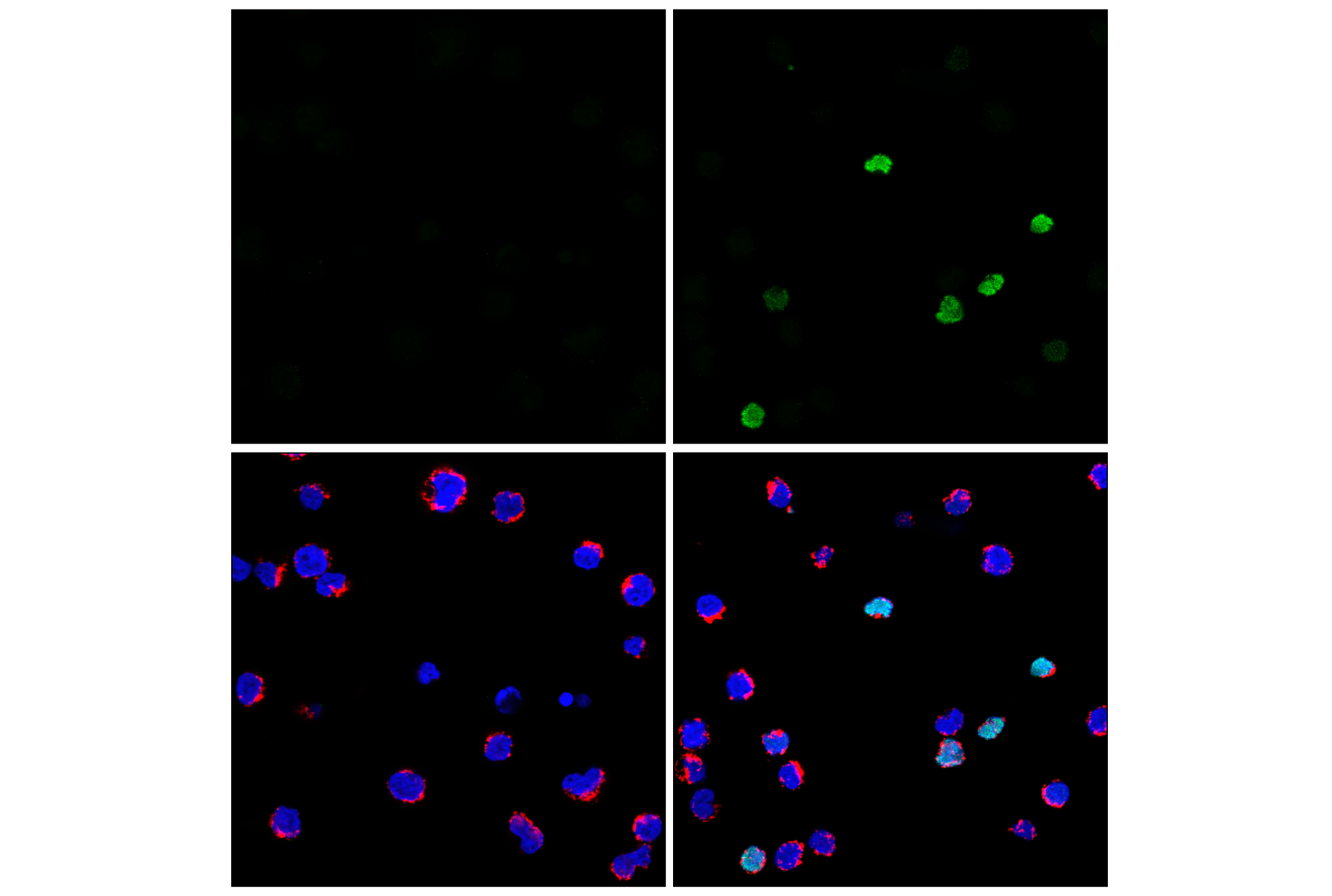
| Immunofluorescence (Immunocytochemistry) | 1:200 - 1:800 |
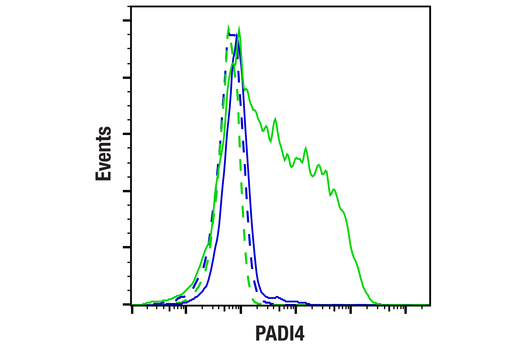
| Flow Cytometry (Fixed/Permeabilized) | 1:50 - 1:200 |
Storage
Specificity / Sensitivity
Species Reactivity:
Human
Source / Purification
Monoclonal antibody is produced by immunizing animals with recombinant human PADI4 protein.
Background
Peptidyl arginine deiminase (PAD) proteins are a family of Ca2+-dependent enzymes that catalyze the post-translational conversion of arginine to citrulline. There are currently five known PAD isozymes in humans, referred to as PADI1-4 and PADI6 (1). PADI4 is a nuclear member of the PAD family and is responsible for deiminating R2, R8, R17, and R26 on the histone H3 (2-4). Deimination of histone residues prevents CARM1-mediated arginine methylation and results in transcriptional repression (2,3). Expression of PADI4 is increased during myeloid differentiation and various polymorphisms have been implicated in rheumatoid arthritis (5-8). PADI4 can play a role in apoptosis and cell cycle arrest upon DNA damage, as it is recruited to the p21 promoter by p53 and also regulates a subset of p53 target genes (9,10). Hypercitrullination of histones by PADI4 contributes to chromatin decondensation and the formation of neutrophil extracellular traps (NETs) to combat bacterial infection in a process called NETosis (11,12). Inhibition of PADI4 reduces NET formation, making it a potential therapeutic target (13).
- Jones, J.E. et al. (2009) Curr Opin Drug Discov Devel 12, 616-27.
- Cuthbert, G.L. et al. (2004) Cell 118, 545-53.
- Wang, Y. et al. (2004) Science 306, 279-83.
- Nakashima, K. et al. (2002) J Biol Chem 277, 49562-8.
- Nakashima, K. et al. (1999) J Biol Chem 274, 27786-92.
- Suzuki, A. et al. (2003) Nat Genet 34, 395-402.
- Worthington, J. and John, S. (2003) Trends Mol Med 9, 405-7.
- Yamada, R. et al. (2003) Trends Mol Med 9, 503-8.
- Li, P. et al. (2008) Mol Cell Biol 28, 4745-58.
- Yao, H. et al. (2008) J Biol Chem 283, 20060-8.
- Wang, Y. et al. (2009) J Cell Biol 184, 205-13.
- Brinkmann, V. et al. (2004) Science 303, 1532-5.
- Lewis, H.D. et al. (2015) Nat Chem Biol 11, 189-91.
Species Reactivity
Species reactivity is determined by testing in at least one approved application (e.g., western blot).
Western Blot Buffer
IMPORTANT: For western blots, incubate membrane with diluted primary antibody in 5% w/v BSA, 1X TBS, 0.1% Tween® 20 at 4°C with gentle shaking, overnight.
Applications Key
WB: Western Blotting IP: Immunoprecipitation IF-IC: Immunofluorescence (Immunocytochemistry) FC-FP: Flow Cytometry (Fixed/Permeabilized)
Cross-Reactivity Key
H: human M: mouse R: rat Hm: hamster Mk: monkey Vir: virus Mi: mink C: chicken Dm: D. melanogaster X: Xenopus Z: zebrafish B: bovine Dg: dog Pg: pig Sc: S. cerevisiae Ce: C. elegans Hr: horse GP: Guinea Pig Rab: rabbit All: all species expected
Trademarks and Patents
Limited Uses
Except as otherwise expressly agreed in a writing signed by a legally authorized representative of CST, the following terms apply to Products provided by CST, its affiliates or its distributors. Any Customer's terms and conditions that are in addition to, or different from, those contained herein, unless separately accepted in writing by a legally authorized representative of CST, are rejected and are of no force or effect.
Products are labeled with For Research Use Only or a similar labeling statement and have not been approved, cleared, or licensed by the FDA or other regulatory foreign or domestic entity, for any purpose. Customer shall not use any Product for any diagnostic or therapeutic purpose, or otherwise in any manner that conflicts with its labeling statement. Products sold or licensed by CST are provided for Customer as the end-user and solely for research and development uses. Any use of Product for diagnostic, prophylactic or therapeutic purposes, or any purchase of Product for resale (alone or as a component) or other commercial purpose, requires a separate license from CST. Customer shall (a) not sell, license, loan, donate or otherwise transfer or make available any Product to any third party, whether alone or in combination with other materials, or use the Products to manufacture any commercial products, (b) not copy, modify, reverse engineer, decompile, disassemble or otherwise attempt to discover the underlying structure or technology of the Products, or use the Products for the purpose of developing any products or services that would compete with CST products or services, (c) not alter or remove from the Products any trademarks, trade names, logos, patent or copyright notices or markings, (d) use the Products solely in accordance with CST Product Terms of Sale and any applicable documentation, and (e) comply with any license, terms of service or similar agreement with respect to any third party products or services used by Customer in connection with the Products.