| Product Includes | Product # | Quantity | Mol. Wt | Isotype/Source |
|---|---|---|---|---|
| Insulin (C27C9) Rabbit mAb | 3014 | 20 µl | Rabbit IgG | |
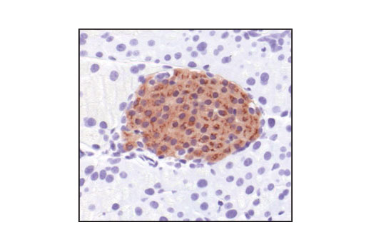
| Proglucagon (D16G10) XP® Rabbit mAb | 8233 | 20 µl | Rabbit IgG | |
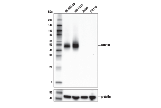
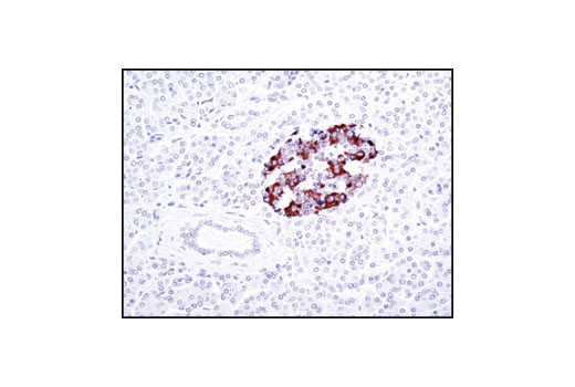
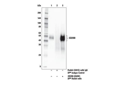
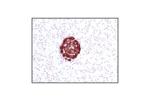
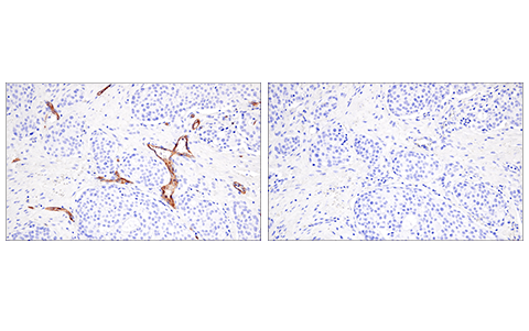
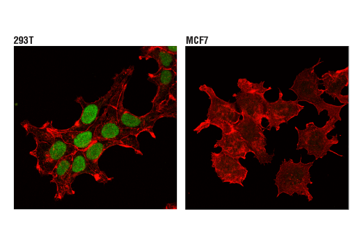
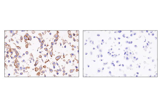
| CD200 (E5I9V) XP® Rabbit mAb | 23451 | 20 µl | 45-50 kDa | Rabbit IgG |
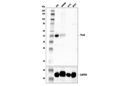
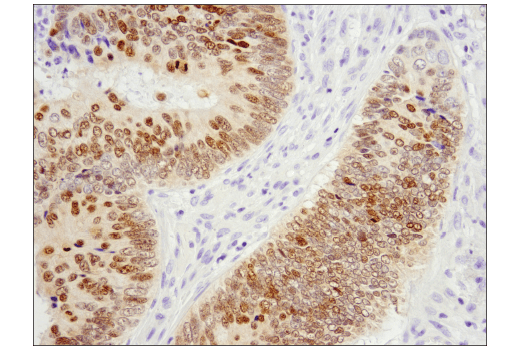
| Pax6 (D3A9V) XP® Rabbit mAb | 60433 | 20 µl | 50 kDa | Rabbit IgG |
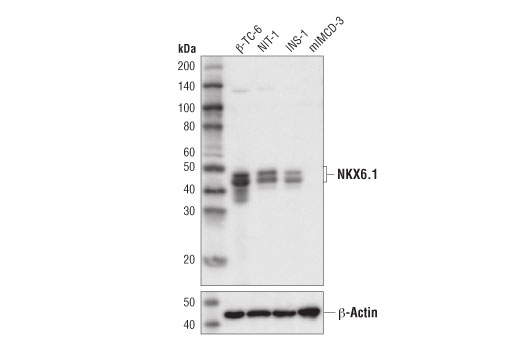
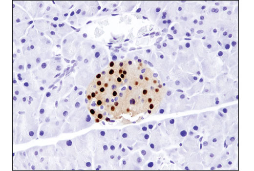
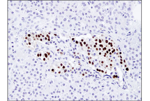
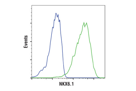
| NKX6.1 (D8O4R) Rabbit mAb | 54551 | 20 µl | 44, 46 kDa | Rabbit IgG |
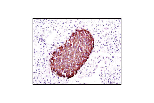
| C-Peptide Antibody | 4593 | 20 µl | 4 kDa | Rabbit |
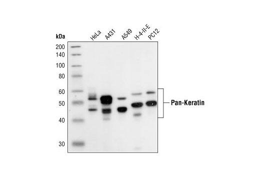
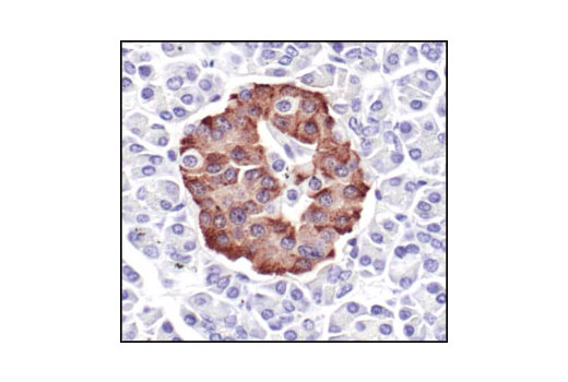
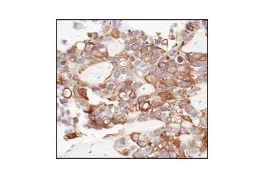
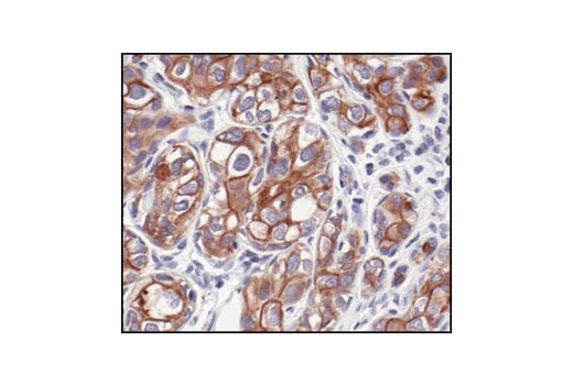
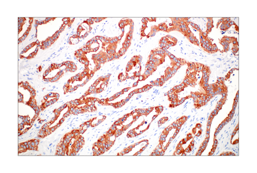
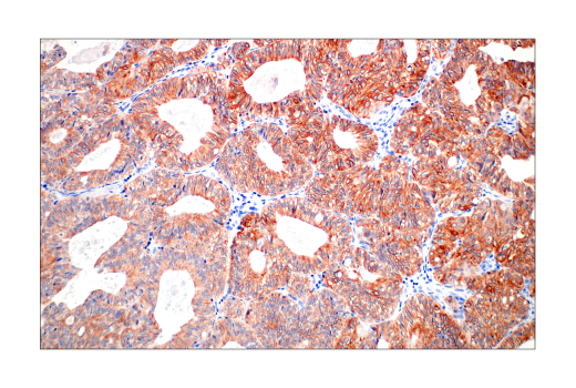
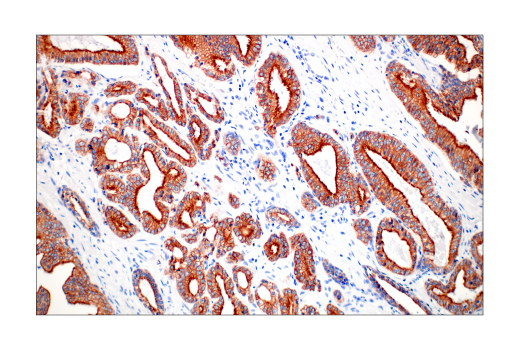
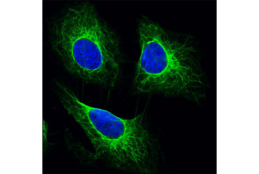
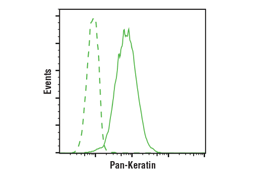
| Pan-Keratin (C11) Mouse mAb | 4545 | 20 µl | 46-58 kDa | Mouse IgG1 |
| Anti-rabbit IgG, HRP-linked Antibody | 7074 | 100 µl | Goat | |
| Anti-mouse IgG, HRP-linked Antibody | 7076 | 100 µl | Horse |
Please visit cellsignal.com for individual component applications, species cross-reactivity, dilutions, protocols, and additional product information.
Description
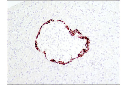
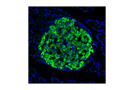
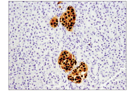
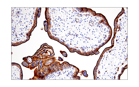
The Pancreatic Marker IHC Antibody Sampler Kit provides a useful selection of markers to distinguish pancreatic cell types that perform important functions to maintain glucose homeostasis. The kit also includes antibodies that differentiate pancreatic tumor subtypes. The sampler kit is designed for use on formalin-fixed, paraffin-embedded tissue samples.
Storage
Background
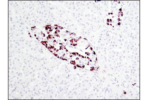
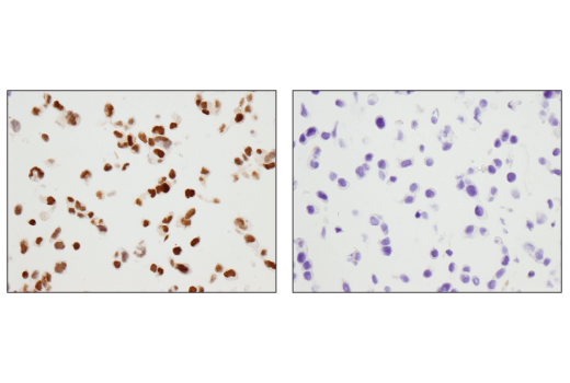
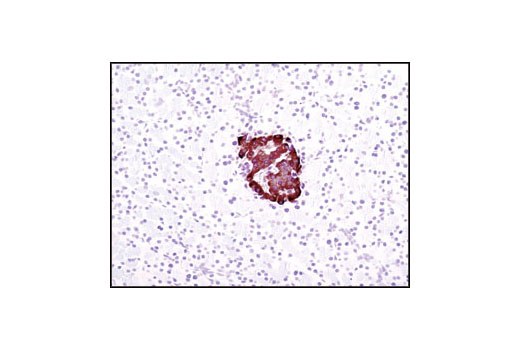
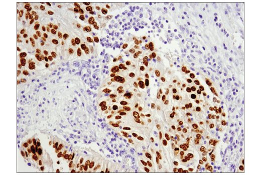
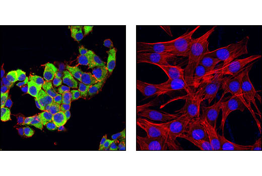
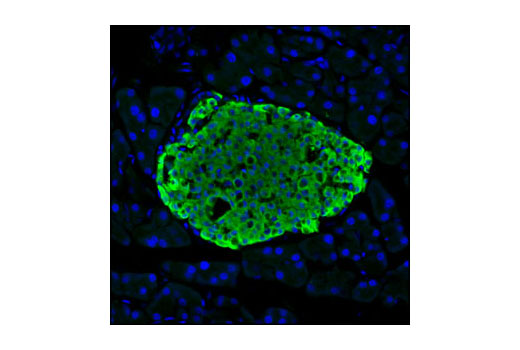
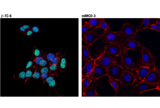
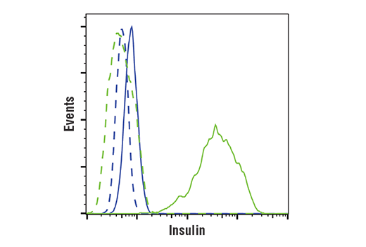
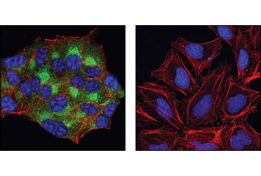
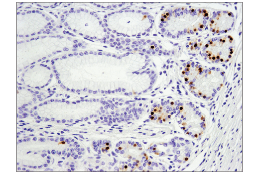
Insulin is a hormone that is produced and released from pancreatic β cells through a glucose sensing pathway. Proinsulin is the precursor molecule to insulin and is processed prior to its secretion. Insulin is composed of A-peptide and B-peptide which are joined by a disulfide bond. The center one-third of the molecule is cleaved and released as C-peptide, which has a longer half-life than insulin (1). Antibodies to both insulin and C-peptide are useful markers for β cells.
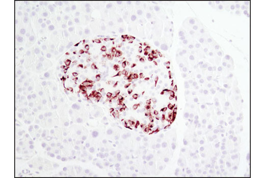
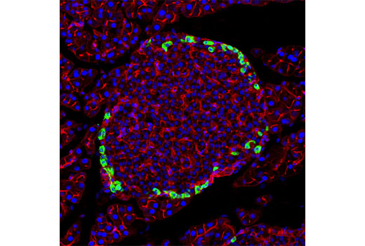
Glucose homeostasis is regulated by a variety of hormones including glucagon. Glucagon is synthesized as the precursor molecule proglucagon and is proteolytically processed to yield the mature peptide in α cells of the pancreatic islets. Glucagon causes the release of glucose from glycogen and stimulates gluconeogenesis in the liver (2). Antibodies to glucagon and proglucagon are useful markers for pancreatic α cells.
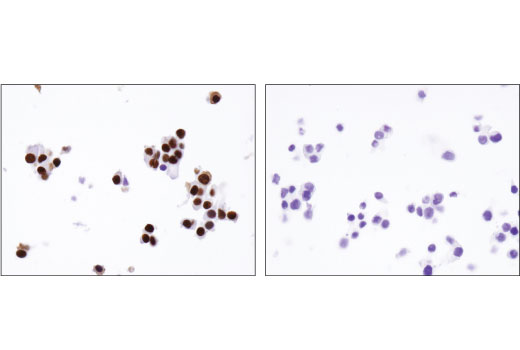
NKX6.1 is an important transcription factor in a network of transcription factors that are critical for pancreatic β cell development and maintenance (3). Antibodies to NKX6.1 are useful markers for β cells.
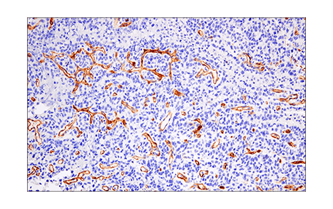
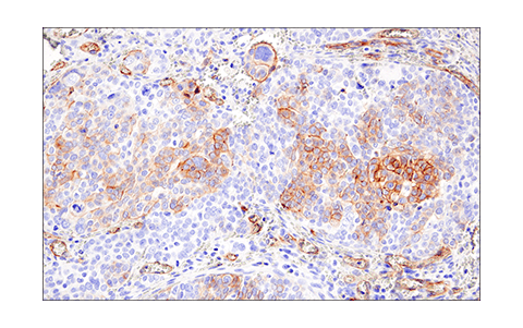
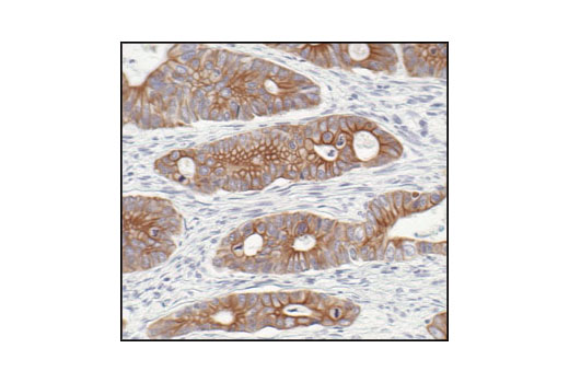
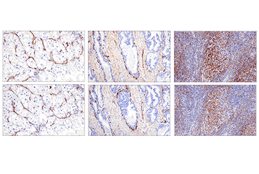
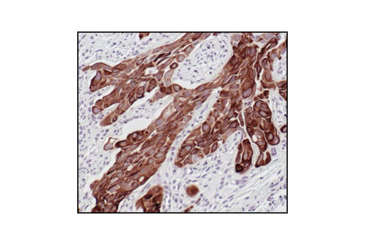
Pan-Keratin and CD200 antibodies are useful to mark and differentiate some pancreatic tumor subtypes. Pan-Keratin antibodies mark epithelial cells in pancreatic adenocarcinomas while CD200 is a useful marker for neuroendocrine pancreatic tumors (4).
Background References
Trademarks and Patents
Limited Uses
Except as otherwise expressly agreed in a writing signed by a legally authorized representative of CST, the following terms apply to Products provided by CST, its affiliates or its distributors. Any Customer's terms and conditions that are in addition to, or different from, those contained herein, unless separately accepted in writing by a legally authorized representative of CST, are rejected and are of no force or effect.
Products are labeled with For Research Use Only or a similar labeling statement and have not been approved, cleared, or licensed by the FDA or other regulatory foreign or domestic entity, for any purpose. Customer shall not use any Product for any diagnostic or therapeutic purpose, or otherwise in any manner that conflicts with its labeling statement. Products sold or licensed by CST are provided for Customer as the end-user and solely for research and development uses. Any use of Product for diagnostic, prophylactic or therapeutic purposes, or any purchase of Product for resale (alone or as a component) or other commercial purpose, requires a separate license from CST. Customer shall (a) not sell, license, loan, donate or otherwise transfer or make available any Product to any third party, whether alone or in combination with other materials, or use the Products to manufacture any commercial products, (b) not copy, modify, reverse engineer, decompile, disassemble or otherwise attempt to discover the underlying structure or technology of the Products, or use the Products for the purpose of developing any products or services that would compete with CST products or services, (c) not alter or remove from the Products any trademarks, trade names, logos, patent or copyright notices or markings, (d) use the Products solely in accordance with CST Product Terms of Sale and any applicable documentation, and (e) comply with any license, terms of service or similar agreement with respect to any third party products or services used by Customer in connection with the Products.