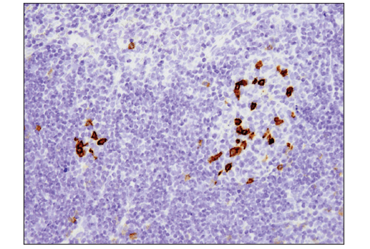
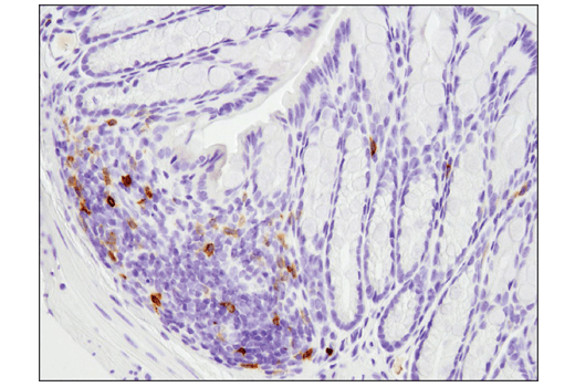
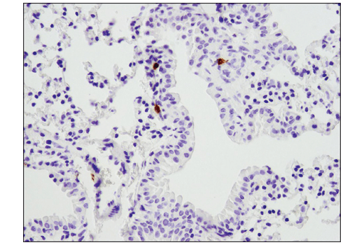
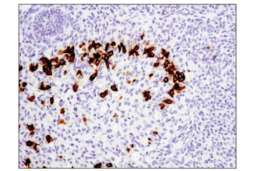
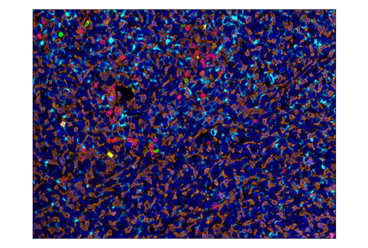
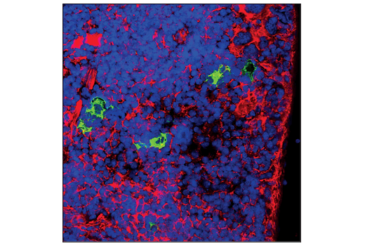
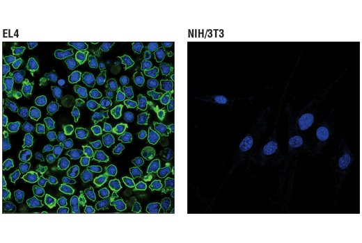
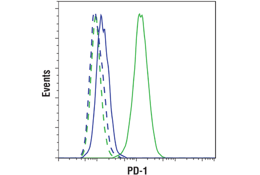
WB, IP, IHC-Bond, IHC-P, IF-F, IF-IC, FC-FP
M
Endogenous
40-75
Rabbit IgG
#Q02242
18566
Product Information
Product Usage Information
| Application | Dilution |
|---|---|
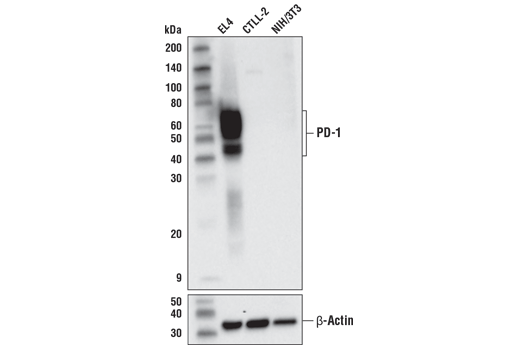
| Western Blotting | 1:1000 |
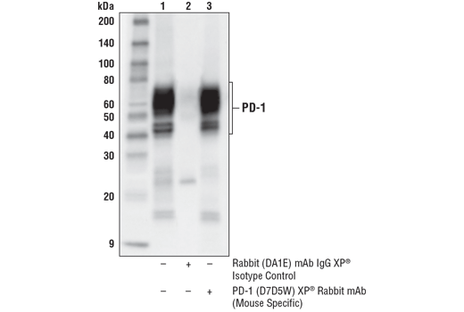
| Immunoprecipitation | 1:200 |
| IHC Leica Bond | 1:50 - 1:200 |
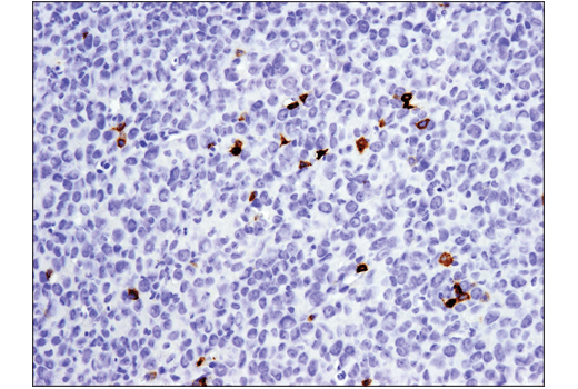
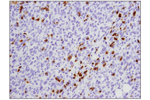
| Immunohistochemistry (Paraffin) | 1:100 - 1:400 |
| Immunofluorescence (Frozen) | 1:100 - 1:400 |
| Immunofluorescence (Immunocytochemistry) | 1:100 - 1:400 |
| Flow Cytometry (Fixed/Permeabilized) | 1:100 - 1:400 |
Storage
For a carrier free (BSA and azide free) version of this product see product #55789.
Specificity / Sensitivity
Species Reactivity:
Mouse
Species predicted to react based on 100% sequence homology
The antigen sequence used to produce this antibody shares
100% sequence homology with the species listed here, but
reactivity has not been tested or confirmed to work by CST.
Use of this product with these species is not covered under
our
Product Performance Guarantee.
Rat, Hamster
Source / Purification
Monoclonal antibody is produced by immunizing animals with a synthetic peptide corresponding to residues surrounding Ala242 of mouse PD-1 protein.
Background
The programmed cell death 1 protein (PD-1, PDCD1, CD279) is a member of the CD28 family of immunoreceptors that regulate T cell activation and immune responses (1-3). The PD-1 protein contains an extracellular Ig V domain, a transmembrane domain, and a cytoplasmic tail that includes an immunoreceptor tyrosine-based inhibitory motif (ITIM) and an immunoreceptor tyrosine-based switch motif (ITSM). PD-1 is activated by the cell surface ligands PD-L1 and PD-L2 (4). Upon activation, PD-1 ITIM and ITSM phosphorylation leads to the recruitment of the protein tyrosine phosphatases SHP-1 and SHP-2, which suppress TCR signaling (5-7). In addition to activated T cells, PD-1 is expressed in activated B cells and monocytes, although its function in these cell types has not been fully characterized (8). The PD-1 pathway plays an important role in immune tolerance (3); however, research studies show that cancer cells often adopt this pathway to escape immune surveillance (9). Consequently, blockade of PD-1 and its ligands is proving to be a sound strategy for neoplastic intervention (10).
- Ishida, Y. et al. (1992) EMBO J 11, 3887-95.
- Shinohara, T. et al. (1994) Genomics 23, 704-6.
- Nishimura, H. et al. (1999) Immunity 11, 141-51.
- Freeman, G.J. et al. (2000) J Exp Med 192, 1027-34.
- Yokosuka, T. et al. (2012) J Exp Med 209, 1201-17.
- Sheppard, K.A. et al. (2004) FEBS Lett 574, 37-41.
- Chemnitz, J.M. et al. (2004) J Immunol 173, 945-54.
- Thibult, M.L. et al. (2013) Int Immunol 25, 129-37.
- Dong, H. et al. (2002) Nat Med 8, 793-800.
- Topalian, S.L. et al. (2012) Curr Opin Immunol 24, 207-12.
Species Reactivity
Species reactivity is determined by testing in at least one approved application (e.g., western blot).
Western Blot Buffer
IMPORTANT: For western blots, incubate membrane with diluted primary antibody in 5% w/v BSA, 1X TBS, 0.1% Tween® 20 at 4°C with gentle shaking, overnight.
Applications Key
WB: Western Blotting IP: Immunoprecipitation IHC-Bond: IHC Leica Bond IHC-P: Immunohistochemistry (Paraffin) IF-F: Immunofluorescence (Frozen) IF-IC: Immunofluorescence (Immunocytochemistry) FC-FP: Flow Cytometry (Fixed/Permeabilized)
Cross-Reactivity Key
H: human M: mouse R: rat Hm: hamster Mk: monkey Vir: virus Mi: mink C: chicken Dm: D. melanogaster X: Xenopus Z: zebrafish B: bovine Dg: dog Pg: pig Sc: S. cerevisiae Ce: C. elegans Hr: horse GP: Guinea Pig Rab: rabbit All: all species expected
Trademarks and Patents
Limited Uses
Except as otherwise expressly agreed in a writing signed by a legally authorized representative of CST, the following terms apply to Products provided by CST, its affiliates or its distributors. Any Customer's terms and conditions that are in addition to, or different from, those contained herein, unless separately accepted in writing by a legally authorized representative of CST, are rejected and are of no force or effect.
Products are labeled with For Research Use Only or a similar labeling statement and have not been approved, cleared, or licensed by the FDA or other regulatory foreign or domestic entity, for any purpose. Customer shall not use any Product for any diagnostic or therapeutic purpose, or otherwise in any manner that conflicts with its labeling statement. Products sold or licensed by CST are provided for Customer as the end-user and solely for research and development uses. Any use of Product for diagnostic, prophylactic or therapeutic purposes, or any purchase of Product for resale (alone or as a component) or other commercial purpose, requires a separate license from CST. Customer shall (a) not sell, license, loan, donate or otherwise transfer or make available any Product to any third party, whether alone or in combination with other materials, or use the Products to manufacture any commercial products, (b) not copy, modify, reverse engineer, decompile, disassemble or otherwise attempt to discover the underlying structure or technology of the Products, or use the Products for the purpose of developing any products or services that would compete with CST products or services, (c) not alter or remove from the Products any trademarks, trade names, logos, patent or copyright notices or markings, (d) use the Products solely in accordance with CST Product Terms of Sale and any applicable documentation, and (e) comply with any license, terms of service or similar agreement with respect to any third party products or services used by Customer in connection with the Products.