| Product Includes | Product # | Quantity | Mol. Wt | Isotype/Source |
|---|---|---|---|---|
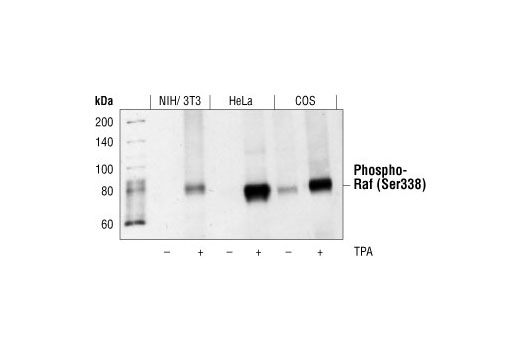
| Phospho-c-Raf (Ser338) (56A6) Rabbit mAb | 9427 | 20 µl | 74 kDa | Rabbit IgG |
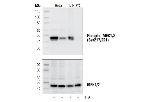
| Phospho-MEK1/2 (Ser217/221) (41G9) Rabbit mAb | 9154 | 20 µl | 45 kDa | Rabbit IgG |
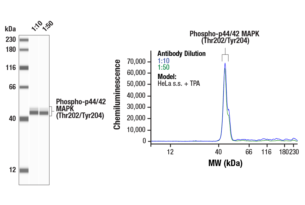
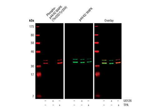
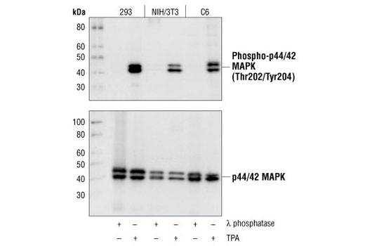
| Phospho-p44/42 MAPK (Erk1/2) (Thr202/Tyr204) (D13.14.4E) XP® Rabbit mAb | 4370 | 20 µl | 44, 42 kDa | Rabbit IgG |
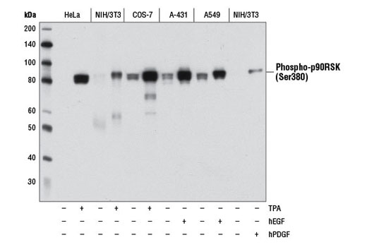
| Phospho-p90RSK (Ser380) (D3H11) Rabbit mAb | 11989 | 20 µl | 90 kDa | Rabbit IgG |
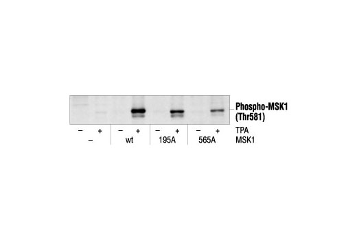
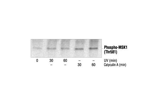
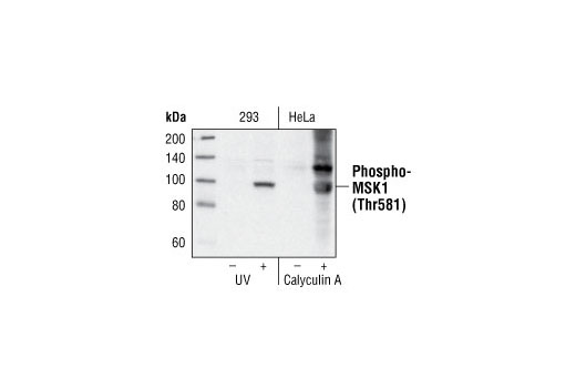
| Phospho-MSK1 (Thr581) Antibody | 9595 | 20 µl | 90 kDa | Rabbit |
| Anti-rabbit IgG, HRP-linked Antibody | 7074 | 100 µl | Goat |
Please visit cellsignal.com for individual component applications, species cross-reactivity, dilutions, protocols, and additional product information.
Description
The Phospho-Erk1/2 Pathway Antibody Sampler Kit provides an economical means of evaluating multiple members of the Erk pathway as well as their activation state. The kit contains enough primary and secondary antibodies to perform two Western blot experiments.
Storage
Background
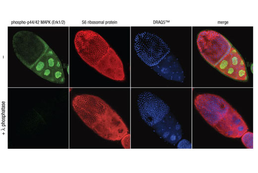
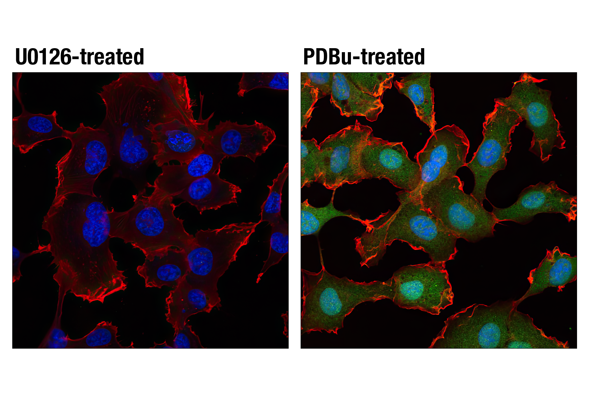
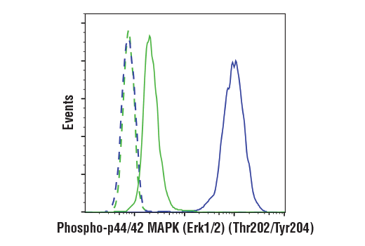
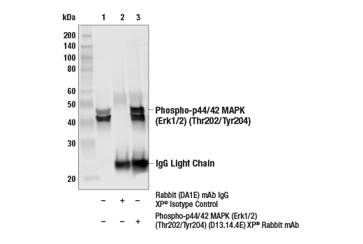
Mitogen-activated protein kinases (MAPKs) are a widely conserved family of serine/threonine protein kinases involved in many cellular programs, such as cell proliferation, differentiation, motility, and death. The p44/42 MAPK (Erk1/2) signaling pathway can be activated in response to a diverse range of extracellular stimuli, including mitogens, growth factors, and cytokines (1-3), and research investigators consider it an important target in the diagnosis and treatment of cancer (4). Upon stimulation, a sequential three-part protein kinase cascade is initiated, consisting of a MAP kinase kinase kinase (MAPKKK or MAP3K), a MAP kinase kinase (MAPKK or MAP2K), and a MAP kinase (MAPK). Multiple p44/42 MAP3Ks have been identified, including members of the Raf family, as well as Mos and Tpl2/COT. MEK1 and MEK2 are the primary MAPKKs in this pathway (5,6). MEK1 and MEK2 activate p44 and p42 through phosphorylation of activation loop residues Thr202/Tyr204 and Thr185/Tyr187, respectively. Several downstream targets of p44/42 have been identified, including p90RSK (7) and the transcription factor Elk-1 (8,9). p44/42 are negatively regulated by a family of dual-specificity (Thr/Tyr) MAPK phosphatases, known as DUSPs or MKPs (10), along with MEK inhibitors, such as U0126 and PD98059.
- Roux, P.P. and Blenis, J. (2004) Microbiol Mol Biol Rev 68, 320-44.
- Baccarini, M. (2005) FEBS Lett 579, 3271-7.
- Meloche, S. and Pouysségur, J. (2007) Oncogene 26, 3227-39.
- Roberts, P.J. and Der, C.J. (2007) Oncogene 26, 3291-310.
- Rubinfeld, H. and Seger, R. (2005) Mol Biotechnol 31, 151-74.
- Murphy, L.O. and Blenis, J. (2006) Trends Biochem Sci 31, 268-75.
- Dalby, K.N. et al. (1998) J Biol Chem 273, 1496-505.
- Marais, R. et al. (1993) Cell 73, 381-93.
- Kortenjann, M. et al. (1994) Mol Cell Biol 14, 4815-24.
- Owens, D.M. and Keyse, S.M. (2007) Oncogene 26, 3203-13.
Background References
Trademarks and Patents
Limited Uses
Except as otherwise expressly agreed in a writing signed by a legally authorized representative of CST, the following terms apply to Products provided by CST, its affiliates or its distributors. Any Customer's terms and conditions that are in addition to, or different from, those contained herein, unless separately accepted in writing by a legally authorized representative of CST, are rejected and are of no force or effect.
Products are labeled with For Research Use Only or a similar labeling statement and have not been approved, cleared, or licensed by the FDA or other regulatory foreign or domestic entity, for any purpose. Customer shall not use any Product for any diagnostic or therapeutic purpose, or otherwise in any manner that conflicts with its labeling statement. Products sold or licensed by CST are provided for Customer as the end-user and solely for research and development uses. Any use of Product for diagnostic, prophylactic or therapeutic purposes, or any purchase of Product for resale (alone or as a component) or other commercial purpose, requires a separate license from CST. Customer shall (a) not sell, license, loan, donate or otherwise transfer or make available any Product to any third party, whether alone or in combination with other materials, or use the Products to manufacture any commercial products, (b) not copy, modify, reverse engineer, decompile, disassemble or otherwise attempt to discover the underlying structure or technology of the Products, or use the Products for the purpose of developing any products or services that would compete with CST products or services, (c) not alter or remove from the Products any trademarks, trade names, logos, patent or copyright notices or markings, (d) use the Products solely in accordance with CST Product Terms of Sale and any applicable documentation, and (e) comply with any license, terms of service or similar agreement with respect to any third party products or services used by Customer in connection with the Products.