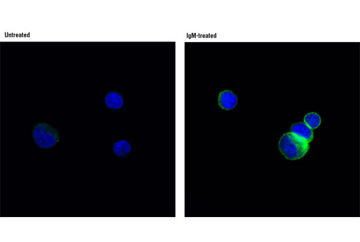
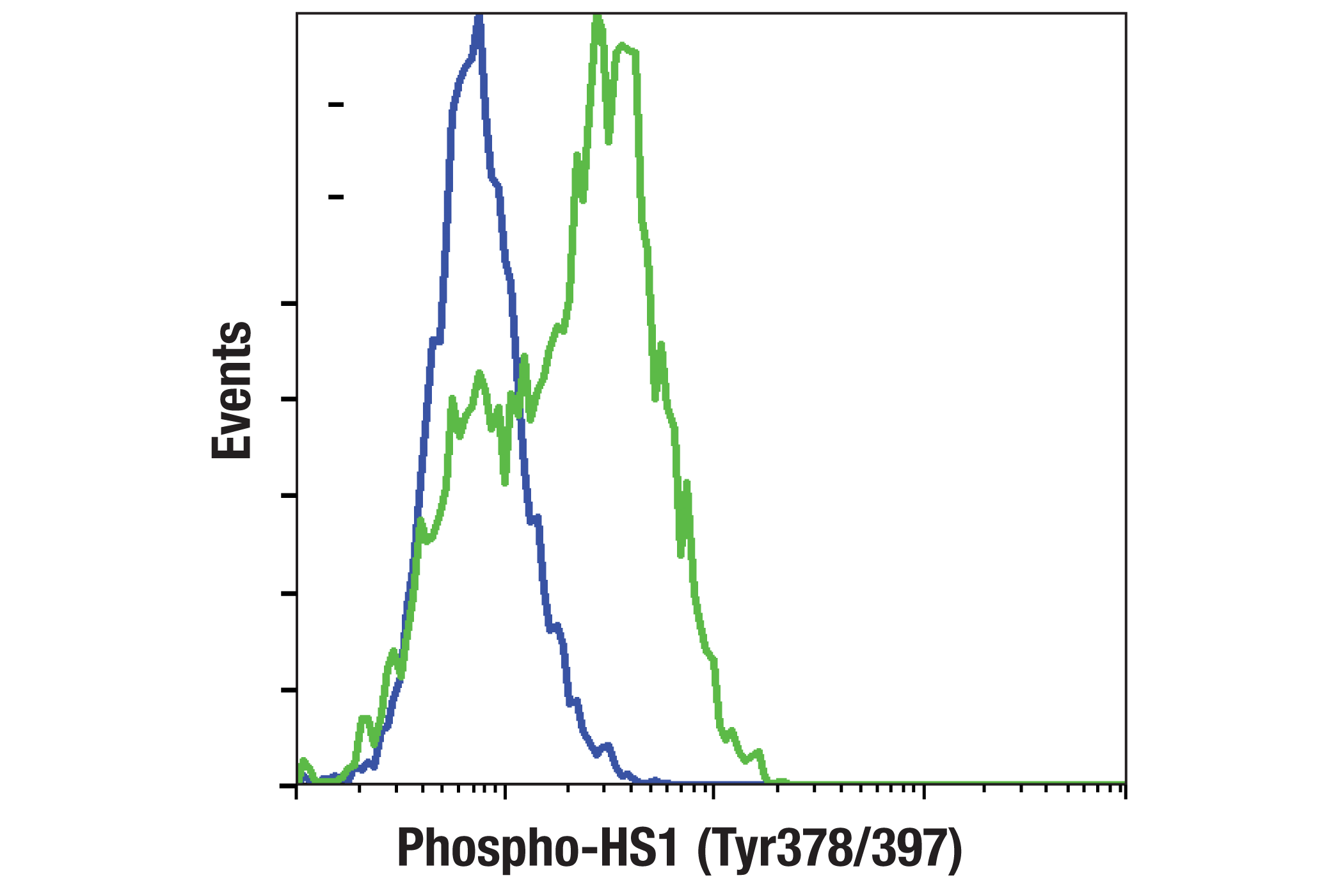
IF-IC, FC-FP
H
Endogenous
80
Rabbit IgG
#P14317
3059
Product Information
Product Usage Information
| Application | Dilution |
|---|---|
| Immunofluorescence (Immunocytochemistry) | 1:50 |
| Flow Cytometry (Fixed/Permeabilized) | 1:200 |
Storage
Specificity / Sensitivity
Species Reactivity:
Human
Species predicted to react based on 100% sequence homology
The antigen sequence used to produce this antibody shares
100% sequence homology with the species listed here, but
reactivity has not been tested or confirmed to work by CST.
Use of this product with these species is not covered under
our
Product Performance Guarantee.
Mouse, Rat
Source / Purification
Monoclonal antibody is produced by immunizing animals with a synthetic phosphopeptide corresponding to residues surrounding Tyr405 of mouse HS1 protein. This site corresponds to Tyr397 of human HS1 protein.
Background
HS1 (HCLS1, LckBP1, p75) is a protein kinase substrate that is expressed only in tissues and cells of hematopoietic origin (1,2). HS1 contains four cortactin repeats and a single SH3 domain (2). This intracellular protein is phosphorylated following immune receptor activation, which promotes recruitment of HS1 to the immune synapse (3-5). Phosphorylation of HS1 is required to regulate actin dynamics and provide docking sites for many other signaling molecules, such as Vav1 and PLCγ1 (6). HS1 also plays an important role in platelet activation (7).
HS1 is rapidly phosphorylated at Tyr397 by Syk and/or Lyn kinases following immune receptor stimulation and thrombin-mediated platelet stimulation. This phosphorylation is an important step in cytoskeletal rearrangement and signaling complex formation (6-10).
- Kitamura, D. et al. (1989) Nucleic Acids Res 17, 9367-79.
- Kitamura, D. et al. (1995) Biochem Biophys Res Commun 208, 1137-46.
- Suzuki, H. et al. (1997) J Immunol 159, 5881-8.
- Hata, D. et al. (1994) Immunol Lett 40, 65-71.
- Yamanashi, Y. et al. (1993) Proc Natl Acad Sci USA 90, 3631-5.
- Gomez, T.S. et al. (2006) Immunity 24, 741-52.
- Kahner, B.N. et al. (2007) Blood 110, 2449-56.
- Yamanashi, Y. et al. (1997) J Exp Med 185, 1387-92.
- Hao, J.J. et al. (2004) J Biol Chem 279, 33413-20.
- Brunati, A.M. et al. (2005) J Biol Chem 280, 21029-35.
Species Reactivity
Species reactivity is determined by testing in at least one approved application (e.g., western blot).
Applications Key
IF-IC: Immunofluorescence (Immunocytochemistry) FC-FP: Flow Cytometry (Fixed/Permeabilized)
Cross-Reactivity Key
H: human M: mouse R: rat Hm: hamster Mk: monkey Vir: virus Mi: mink C: chicken Dm: D. melanogaster X: Xenopus Z: zebrafish B: bovine Dg: dog Pg: pig Sc: S. cerevisiae Ce: C. elegans Hr: horse GP: Guinea Pig Rab: rabbit All: all species expected
Trademarks and Patents
Limited Uses
Except as otherwise expressly agreed in a writing signed by a legally authorized representative of CST, the following terms apply to Products provided by CST, its affiliates or its distributors. Any Customer's terms and conditions that are in addition to, or different from, those contained herein, unless separately accepted in writing by a legally authorized representative of CST, are rejected and are of no force or effect.
Products are labeled with For Research Use Only or a similar labeling statement and have not been approved, cleared, or licensed by the FDA or other regulatory foreign or domestic entity, for any purpose. Customer shall not use any Product for any diagnostic or therapeutic purpose, or otherwise in any manner that conflicts with its labeling statement. Products sold or licensed by CST are provided for Customer as the end-user and solely for research and development uses. Any use of Product for diagnostic, prophylactic or therapeutic purposes, or any purchase of Product for resale (alone or as a component) or other commercial purpose, requires a separate license from CST. Customer shall (a) not sell, license, loan, donate or otherwise transfer or make available any Product to any third party, whether alone or in combination with other materials, or use the Products to manufacture any commercial products, (b) not copy, modify, reverse engineer, decompile, disassemble or otherwise attempt to discover the underlying structure or technology of the Products, or use the Products for the purpose of developing any products or services that would compete with CST products or services, (c) not alter or remove from the Products any trademarks, trade names, logos, patent or copyright notices or markings, (d) use the Products solely in accordance with CST Product Terms of Sale and any applicable documentation, and (e) comply with any license, terms of service or similar agreement with respect to any third party products or services used by Customer in connection with the Products.