WB, IF-IC, FC-FP
H
Endogenous
32
Rabbit IgG
#P15927
6118
Product Information
Product Usage Information
| Application | Dilution |
|---|---|
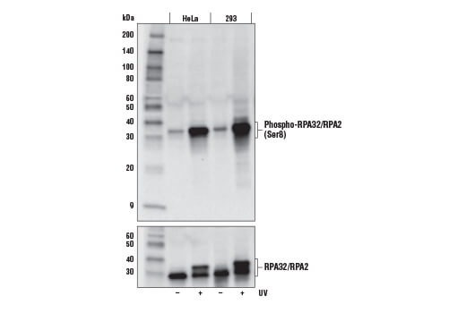
| Western Blotting | 1:1000 |
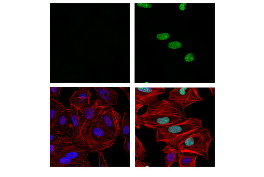
| Immunofluorescence (Immunocytochemistry) | 1:200 - 1:800 |
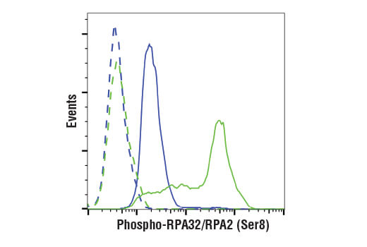
| Flow Cytometry (Fixed/Permeabilized) | 1:400 - 1:1600 |
Storage
Specificity / Sensitivity
Species Reactivity:
Human
Source / Purification
Monoclonal antibody is produced by immunizing animals with a synthetic peptide corresponding to residues surrounding Ser8 of human RPA32/RPA2 protein.
Background
RPA70 (HSSB, REPA1, RF-A, RP-A, p70) is a component of a heterotrimeric complex, composed of 70, 32/30, and 14 kDa subunits, collectively known as RPA. RPA is a single-stranded DNA binding protein, whose DNA binding activity is believed to reside entirely in the 70 kDa subunit. The complex is required for almost all aspects of cellular DNA metabolism such as DNA replication (1-3), recombination, cell cycle and DNA damage checkpoints, and all major types of DNA repair including nucleotide excision, base excision, mismatch, and double-strand break repairs (4-7). In response to genotoxic stress in eukaryotic cells, RPA has been shown to associate with the Rad9/Rad1/Hus1 (9-1-1) checkpoint complex (8). RPA is hyperphosphorylated upon DNA damage or replication stress by checkpoint kinases including ataxia telangiectasia mutated (ATM), ATM and Rad3-related (ATR), and DNA-dependent protein kinase (DNA-PK) (9-11). Phosphorylation of RPA32 occurs at serines 4, 8, and 33 (11). Hyperphosphorylation may alter RPA-DNA and RPA-protein interactions. In addition to the checkpoint partners, RPA interacts with a wide variety of protein partners, including proteins required for normal replication such as RCF, PCNA, and Pol α, and also proteins involved in SV40 replication, such as DNA polymerase I and SV40 large T antigen (10,12).
- Liu, V.F. and Weaver, D.T. (1993) Mol. Cell Biol. 13, 7222-31.
- Wobbe, C.R. et al. (1987) Proc. Natl. Acad. Sci. USA 84, 1834-8.
- Fairman, M.P. and Stillman, B. (1988) EMBO J. 7, 1211-8.
- Wold, M.S. and Kelly, T. (1988) Proc. Natl. Acad. Sci. USA 85, 2523-7.
- Zhou, B.B. and Elledge, S.J. (2000) Nature 408, 433-9.
- Kastan, M.B. and Bartek, J. (2004) Nature 432, 316-23.
- Sancar, A. et al. (2004) Annu. Rev. Biochem. 73, 39-85.
- Guo, S. et al. (2006) J Biol Chem 281, 21607-16.
- Wu, X. et al. (2005) Oncogene 24, 4728-35.
- Binz, S.K. et al. DNA Repair (Amst) 3, 1015-24.
- Nuss, J.E. et al. (2005) Biochemistry 44, 8428-37.
- Yuzhakov, A. et al. (1999) EMBO J. 18, 6189-99.
Species Reactivity
Species reactivity is determined by testing in at least one approved application (e.g., western blot).
Western Blot Buffer
IMPORTANT: For western blots, incubate membrane with diluted primary antibody in 5% w/v BSA, 1X TBS, 0.1% Tween® 20 at 4°C with gentle shaking, overnight.
Applications Key
WB: Western Blotting IF-IC: Immunofluorescence (Immunocytochemistry) FC-FP: Flow Cytometry (Fixed/Permeabilized)
Cross-Reactivity Key
H: human M: mouse R: rat Hm: hamster Mk: monkey Vir: virus Mi: mink C: chicken Dm: D. melanogaster X: Xenopus Z: zebrafish B: bovine Dg: dog Pg: pig Sc: S. cerevisiae Ce: C. elegans Hr: horse GP: Guinea Pig Rab: rabbit All: all species expected
Trademarks and Patents
Limited Uses
Except as otherwise expressly agreed in a writing signed by a legally authorized representative of CST, the following terms apply to Products provided by CST, its affiliates or its distributors. Any Customer's terms and conditions that are in addition to, or different from, those contained herein, unless separately accepted in writing by a legally authorized representative of CST, are rejected and are of no force or effect.
Products are labeled with For Research Use Only or a similar labeling statement and have not been approved, cleared, or licensed by the FDA or other regulatory foreign or domestic entity, for any purpose. Customer shall not use any Product for any diagnostic or therapeutic purpose, or otherwise in any manner that conflicts with its labeling statement. Products sold or licensed by CST are provided for Customer as the end-user and solely for research and development uses. Any use of Product for diagnostic, prophylactic or therapeutic purposes, or any purchase of Product for resale (alone or as a component) or other commercial purpose, requires a separate license from CST. Customer shall (a) not sell, license, loan, donate or otherwise transfer or make available any Product to any third party, whether alone or in combination with other materials, or use the Products to manufacture any commercial products, (b) not copy, modify, reverse engineer, decompile, disassemble or otherwise attempt to discover the underlying structure or technology of the Products, or use the Products for the purpose of developing any products or services that would compete with CST products or services, (c) not alter or remove from the Products any trademarks, trade names, logos, patent or copyright notices or markings, (d) use the Products solely in accordance with CST Product Terms of Sale and any applicable documentation, and (e) comply with any license, terms of service or similar agreement with respect to any third party products or services used by Customer in connection with the Products.