| Product Includes | Product # | Quantity | Mol. Wt | Isotype/Source |
|---|---|---|---|---|
| Phospho-MAPK/CDK Substrates (PXS*P or S*PXR/K) (34B2) Rabbit mAb | 2325 | 20 µl | Rabbit IgG | |
| Phospho-(Ser) Arg-X-Tyr/Phe-X-pSer Motif Antibody | 2981 | 20 µl | Rabbit | |
| Phospho-(Ser) 14-3-3 Binding Motif (4E2) Mouse mAb | 9606 | 20 µl | Mouse IgG1 | |
| Phospho-CDK Substrate Motif [(K/H)pSP] MultiMab® Rabbit mAb mix | 9477 | 20 µl | Rabbit IgG | |
| Anti-rabbit IgG, HRP-linked Antibody | 7074 | 100 µl | Goat | |
| Anti-mouse IgG, HRP-linked Antibody | 7076 | 100 µl | Horse |
Please visit cellsignal.com for individual component applications, species cross-reactivity, dilutions, protocols, and additional product information.
Description
The Phospho-(Ser) Kinase Substrate Antibody Sampler Kit provides a fast and economical means of evaluating several phospho-Ser kinase substrates. The kit contains enough primary and secondary antibody to perform two Western blot experiments.
Storage
Background
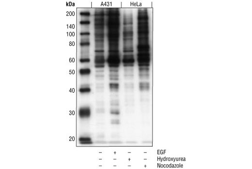
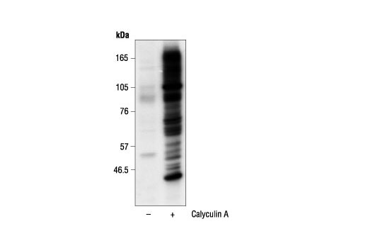
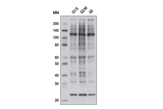
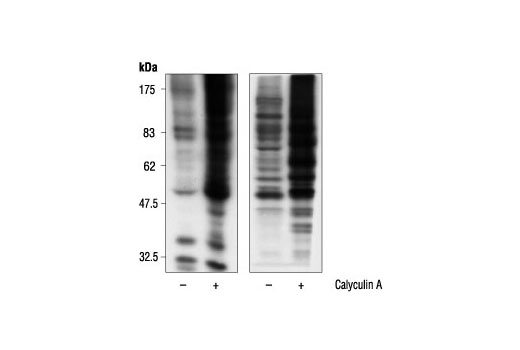
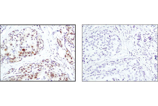
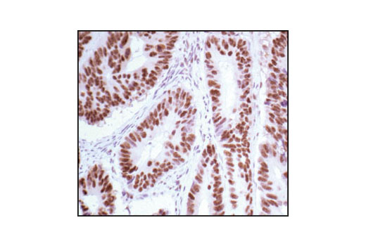
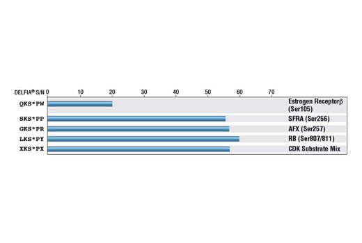
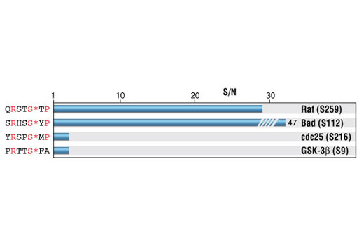
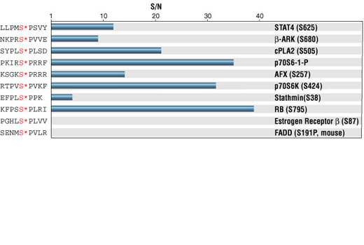
Phospho-(Ser) kinases and phosphatases play critical roles in a wide range of biological processes. Each phospho-(Ser) kinase phosphorylates serine within a specific motif. The MAPK and CDK families of serine protein kinases phosphorylate serine followed by proline residue (1-3). The consensus amino acid sequence for CDK substrate is (K/R)(S*)PX(K/R), where X denotes any one of the 20 amino acids and S* is the phosphorylation site (4-6). MAPK phosphorylates substrates with the concensus sequence PX(S*)P. The 14-3-3 proteins are a highly conserved family of proteins involved in the regulation of cell survival, apoptosis, proliferation and checkpoint control (7-11). Binding of 14-3-3 is mediated through phospho-serine-containing proteins (12). Two different phospho-serine containing motifs are found using a degenerate phospho-serine-oriented peptide library technique, RSXS*XP and RXY/FXS*XP (12). Motif 1 (Arg/Lys and Ser at positions -3 and -2, phospho-Ser at position 0, and Pro at position +2) is found in critical regulatory proteins including Bad, cdc25C, FoxO3A, PKC and c-Raf (11, 13). Motif 2 (RXY/FXS*XP) is found in critical regulatory proteins including cdc25A, cdc25B, PKCγ, IRS-1 and BCR (12). Although Phospho-(Ser) Arg-X-Tyr/Phe-X-pSer Motif Antibody binds 14-3-3 binding motif 2 with no requirement for proline in the +2 position, it provides a powerful tool for the discovery and characterization of potential 14-3-3 binding motif 2-containing proteins or other proteins with the RXY/FXS* motif. Antibodies specific to particular kinase substrates are invaluable reagents in determining kinase activity and identifying potential new kinase substrates. CST has developed antibodies that recognize phosphorylated serine within the context of a protein motif that is phosphorylated by MAPK/CDK, CDKs or 14-3-3. As shown by DELFIA or ELISA, each phospho-(Ser) kinase substrate antibody in this sampler kit is specific to its kinase substrate motif.
- Pearson, R.B. and Kemp, B.E. (1991) Methods Enzymol 200, 62-81.
- Karin, M. (1994) Curr Opin Cell Biol 6, 415-24.
- Lewis, T.S. et al. (1998) Adv Cancer Res 74, 49-139.
- Songyang, Z. et al. (1996) Mol Cell Biol 16, 6486-93.
- Songyang, Z. (1999) Prog Biophys Mol Biol 71, 359-72.
- Holmes, J.K. and Solomon, M.J. (1996) J Biol Chem 271, 25240-6.
- Aitken, A. (1995) Trends Biochem Sci 20, 95-7.
- Zha, J. et al. (1996) Cell 87, 619-28.
- Piwnica-Worms, H. (1999) Nature 401, 535, 537.
- Tzivion, G. et al. (1998) Nature 394, 88-92.
- Xing, H. et al. (2000) EMBO J 19, 349-58.
- Muslin, A.J. et al. (1996) Cell 84, 889-97.
- Yaffe, M.B. et al. (1997) Cell 91, 961-71.
Background References
Trademarks and Patents
Limited Uses
Except as otherwise expressly agreed in a writing signed by a legally authorized representative of CST, the following terms apply to Products provided by CST, its affiliates or its distributors. Any Customer's terms and conditions that are in addition to, or different from, those contained herein, unless separately accepted in writing by a legally authorized representative of CST, are rejected and are of no force or effect.
Products are labeled with For Research Use Only or a similar labeling statement and have not been approved, cleared, or licensed by the FDA or other regulatory foreign or domestic entity, for any purpose. Customer shall not use any Product for any diagnostic or therapeutic purpose, or otherwise in any manner that conflicts with its labeling statement. Products sold or licensed by CST are provided for Customer as the end-user and solely for research and development uses. Any use of Product for diagnostic, prophylactic or therapeutic purposes, or any purchase of Product for resale (alone or as a component) or other commercial purpose, requires a separate license from CST. Customer shall (a) not sell, license, loan, donate or otherwise transfer or make available any Product to any third party, whether alone or in combination with other materials, or use the Products to manufacture any commercial products, (b) not copy, modify, reverse engineer, decompile, disassemble or otherwise attempt to discover the underlying structure or technology of the Products, or use the Products for the purpose of developing any products or services that would compete with CST products or services, (c) not alter or remove from the Products any trademarks, trade names, logos, patent or copyright notices or markings, (d) use the Products solely in accordance with CST Product Terms of Sale and any applicable documentation, and (e) comply with any license, terms of service or similar agreement with respect to any third party products or services used by Customer in connection with the Products.