| Product Includes | Product # | Quantity | Mol. Wt | Isotype/Source |
|---|---|---|---|---|
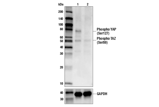
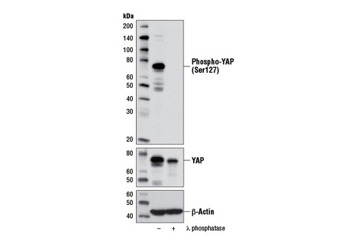
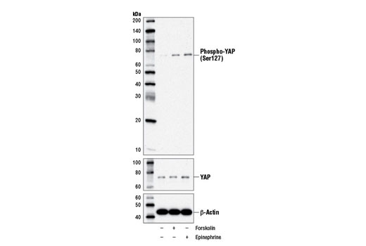
| Phospho-YAP (Ser127) (D9W2I) Rabbit mAb | 13008 | 20 µl | 65-78 kDa | Rabbit IgG |
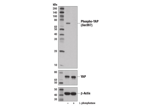
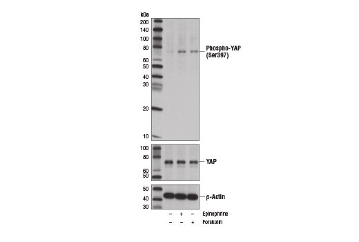
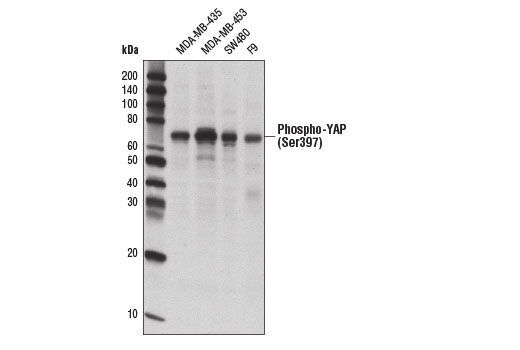
| Phospho-YAP (Ser397) (D1E7Y) Rabbit mAb | 13619 | 20 µl | 65-78 kDa | Rabbit IgG |
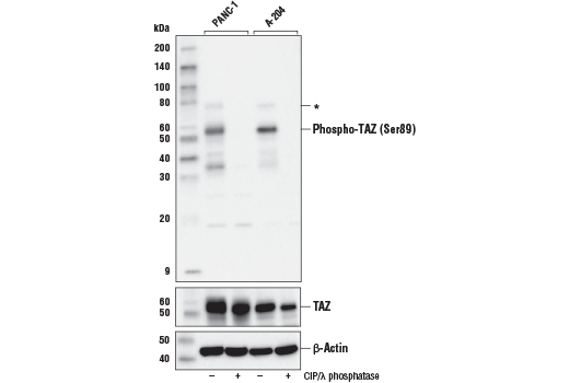
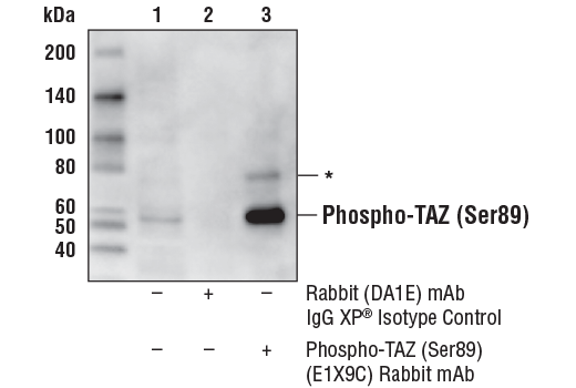
| Phospho-TAZ (Ser89) (E1X9C) Rabbit mAb | 59971 | 20 µl | 55 kDa | Rabbit IgG |
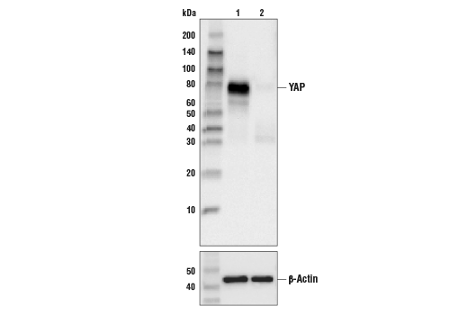
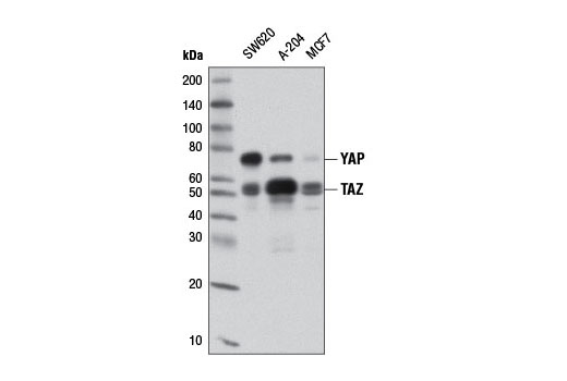
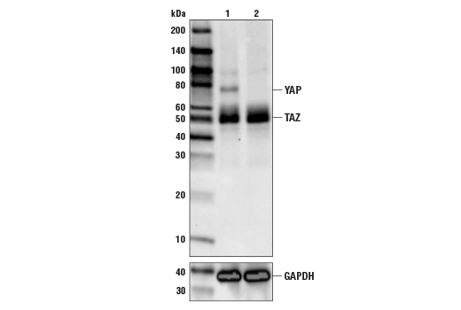
| YAP/TAZ (D24E4) Rabbit mAb | 8418 | 20 µl | 55, 78 kDa | Rabbit IgG |
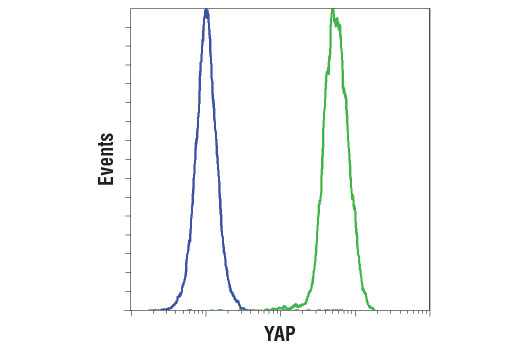
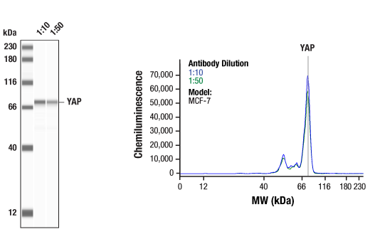
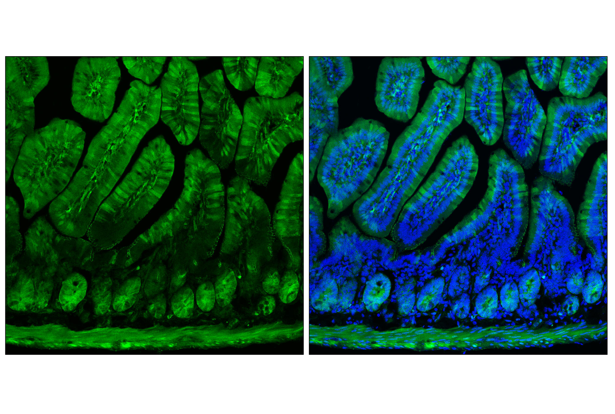
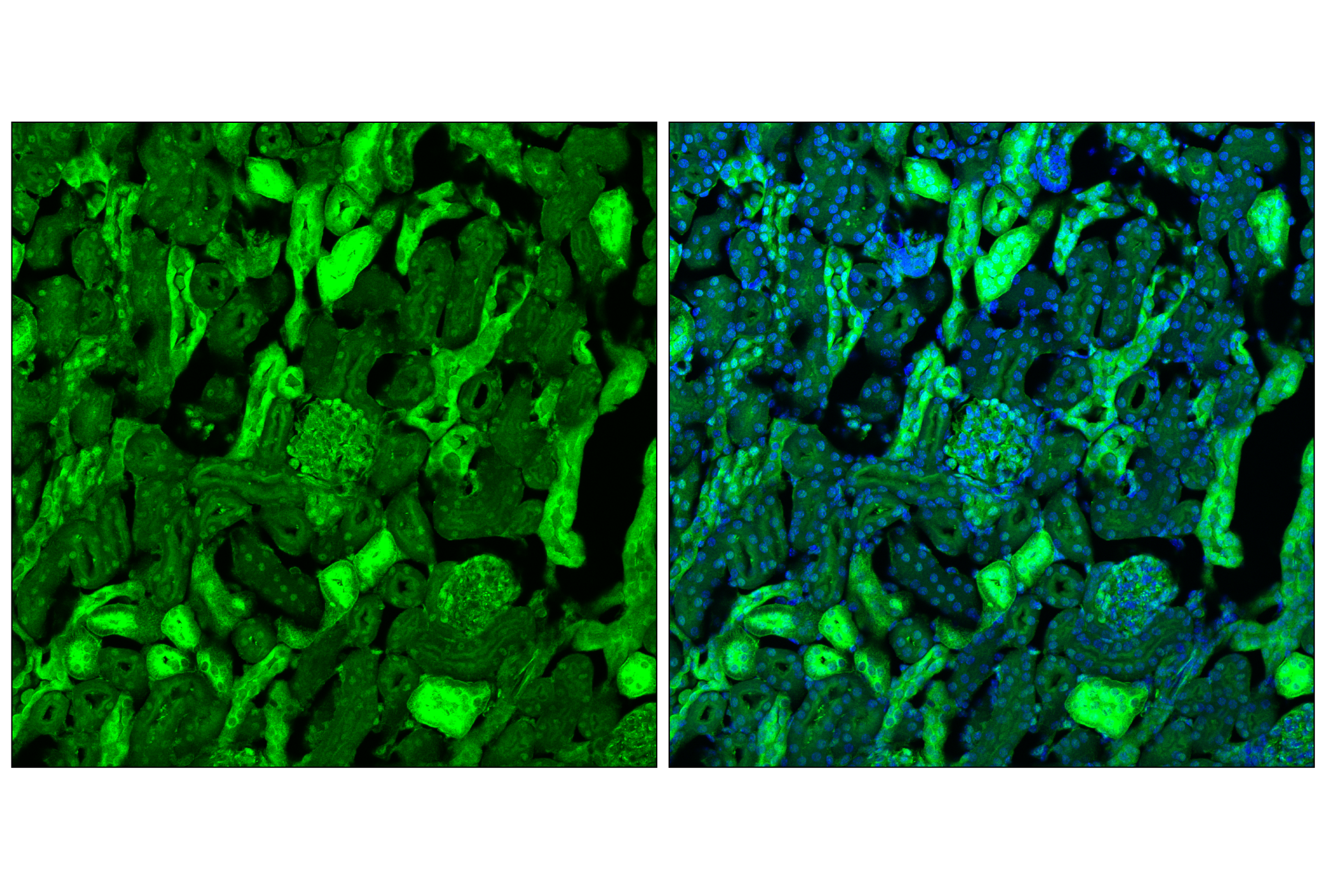
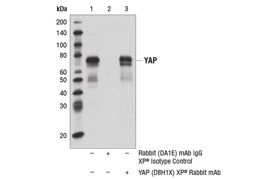
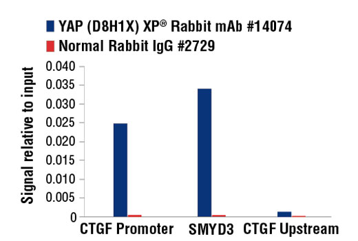
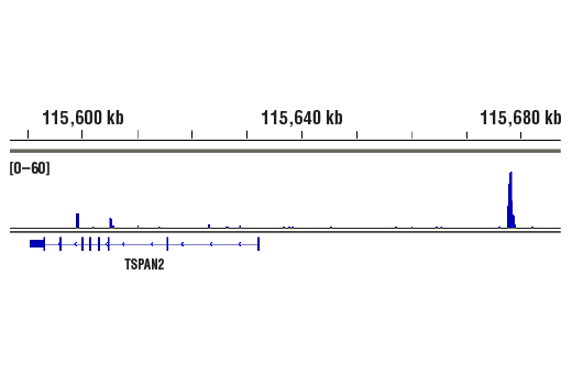
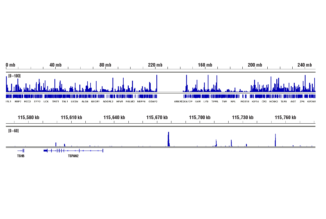
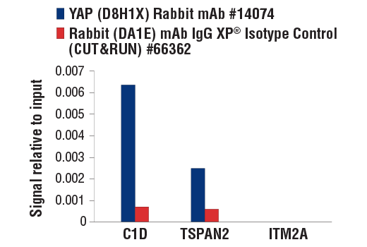
| YAP (D8H1X) XP® Rabbit mAb | 14074 | 20 µl | 65-78 kDa | Rabbit IgG |
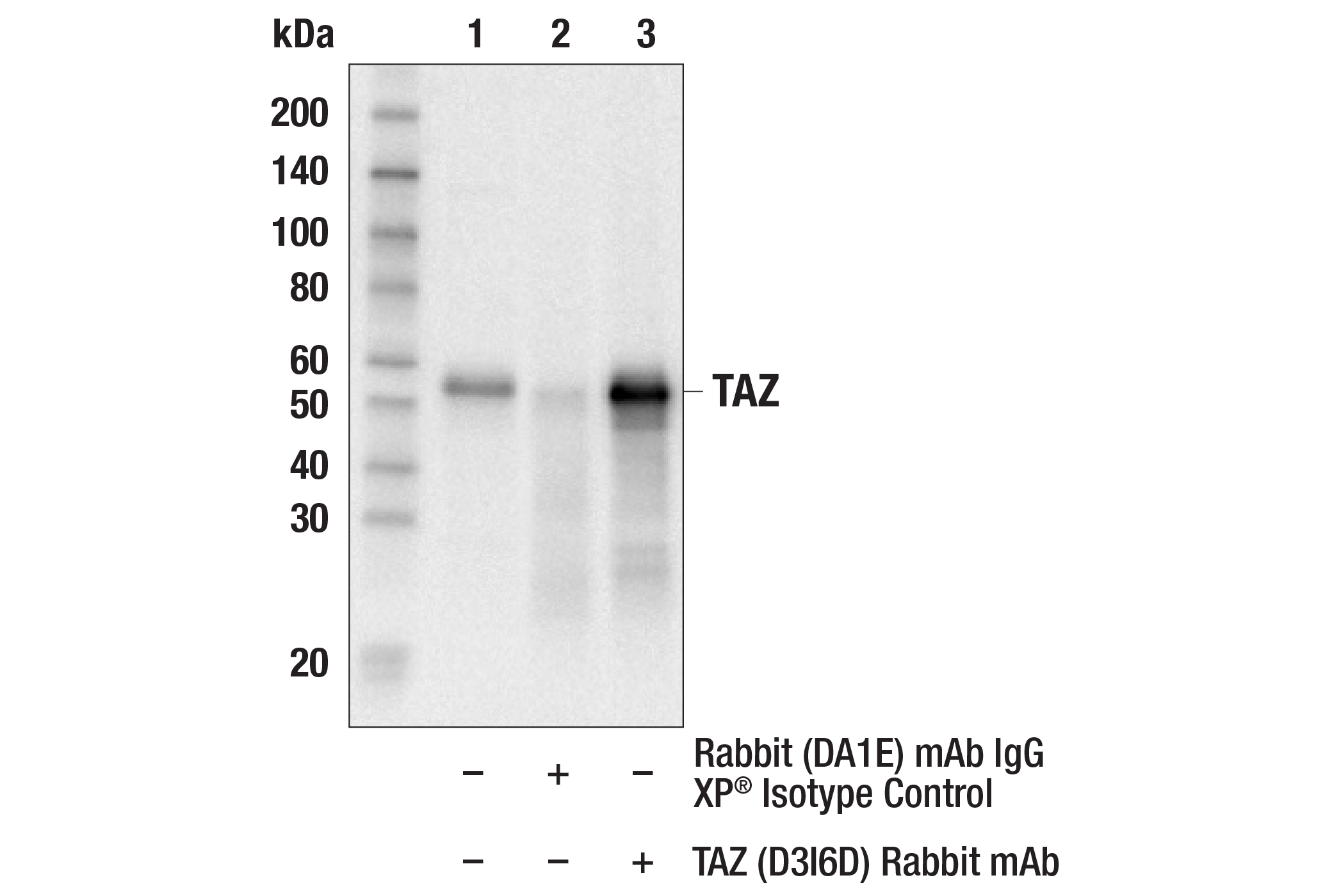
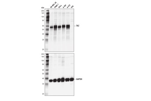
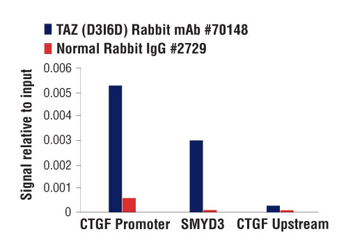
| TAZ (D3I6D) Rabbit mAb | 70148 | 20 µl | 50 kDa | Rabbit IgG |
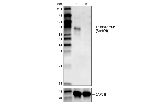
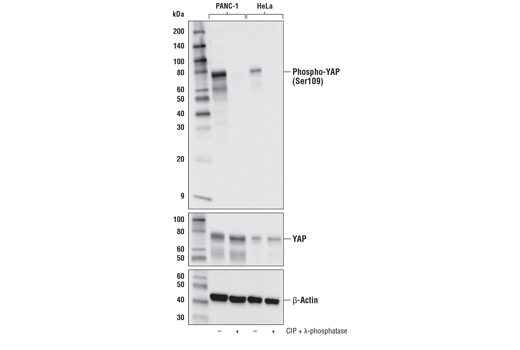
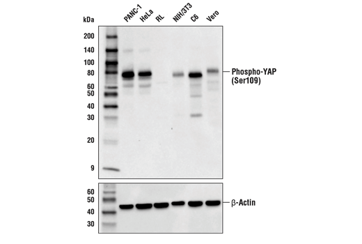
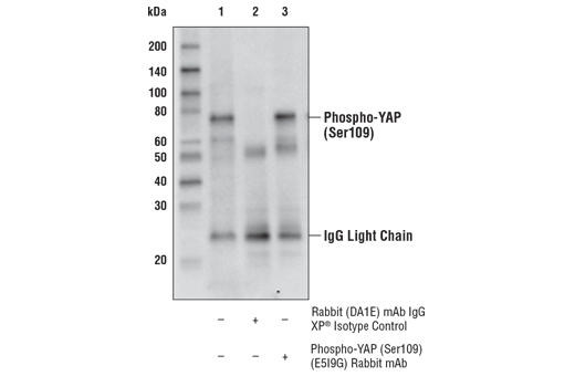
| Phospho-YAP (Ser109) (E5I9G) Rabbit mAb | 53749 | 20 µl | 65-78 kDa | Rabbit IgG |
| Anti-rabbit IgG, HRP-linked Antibody | 7074 | 100 µl | Goat |
Please visit cellsignal.com for individual component applications, species cross-reactivity, dilutions, protocols, and additional product information.
Description
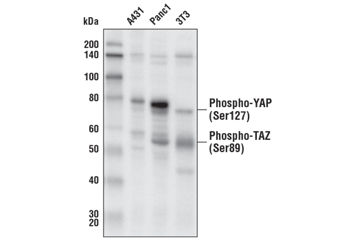
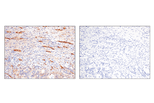
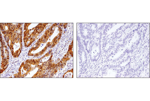
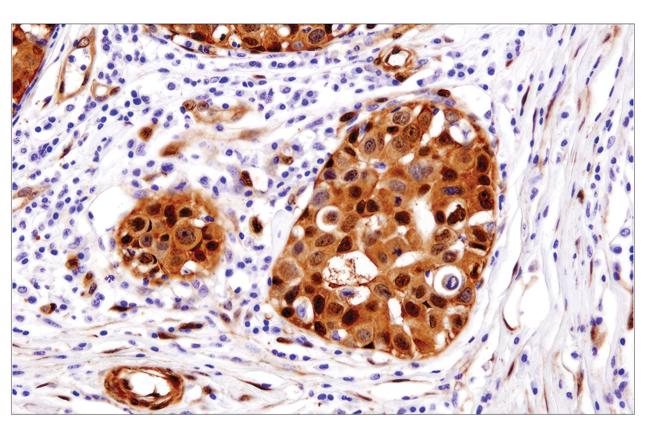
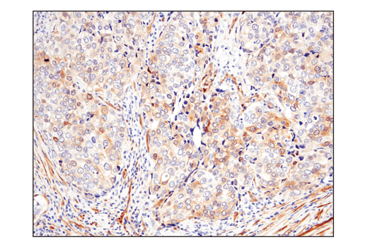
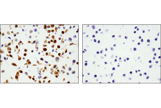
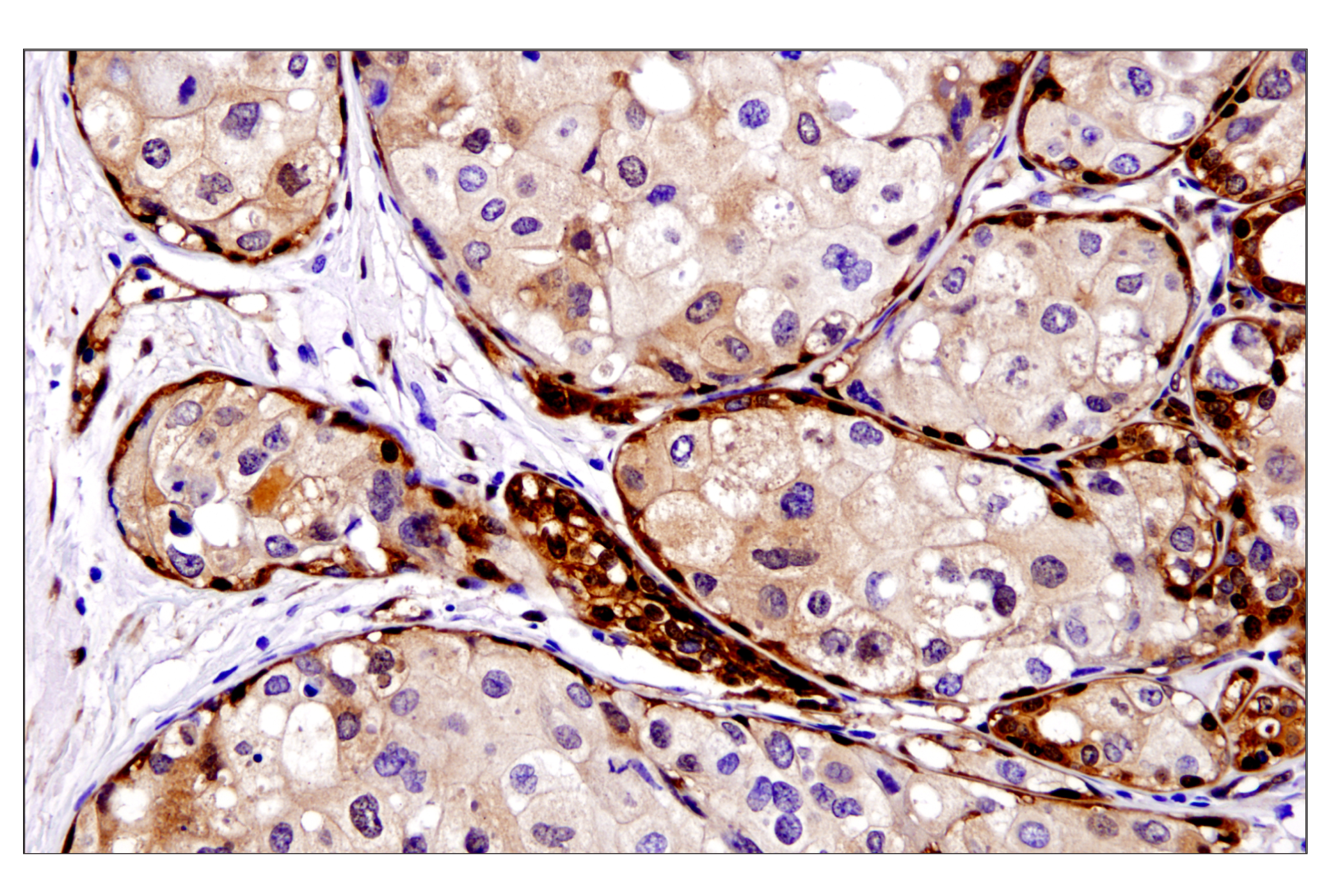
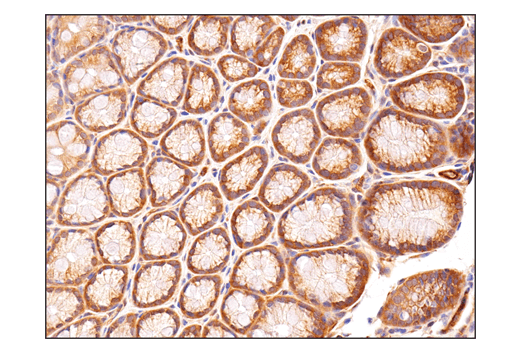
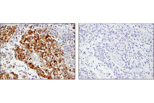
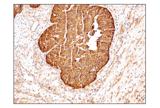
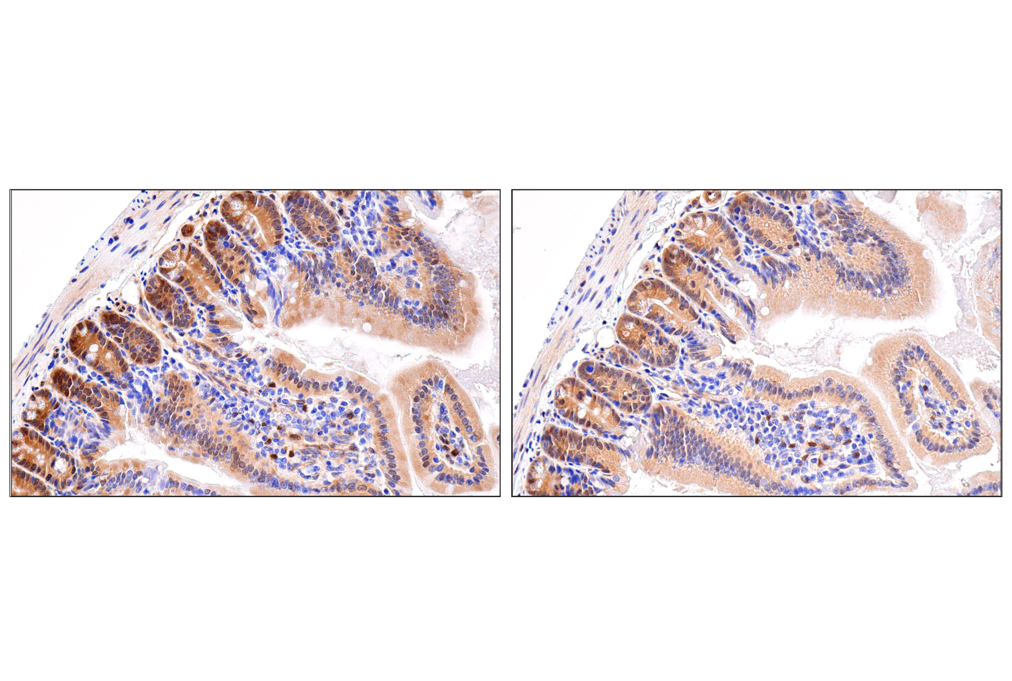
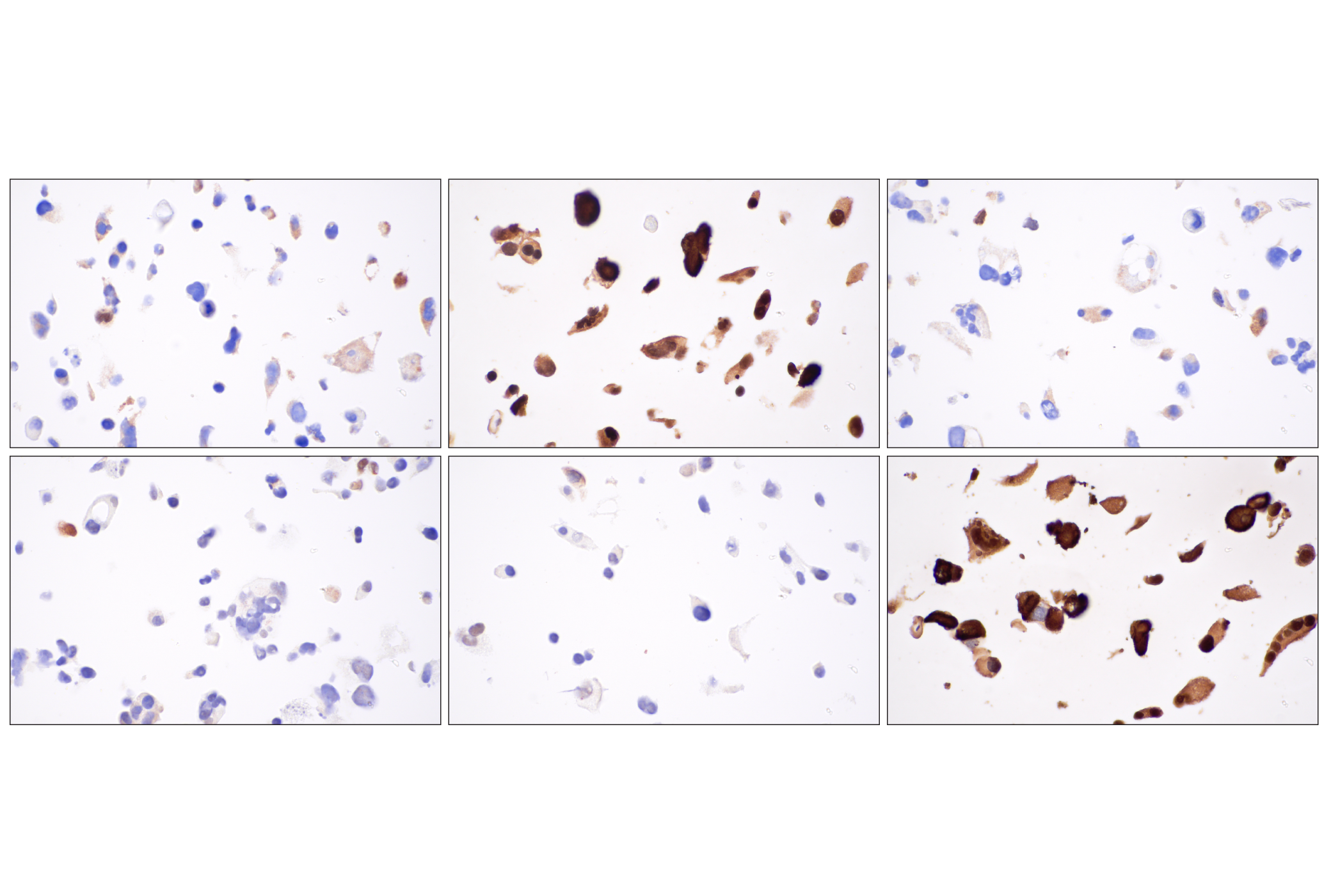
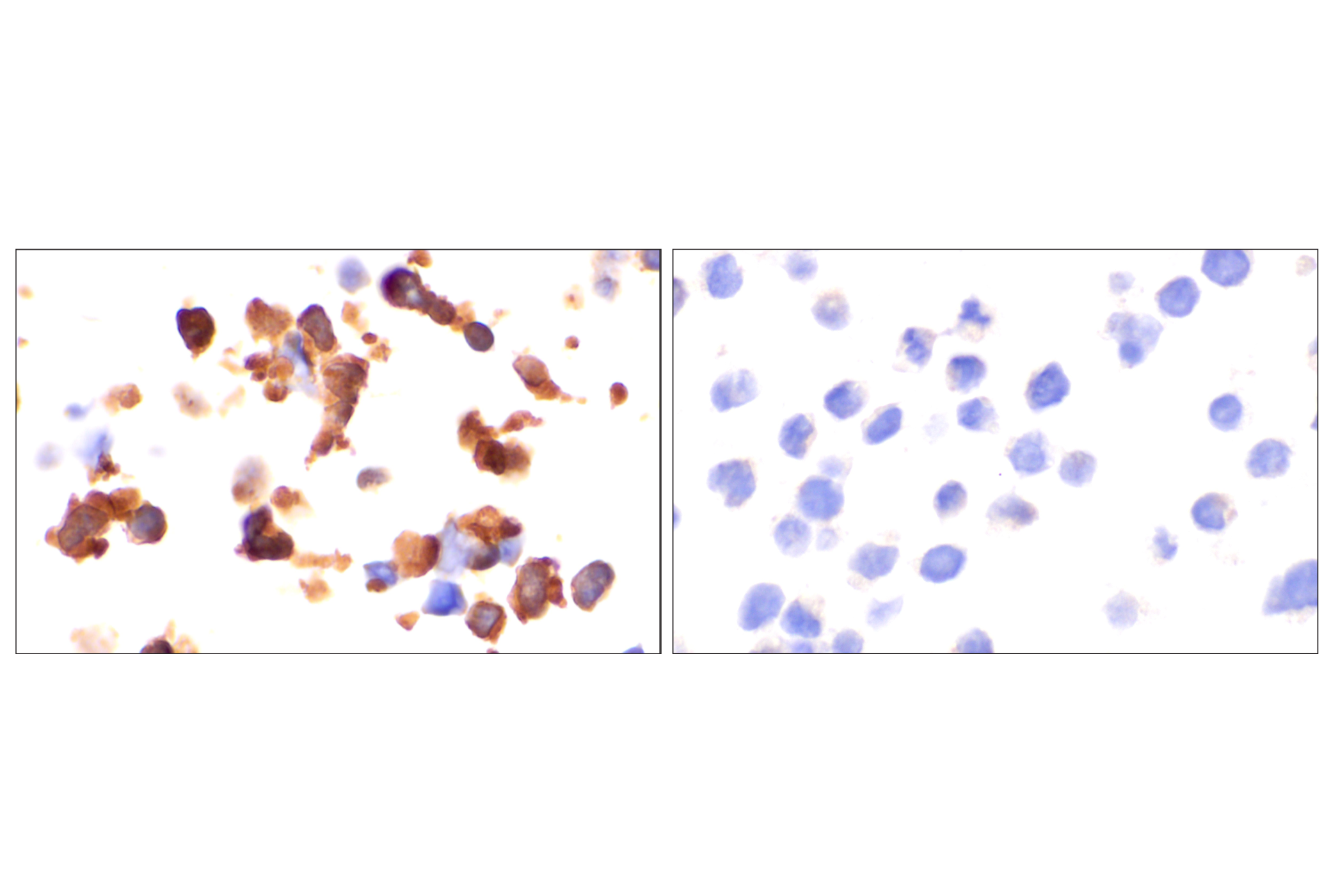
The Phospho-YAP/TAZ Antibody Sampler Kit uses phospho-specific and control antibodies to provide an economical means of detecting the phosphorylation of YAP and TAZ proteins at critical residues that are reported to regulate YAP and TAZ protein stability. The kit includes enough antibody to perform two western blot experiments with each primary antibody.
Storage
Background
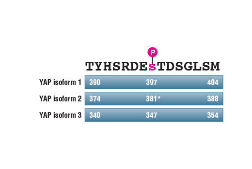
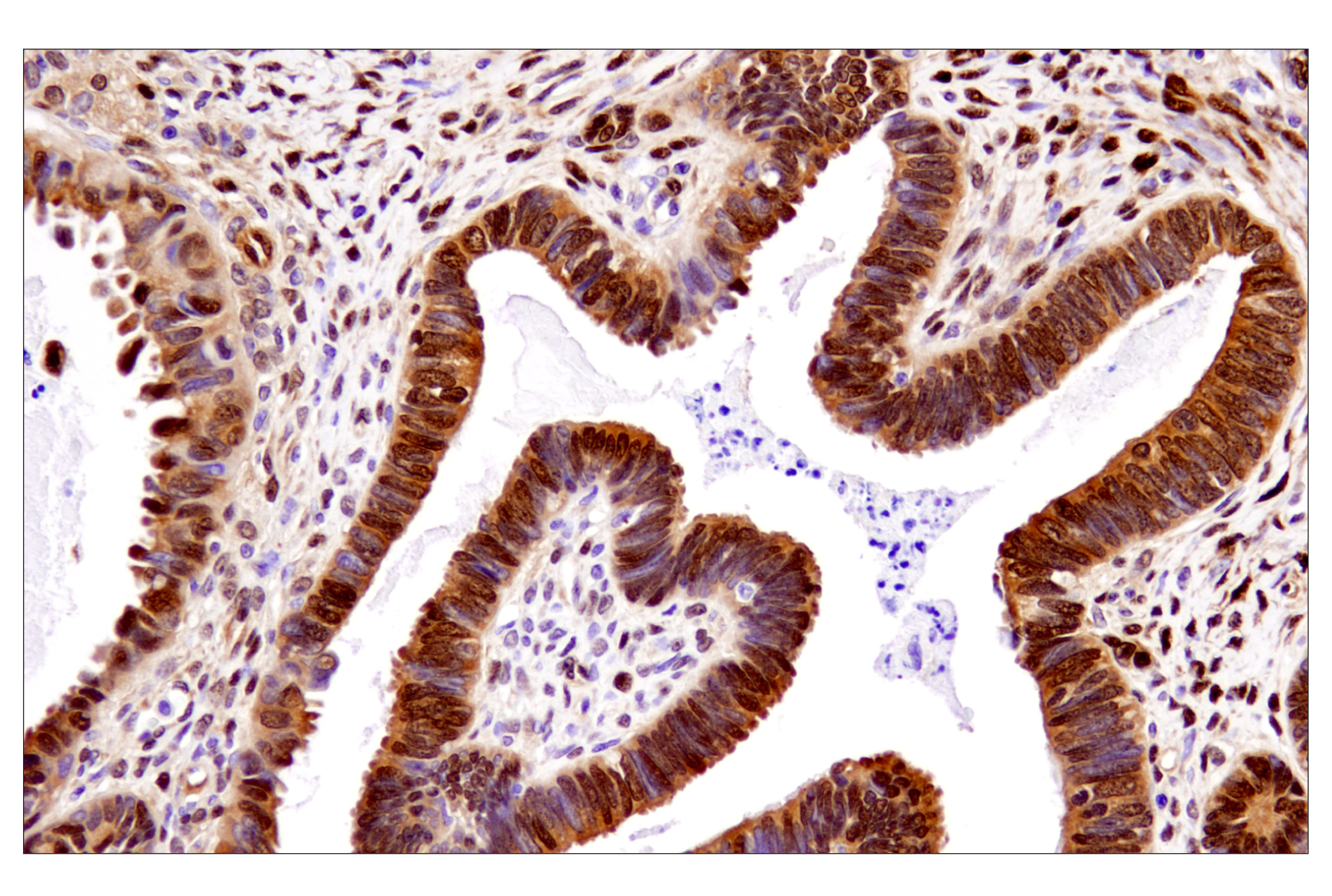
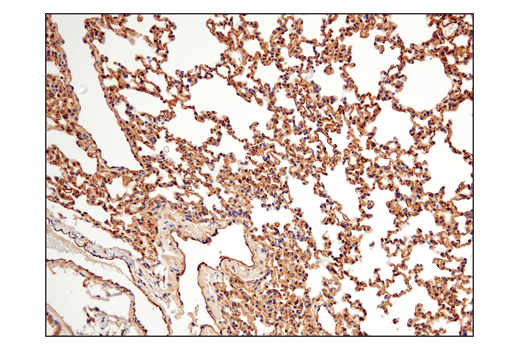
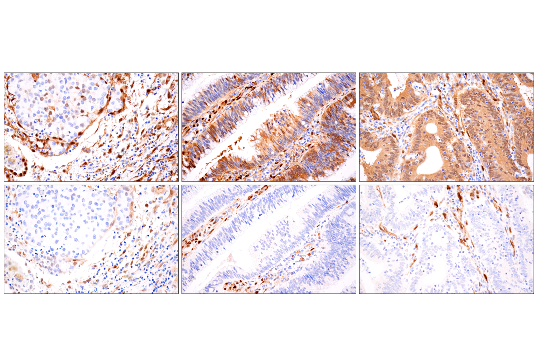
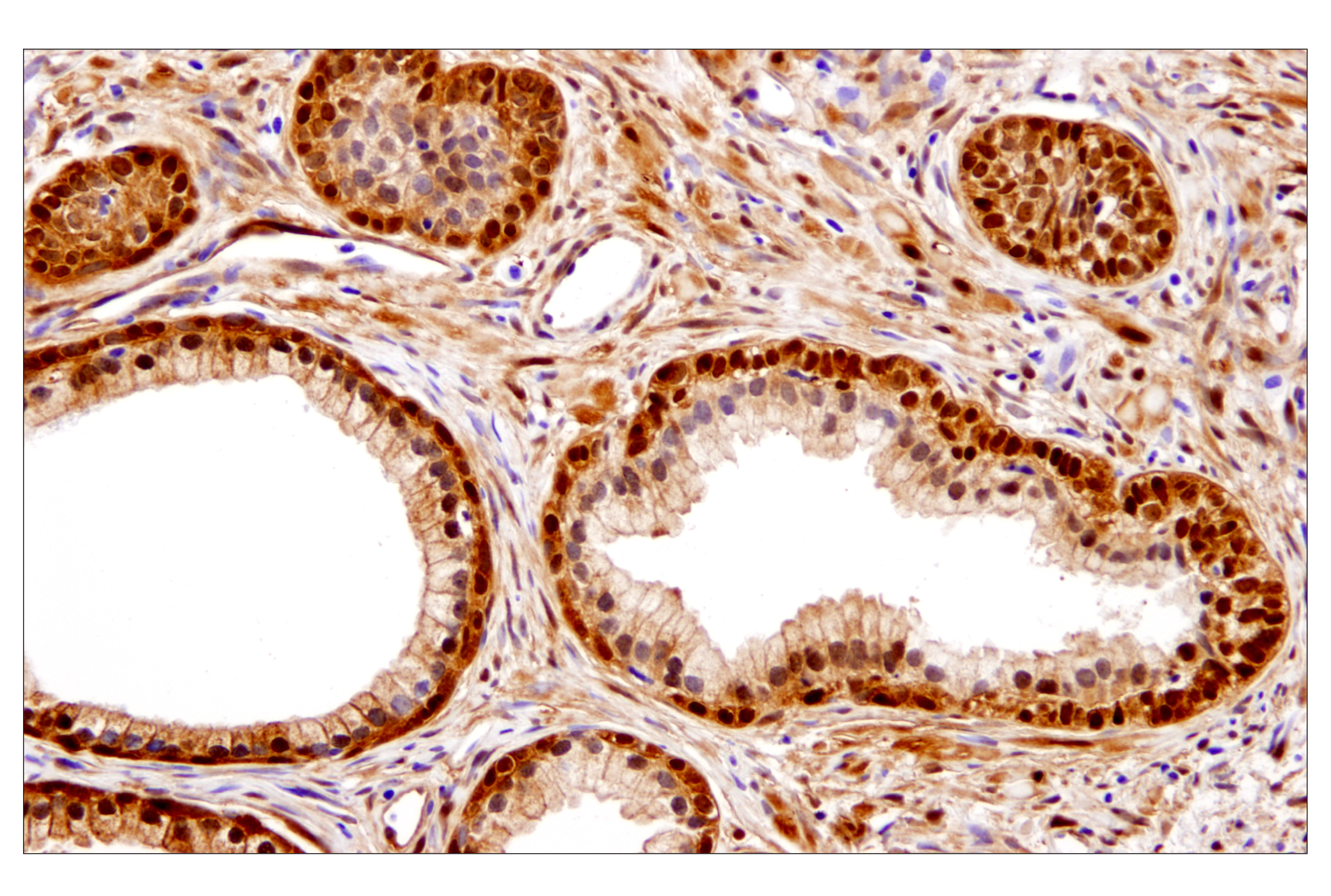
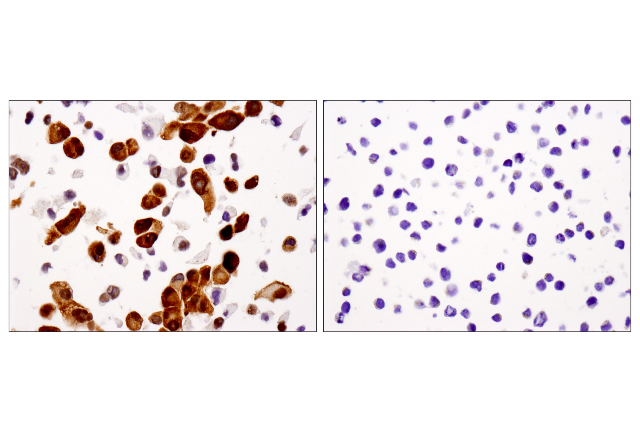
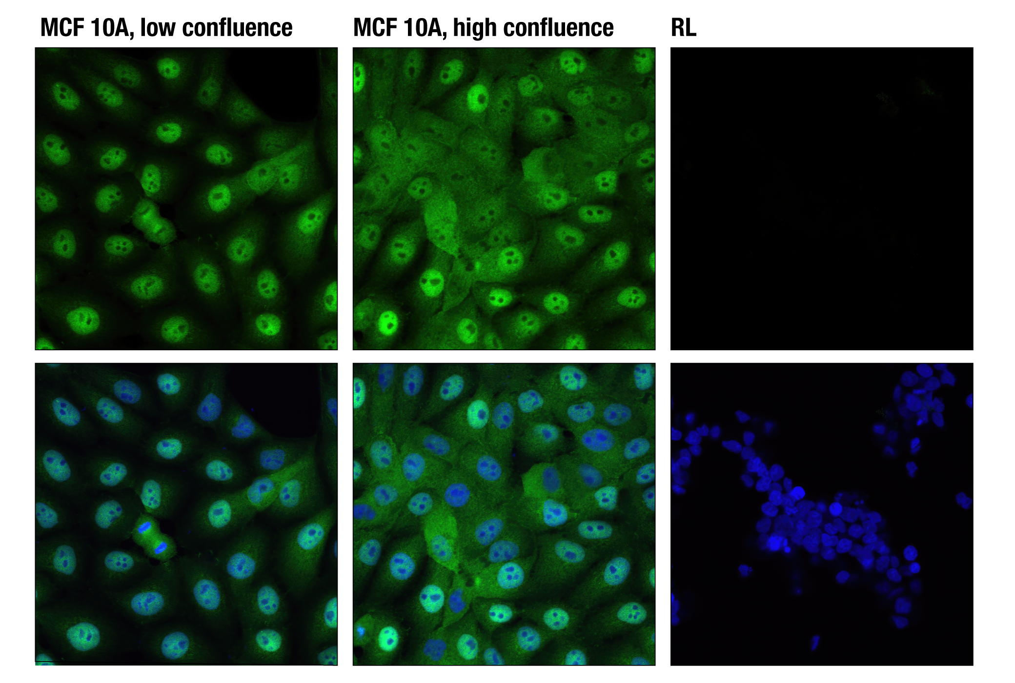
YAP and TAZ (WWTR1) are transcriptional co-activators that play a central role in the Hippo Signaling pathway that regulates cell, tissue and organ growth. YAP and TAZ are structurally and functionally similar, but exhibit differential patterns of expression among cells and tissues that suggest partially non-redundant functions (1). YAP and TAZ are dynamically regulated in response to internal and external cellular signals. Under growth conditions, YAP and TAZ are translocated to the nucleus, where they interact with transcription factors (e.g., TEA domain proteins) that regulate the transcription of genes that control proliferation and cell survival (2). The subcellular localization of YAP and TAZ is dynamically regulated by a kinase cascade that regulates the phosphorylation status of key residues within YAP and TAZ. Phosphorylation of YAP and TAZ (e.g., Ser109, Ser127, Ser397 in YAP; Ser89 in TAZ) results in their cytoplasmic translocation, sequestration by 14-3-3 proteins, and recruitment of the β-TrCP (SCF) ubiquitin ligase complex (3,4). This complex ubiquitinates YAP and TAZ, triggering their proteolytic degradation in the proteasome, thereby altering the transcription of genes that control proliferation and cell survival (3-5).
Background References
Trademarks and Patents
Limited Uses
Except as otherwise expressly agreed in a writing signed by a legally authorized representative of CST, the following terms apply to Products provided by CST, its affiliates or its distributors. Any Customer's terms and conditions that are in addition to, or different from, those contained herein, unless separately accepted in writing by a legally authorized representative of CST, are rejected and are of no force or effect.
Products are labeled with For Research Use Only or a similar labeling statement and have not been approved, cleared, or licensed by the FDA or other regulatory foreign or domestic entity, for any purpose. Customer shall not use any Product for any diagnostic or therapeutic purpose, or otherwise in any manner that conflicts with its labeling statement. Products sold or licensed by CST are provided for Customer as the end-user and solely for research and development uses. Any use of Product for diagnostic, prophylactic or therapeutic purposes, or any purchase of Product for resale (alone or as a component) or other commercial purpose, requires a separate license from CST. Customer shall (a) not sell, license, loan, donate or otherwise transfer or make available any Product to any third party, whether alone or in combination with other materials, or use the Products to manufacture any commercial products, (b) not copy, modify, reverse engineer, decompile, disassemble or otherwise attempt to discover the underlying structure or technology of the Products, or use the Products for the purpose of developing any products or services that would compete with CST products or services, (c) not alter or remove from the Products any trademarks, trade names, logos, patent or copyright notices or markings, (d) use the Products solely in accordance with CST Product Terms of Sale and any applicable documentation, and (e) comply with any license, terms of service or similar agreement with respect to any third party products or services used by Customer in connection with the Products.