IHC-Bond, IHC-P
H
Endogenous
Mouse IgG1
#P54278
5395
Product Information
Product Usage Information
| Application | Dilution |
|---|---|
| IHC Leica Bond | 1:200 - 1:800 |
| Immunohistochemistry (Paraffin) | 1:200 - 1:800 |
Storage
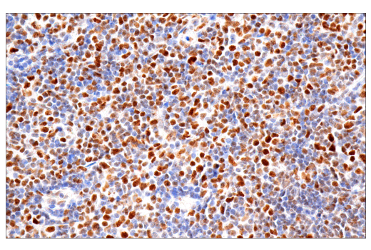
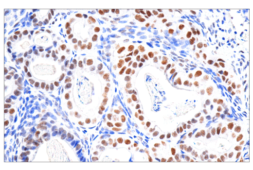
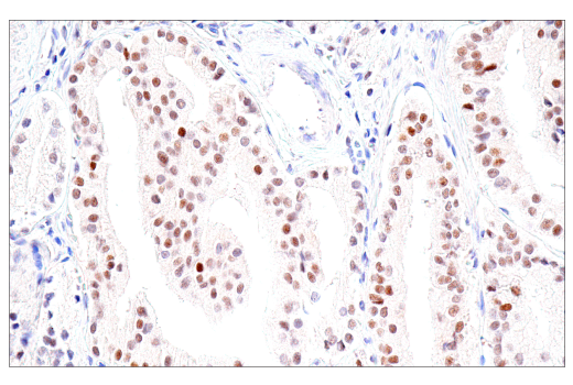
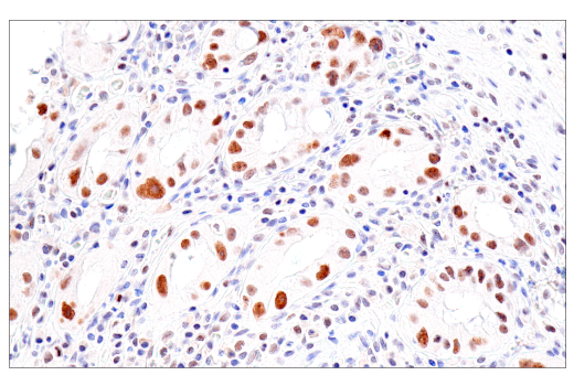
Specificity / Sensitivity
Species Reactivity:
Human
Source / Purification
Monoclonal antibody is produced by immunizing animals with a prokaryotic recombinant protein corresponding to 160 amino acids of the C–terminal region of human PMS2 protein.
Background
DNA mismatch repair (MMR), a conserved process for detecting and correcting errors made during DNA synthesis, is crucial to the maintenance of genomic integrity (1). In prokaryotes, a MutS homodimer recruits a MutL homodimer to sites of DNA mismatches. In eukaryotes, six MutS homologues (MSH1-6) and four MutL homologues (MLH1, PMS2, PMS1, and MLH3) have been identified. Heterodimers composed of two MutL homologues detect distinct DNA mismatch lesions, and heterodimers composed of two MutS homologues perform the repair (2).
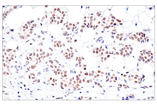
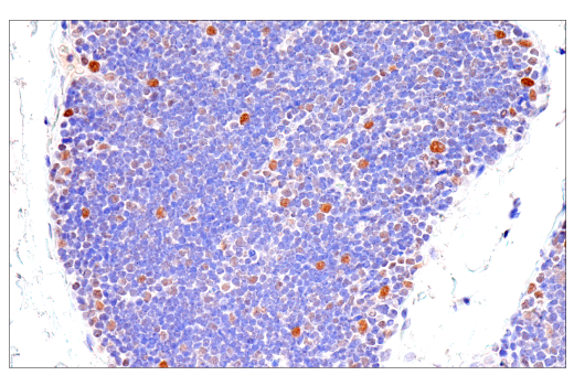
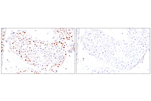
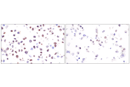
Microsatellite instability (MSI) is a predisposition to genetic mutation resulting from MMR deficiency (dMMR). High MSI (MSI-H) arising from dMMR results in Lynch syndrome, also known as hereditary non-polyposis colorectal cancer (HNPCC). Lynch syndrome is associated with colon cancer, as well as other human cancers (3). MSI and dMMR are strongly associated with tumor responsiveness to immune checkpoint blockade (4,5). MSI status can be determined through PCR amplification of microsatellite markers and/or immunohistochemical detection of MMR proteins MLH1, PMS2, MSH2, and MSH6. The absence of expression of any of these MMR proteins indicates dMMR (3).
Species Reactivity
Species reactivity is determined by testing in at least one approved application (e.g., western blot).
Applications Key
IHC-Bond: IHC Leica Bond IHC-P: Immunohistochemistry (Paraffin)
Cross-Reactivity Key
H: human M: mouse R: rat Hm: hamster Mk: monkey Vir: virus Mi: mink C: chicken Dm: D. melanogaster X: Xenopus Z: zebrafish B: bovine Dg: dog Pg: pig Sc: S. cerevisiae Ce: C. elegans Hr: horse GP: Guinea Pig Rab: rabbit All: all species expected
Trademarks and Patents
Limited Uses
Except as otherwise expressly agreed in a writing signed by a legally authorized representative of CST, the following terms apply to Products provided by CST, its affiliates or its distributors. Any Customer's terms and conditions that are in addition to, or different from, those contained herein, unless separately accepted in writing by a legally authorized representative of CST, are rejected and are of no force or effect.
Products are labeled with For Research Use Only or a similar labeling statement and have not been approved, cleared, or licensed by the FDA or other regulatory foreign or domestic entity, for any purpose. Customer shall not use any Product for any diagnostic or therapeutic purpose, or otherwise in any manner that conflicts with its labeling statement. Products sold or licensed by CST are provided for Customer as the end-user and solely for research and development uses. Any use of Product for diagnostic, prophylactic or therapeutic purposes, or any purchase of Product for resale (alone or as a component) or other commercial purpose, requires a separate license from CST. Customer shall (a) not sell, license, loan, donate or otherwise transfer or make available any Product to any third party, whether alone or in combination with other materials, or use the Products to manufacture any commercial products, (b) not copy, modify, reverse engineer, decompile, disassemble or otherwise attempt to discover the underlying structure or technology of the Products, or use the Products for the purpose of developing any products or services that would compete with CST products or services, (c) not alter or remove from the Products any trademarks, trade names, logos, patent or copyright notices or markings, (d) use the Products solely in accordance with CST Product Terms of Sale and any applicable documentation, and (e) comply with any license, terms of service or similar agreement with respect to any third party products or services used by Customer in connection with the Products.