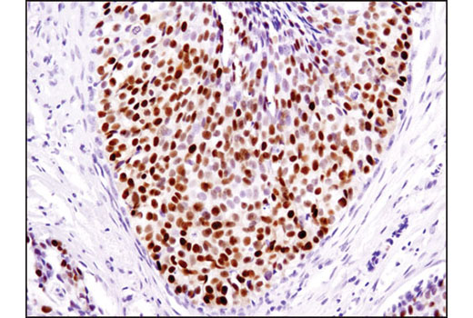
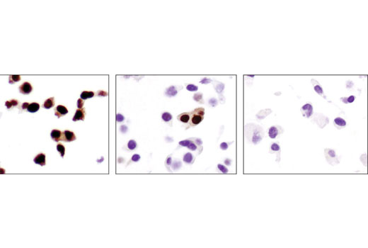
WB, IP, IHC-P, IF-IC, FC-FP, ChIP, ChIP-seq, C&R
H
Endogenous
90 (PR-A), 118 (PR-B)
Rabbit IgG
#P06401
5241
Product Information
Product Usage Information
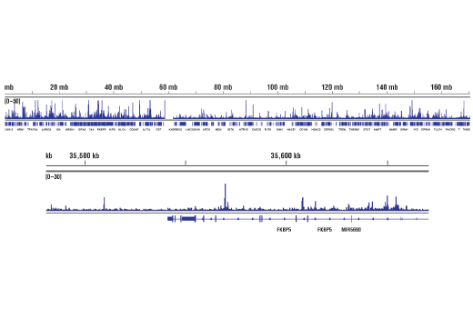
The CUT&RUN dilution was determined using CUT&RUN Assay Kit #86652.
| Application | Dilution |
|---|---|
| Western Blotting | 1:1000 |
| Immunoprecipitation | 1:50 |
| Immunohistochemistry (Paraffin) | 1:500 - 1:2000 |
| Immunofluorescence (Immunocytochemistry) | 1:800 - 1:1600 |
| Flow Cytometry (Fixed/Permeabilized) | 1:400 - 1:1600 |
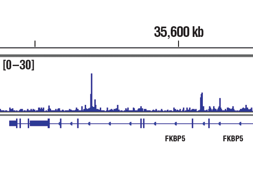
| Chromatin IP | 1:100 |
| Chromatin IP-seq | 1:100 |
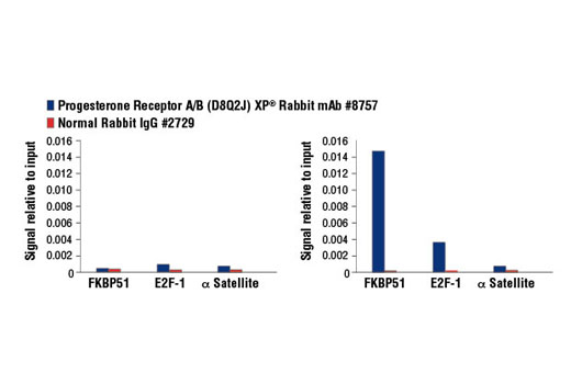
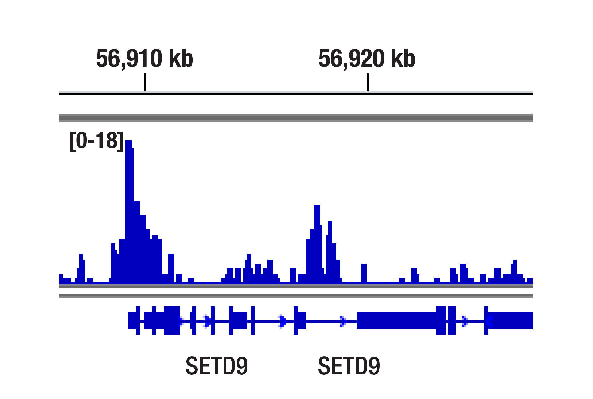
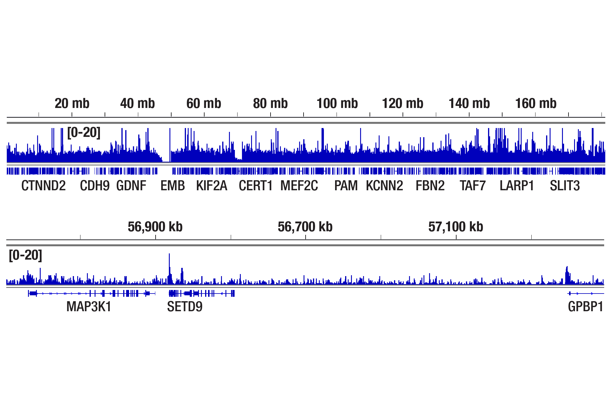
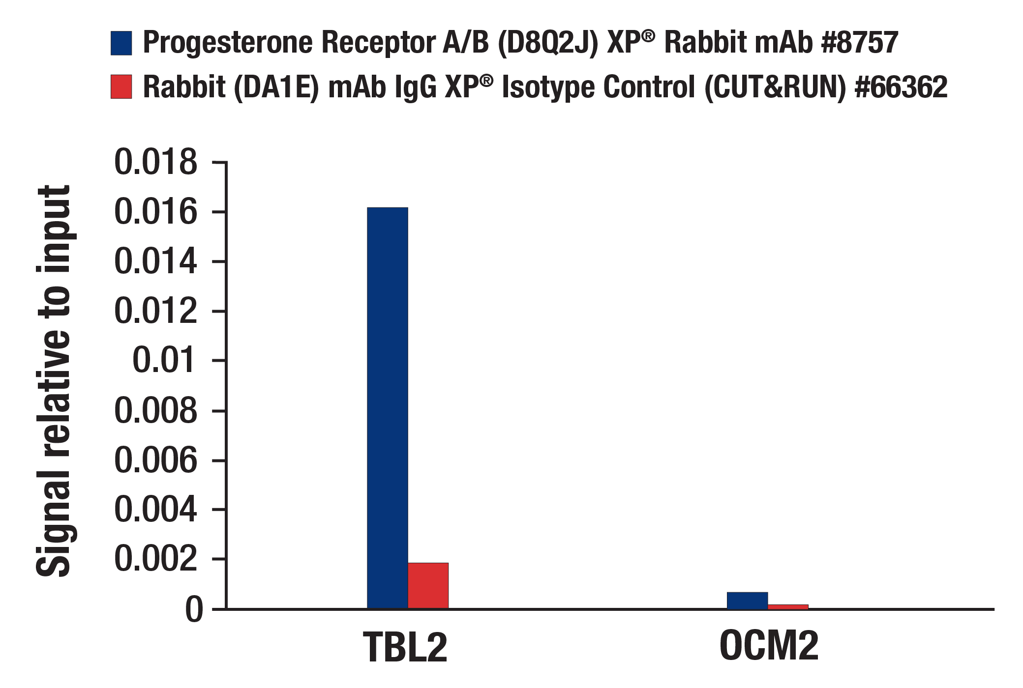
| CUT&RUN | 1:100 |
Storage
For a carrier free (BSA and azide free) version of this product see product #18444.
Specificity / Sensitivity
Species Reactivity:
Human
Species predicted to react based on 100% sequence homology
The antigen sequence used to produce this antibody shares
100% sequence homology with the species listed here, but
reactivity has not been tested or confirmed to work by CST.
Use of this product with these species is not covered under
our
Product Performance Guarantee.
Monkey
Source / Purification
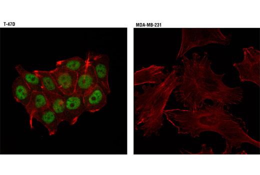
Monoclonal antibody is produced by immunizing animals with a synthetic peptide corresponding to residues surrounding Tyr541 of human progesterone receptor protein.
Background
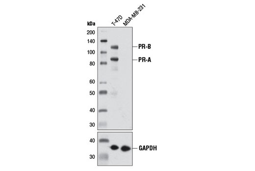
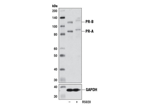
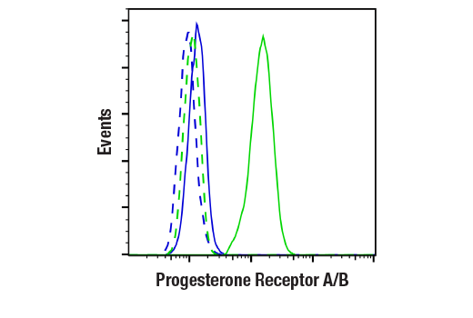
Human progesterone receptor (PR) is expressed as two forms: the full length PR-B and the short form PR-A. PR-A lacks the first 164 amino acid residues of PR-B (1,2). Both PR-A and PR-B are ligand activated, but differ in their relative ability to activate target gene transcription (3,4). The activity of PR is regulated by phosphorylation; at least seven serine residues are phosphorylated in its amino-terminal domain. Three sites (Ser81, Ser102, and Ser162) are unique to full length PR-B, while other sites (Ser190, Ser294, Ser345, and Ser400) are shared by both isoforms (5). Phosphorylation of PR-B at Ser190 (equivalent to Ser26 of PR-A) is catalyzed by CDK2 (6). Mutation of Ser190 results in decreased activity of PR (7), suggesting that the phosphorylation at Ser190 may be critical to its biological function.
- Evans, R.M. (1988) Science 240, 889-895.
- Kastner, P. et al. (1990) EMBO J. 112, 1603-1614.
- Giangrande, P.H. et al. (2000) Mol. Cell. Biol. 20, 3102-3115.
- Wen, D.X. et al. (1994) Mol. Cell. Biol. 14, 8356-8364.
- Clemm, D.L. et al. (2000) Mol. Endocrinol. 14, 52-65.
- Zhang, Y. et al. (1997) Mol. Endocrinol. 11, 823-832.
- Takimoto, G.S. et al. (1996) J. Biol. Chem. 271, 13308-13316.
Species Reactivity
Species reactivity is determined by testing in at least one approved application (e.g., western blot).
Western Blot Buffer
IMPORTANT: For western blots, incubate membrane with diluted primary antibody in 5% w/v nonfat dry milk, 1X TBS, 0.1% Tween® 20 at 4°C with gentle shaking, overnight.
Applications Key
WB: Western Blotting IP: Immunoprecipitation IHC-P: Immunohistochemistry (Paraffin) IF-IC: Immunofluorescence (Immunocytochemistry) FC-FP: Flow Cytometry (Fixed/Permeabilized) ChIP: Chromatin IP ChIP-seq: Chromatin IP-seq C&R: CUT&RUN
Cross-Reactivity Key
H: human M: mouse R: rat Hm: hamster Mk: monkey Vir: virus Mi: mink C: chicken Dm: D. melanogaster X: Xenopus Z: zebrafish B: bovine Dg: dog Pg: pig Sc: S. cerevisiae Ce: C. elegans Hr: horse GP: Guinea Pig Rab: rabbit All: all species expected
Trademarks and Patents
Limited Uses
Except as otherwise expressly agreed in a writing signed by a legally authorized representative of CST, the following terms apply to Products provided by CST, its affiliates or its distributors. Any Customer's terms and conditions that are in addition to, or different from, those contained herein, unless separately accepted in writing by a legally authorized representative of CST, are rejected and are of no force or effect.
Products are labeled with For Research Use Only or a similar labeling statement and have not been approved, cleared, or licensed by the FDA or other regulatory foreign or domestic entity, for any purpose. Customer shall not use any Product for any diagnostic or therapeutic purpose, or otherwise in any manner that conflicts with its labeling statement. Products sold or licensed by CST are provided for Customer as the end-user and solely for research and development uses. Any use of Product for diagnostic, prophylactic or therapeutic purposes, or any purchase of Product for resale (alone or as a component) or other commercial purpose, requires a separate license from CST. Customer shall (a) not sell, license, loan, donate or otherwise transfer or make available any Product to any third party, whether alone or in combination with other materials, or use the Products to manufacture any commercial products, (b) not copy, modify, reverse engineer, decompile, disassemble or otherwise attempt to discover the underlying structure or technology of the Products, or use the Products for the purpose of developing any products or services that would compete with CST products or services, (c) not alter or remove from the Products any trademarks, trade names, logos, patent or copyright notices or markings, (d) use the Products solely in accordance with CST Product Terms of Sale and any applicable documentation, and (e) comply with any license, terms of service or similar agreement with respect to any third party products or services used by Customer in connection with the Products.