WB, IF-IC, FC-FP
H R
Endogenous
23, 28
Mouse IgG1
#P28062
5696
Product Information
Product Usage Information
| Application | Dilution |
|---|---|
| Western Blotting | 1:1000 |
| Immunofluorescence (Immunocytochemistry) | 1:50 |
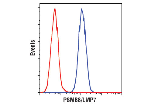
| Flow Cytometry (Fixed/Permeabilized) | 1:50 |
Storage
For a carrier free (BSA and azide free) version of this product see product #60347.
Specificity / Sensitivity
Species Reactivity:
Human, Rat
Source / Purification
Monoclonal antibody is produced by immunizing animals with recombinant protein encompassing the full-length of human PSMB8/LMP7 protein.
Background
The 26S proteasome is a highly abundant proteolytic complex involved in the degradation of ubiquitinated substrate proteins. It consists largely of two sub-complexes, the 20S catalytic core particle (CP) and the 19S/PA700 regulatory particle (RP) that can cap either end of the CP. The CP consists of two stacked heteroheptameric β-rings (β1-7) that contain three catalytic β-subunits and are flanked on either side by two heteroheptameric α-rings (α1-7). The RP includes a base and a lid, each having multiple subunits. The base, in part, is composed of a heterohexameric ring of ATPase subunits belonging to the AAA (ATPases Associated with diverse cellular Activities) family. The ATPase subunits function to unfold the substrate and open the gate formed by the α-subunits, thus exposing the unfolded substrate to the catalytic β-subunits. The lid consists of ubiquitin receptors and DUBs that function in recruitment of ubiquitinated substrates and modification of ubiquitin chain topology (1,2). Other modulators of proteasome activity, such as PA28/11S REG, can also bind to the end of the 20S CP and activate it (1,2).
Constitutively expressed core particle subunits PSMB5, PSMB7, and PSMB6 provide chymotrypsin-like, trypsin-like, and caspase-like activities, respectively (3). In immune cells involved in antigen presentation, these subunits are replaced by highly homologous, induced β-subunits to form the immunoproteasome (4,5).
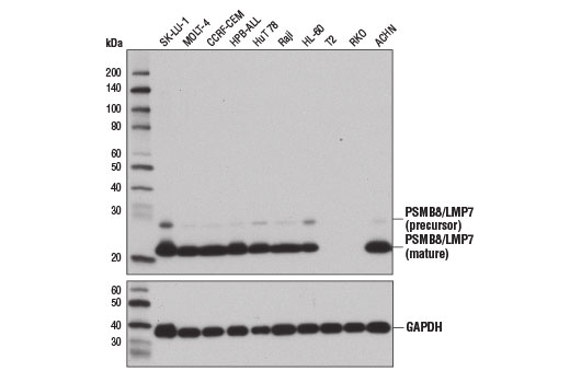
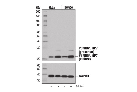
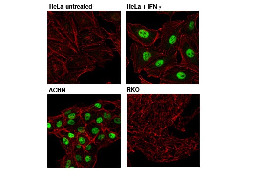
Proteasome subunit beta type-8 (PSMB8, LMP7) is expressed as a proenzyme that is cleaved to form the mature PSMB8 (LMP7) immunoproteasome core particle subunit (6). Interferon-γ induces expression of PSMB8, which functionally replaces the PSMB5 core particle subunit in immunoproteasome processing of MHC class I-restricted peptide antigens (7). Research studies suggest that reduced PSMB8 expression or expression of the non-functional LMP7-E1 isoform may impair immunoproteasome assembly, and that PSMB8 deficiency results in reduced MHC class I molecule expression (8-10). Inhibition of PSMB8 in murine rheumatoid arthritis models attenuates disease indicators, suggesting that PSMB8 is a potential therapeutic target in the treatment of some proinflammatory autoimmune diseases (11). Mutations in the corresponding PSMB8 gene can cause an autoinflammatory syndrome known as CANDLE Syndrome (12).
- Finley, D. (2009) Annu Rev Biochem 78, 477-513.
- Lee, M.J. et al. (2011) Mol Cell Proteomics 10, R110.003871.
- Murata, S. et al. (2009) Nat Rev Mol Cell Biol 10, 104-15.
- Boes, B. et al. (1994) J Exp Med 179, 901-9.
- Cardozo, C. and Kohanski, R.A. (1998) J Biol Chem 273, 16764-70.
- Groettrup, M. et al. (2010) Nat Rev Immunol 10, 73-8.
- Akiyama, K. et al. (1994) FEBS Lett 343, 85-8.
- Heink, S. et al. (2006) Cancer Res 66, 649-52.
- De, M. et al. (2003) J Biol Chem 278, 6153-9.
- Fehling, H.J. et al. (1994) Science 265, 1234-7.
- Muchamuel, T. et al. (2009) Nat Med 15, 781-7.
- Liu, Y. et al. (2012) Arthritis Rheum 64, 895-907.
- Salter, R.D. et al. (1985) Immunogenetics 21, 235-46.
Species Reactivity
Species reactivity is determined by testing in at least one approved application (e.g., western blot).
Western Blot Buffer
IMPORTANT: For western blots, incubate membrane with diluted primary antibody in 5% w/v BSA, 1X TBS, 0.1% Tween® 20 at 4°C with gentle shaking, overnight.
Applications Key
WB: Western Blotting IF-IC: Immunofluorescence (Immunocytochemistry) FC-FP: Flow Cytometry (Fixed/Permeabilized)
Cross-Reactivity Key
H: human M: mouse R: rat Hm: hamster Mk: monkey Vir: virus Mi: mink C: chicken Dm: D. melanogaster X: Xenopus Z: zebrafish B: bovine Dg: dog Pg: pig Sc: S. cerevisiae Ce: C. elegans Hr: horse GP: Guinea Pig Rab: rabbit All: all species expected
Trademarks and Patents
Limited Uses
Except as otherwise expressly agreed in a writing signed by a legally authorized representative of CST, the following terms apply to Products provided by CST, its affiliates or its distributors. Any Customer's terms and conditions that are in addition to, or different from, those contained herein, unless separately accepted in writing by a legally authorized representative of CST, are rejected and are of no force or effect.
Products are labeled with For Research Use Only or a similar labeling statement and have not been approved, cleared, or licensed by the FDA or other regulatory foreign or domestic entity, for any purpose. Customer shall not use any Product for any diagnostic or therapeutic purpose, or otherwise in any manner that conflicts with its labeling statement. Products sold or licensed by CST are provided for Customer as the end-user and solely for research and development uses. Any use of Product for diagnostic, prophylactic or therapeutic purposes, or any purchase of Product for resale (alone or as a component) or other commercial purpose, requires a separate license from CST. Customer shall (a) not sell, license, loan, donate or otherwise transfer or make available any Product to any third party, whether alone or in combination with other materials, or use the Products to manufacture any commercial products, (b) not copy, modify, reverse engineer, decompile, disassemble or otherwise attempt to discover the underlying structure or technology of the Products, or use the Products for the purpose of developing any products or services that would compete with CST products or services, (c) not alter or remove from the Products any trademarks, trade names, logos, patent or copyright notices or markings, (d) use the Products solely in accordance with CST Product Terms of Sale and any applicable documentation, and (e) comply with any license, terms of service or similar agreement with respect to any third party products or services used by Customer in connection with the Products.