WB, IP, IHC-P, eCLIP
H M R Mk Dm
Endogenous
32
Rabbit IgG
#P63244
10399
Product Information
Product Usage Information
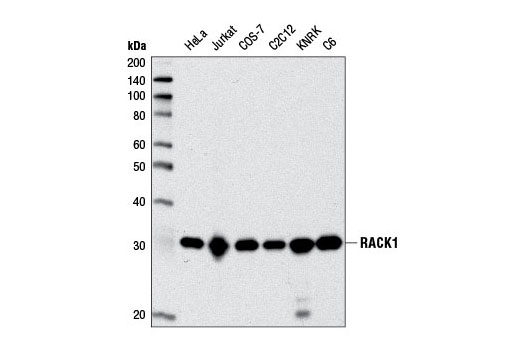
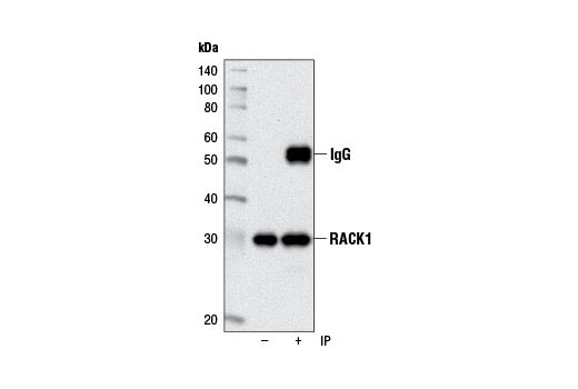
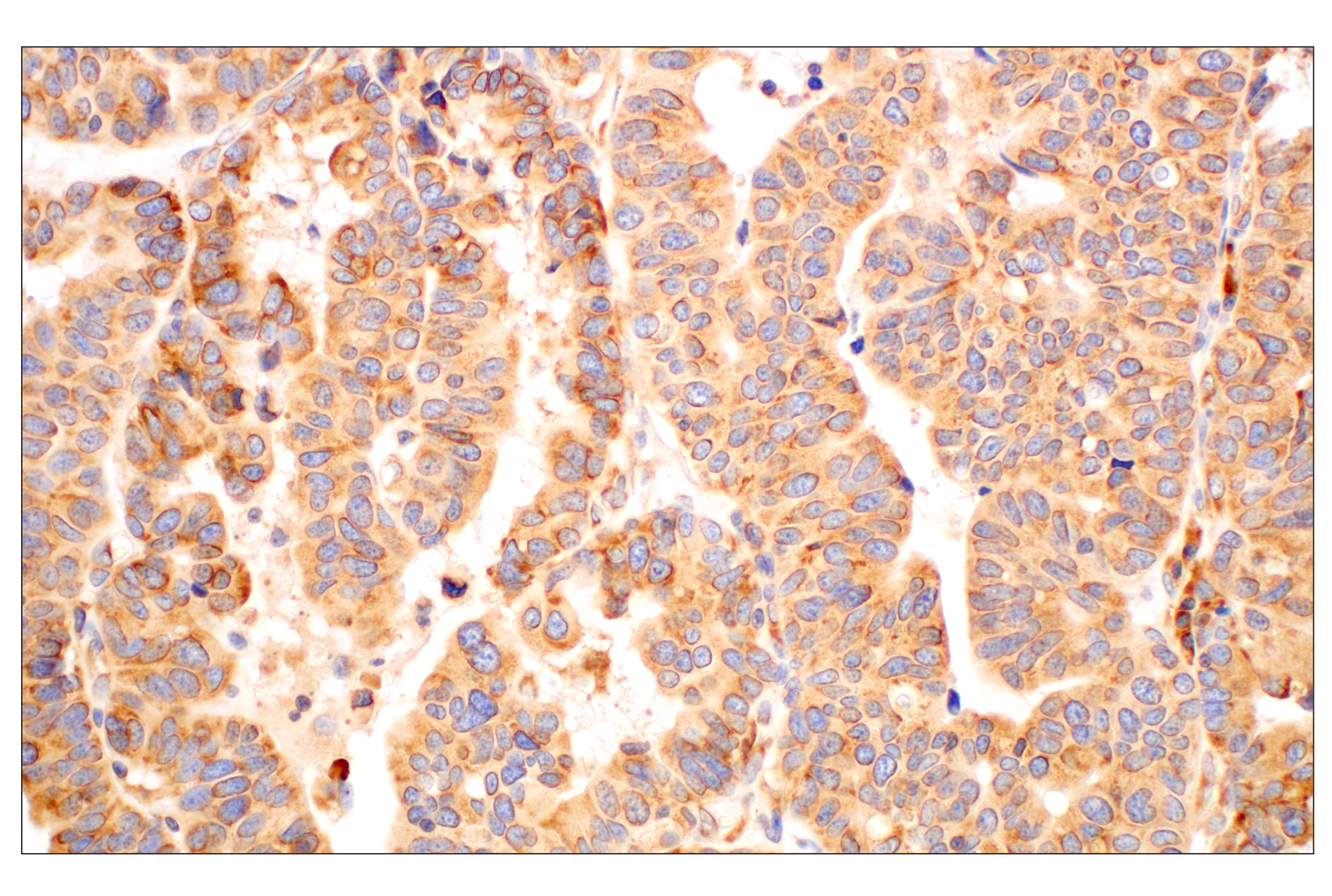
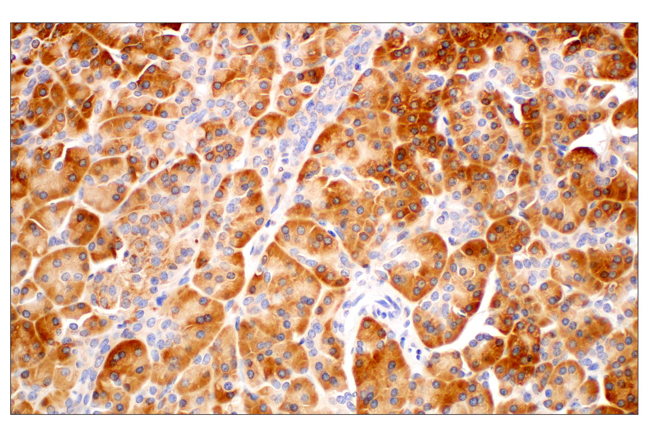
| Application | Dilution |
|---|---|
| Western Blotting | 1:1000 |
| Immunoprecipitation | 1:50 |
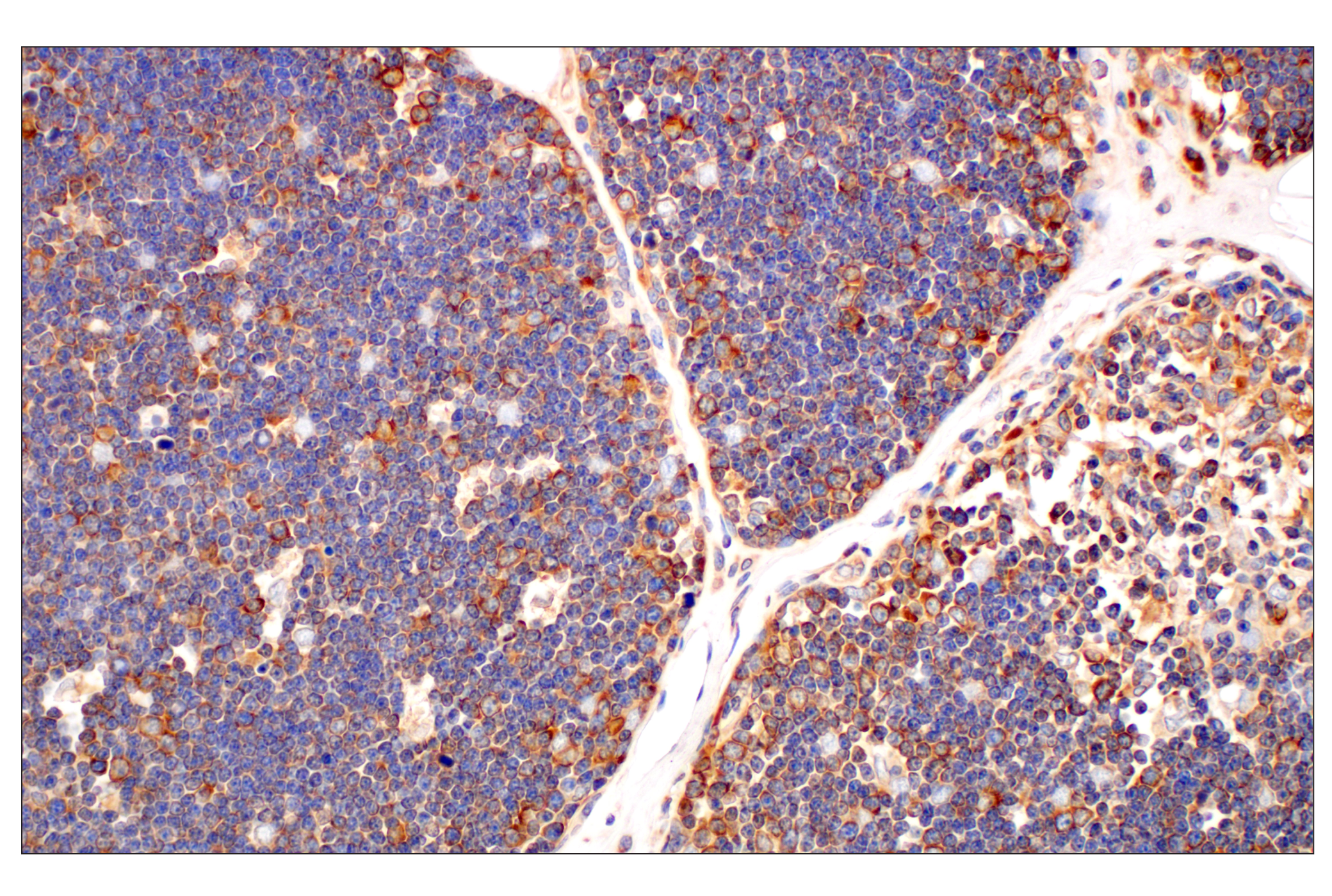
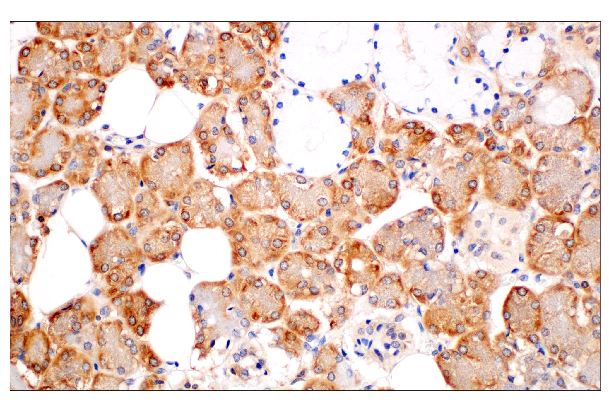
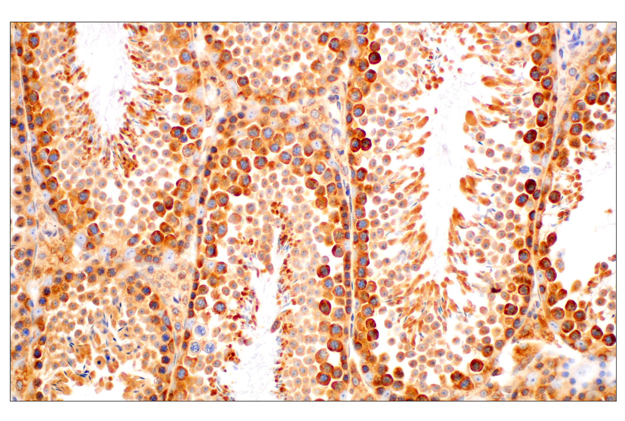
| Immunohistochemistry (Paraffin) | 1:400 - 1:1600 |
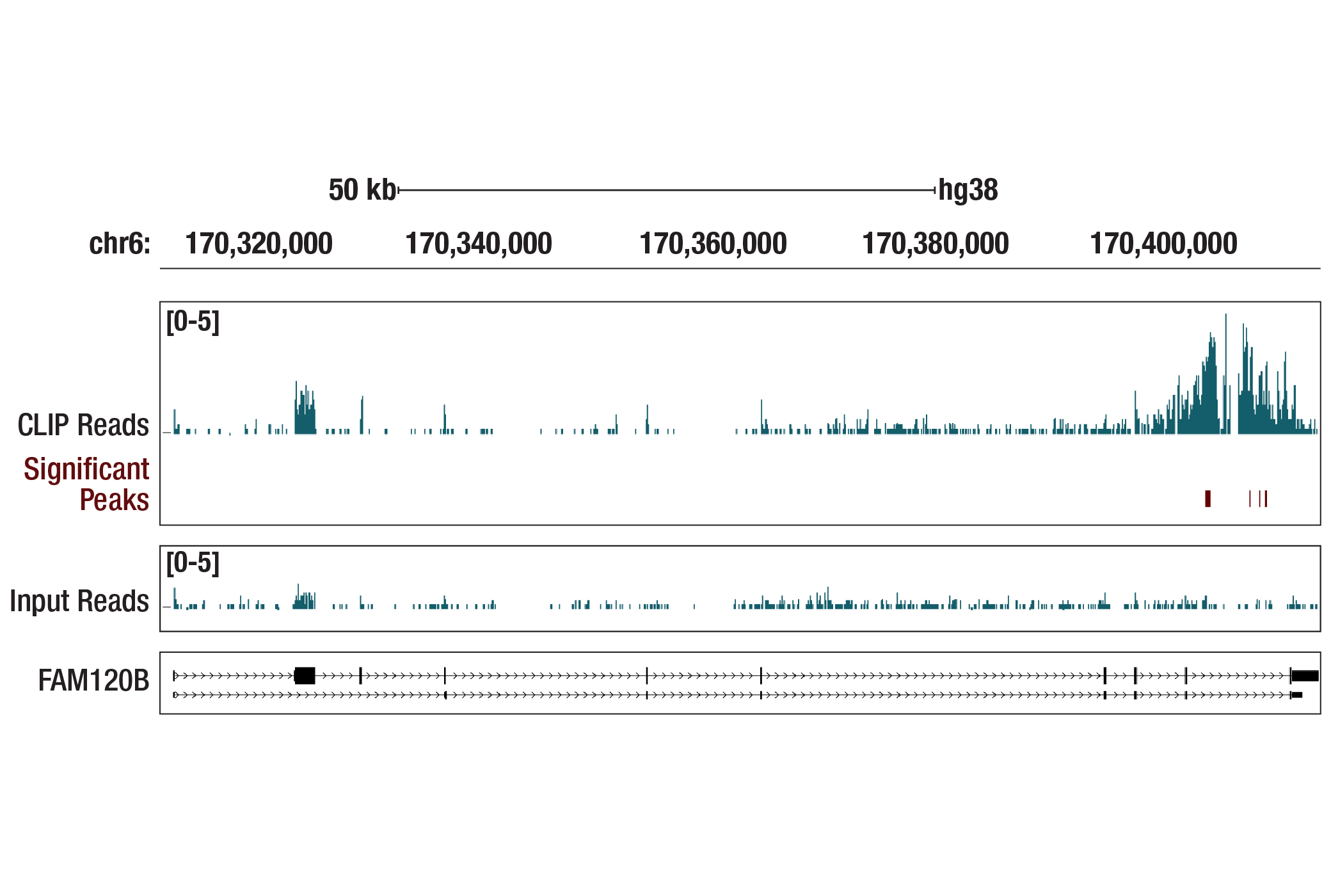
| eCLIP | 1:200 |
For more information about the RBP-eCLIP service please visit Eclipsebio.
Storage
For a carrier free (BSA and azide free) version of this product see product #37124.
Specificity / Sensitivity
Species Reactivity:
Human, Mouse, Rat, Monkey, D. melanogaster
Source / Purification
Monoclonal antibody is produced by immunizing animals with a synthetic peptide corresponding to central residues of human RACK1 protein.
Background
The highly conserved receptor for activated C kinase 1 (RACK1), homologous to the β subunit of heterotrimeric G-proteins, was originally identified through its binding of active PKCβII and other classical PKC isoforms (1). RACK1 is a scaffold protein that recruits PKC and a wide range of other proteins to specific subcellular locations, promoting the formation of multiprotein complexes to induce and integrate various signaling pathways (reviewed in 2). One example of this is its enhancement of PKC-dependent JNK activation (3). RACK1 protein also resides in the eukaryotic ribosome, suggesting the possibility that RACK1 participates in the assembly of signaling complexes that regulate translation as well (reviewed in 4). RACK1 binds the SH2 domain of Src, and phosphorylation of RACK1 by Src occurs at Tyr228 after PKC activation (5).
Species Reactivity
Species reactivity is determined by testing in at least one approved application (e.g., western blot).
Western Blot Buffer
IMPORTANT: For western blots, incubate membrane with diluted primary antibody in 5% w/v nonfat dry milk, 1X TBS, 0.1% Tween® 20 at 4°C with gentle shaking, overnight.
Applications Key
WB: Western Blotting IP: Immunoprecipitation IHC-P: Immunohistochemistry (Paraffin) eCLIP: eCLIP
Cross-Reactivity Key
H: human M: mouse R: rat Hm: hamster Mk: monkey Vir: virus Mi: mink C: chicken Dm: D. melanogaster X: Xenopus Z: zebrafish B: bovine Dg: dog Pg: pig Sc: S. cerevisiae Ce: C. elegans Hr: horse GP: Guinea Pig Rab: rabbit All: all species expected
Trademarks and Patents
Limited Uses
Except as otherwise expressly agreed in a writing signed by a legally authorized representative of CST, the following terms apply to Products provided by CST, its affiliates or its distributors. Any Customer's terms and conditions that are in addition to, or different from, those contained herein, unless separately accepted in writing by a legally authorized representative of CST, are rejected and are of no force or effect.
Products are labeled with For Research Use Only or a similar labeling statement and have not been approved, cleared, or licensed by the FDA or other regulatory foreign or domestic entity, for any purpose. Customer shall not use any Product for any diagnostic or therapeutic purpose, or otherwise in any manner that conflicts with its labeling statement. Products sold or licensed by CST are provided for Customer as the end-user and solely for research and development uses. Any use of Product for diagnostic, prophylactic or therapeutic purposes, or any purchase of Product for resale (alone or as a component) or other commercial purpose, requires a separate license from CST. Customer shall (a) not sell, license, loan, donate or otherwise transfer or make available any Product to any third party, whether alone or in combination with other materials, or use the Products to manufacture any commercial products, (b) not copy, modify, reverse engineer, decompile, disassemble or otherwise attempt to discover the underlying structure or technology of the Products, or use the Products for the purpose of developing any products or services that would compete with CST products or services, (c) not alter or remove from the Products any trademarks, trade names, logos, patent or copyright notices or markings, (d) use the Products solely in accordance with CST Product Terms of Sale and any applicable documentation, and (e) comply with any license, terms of service or similar agreement with respect to any third party products or services used by Customer in connection with the Products.