Revision 1
Cell Signaling Technology

Orders: 877-616-CELL (2355) [email protected]

Support: 877-678-TECH (8324)

Web: [email protected] cellsignal.com

3 Trask LaneDanversMassachusetts01923USA
For Research Use Only. Not for Use in Diagnostic Procedures.
:

:

:

:

UniProt ID:

#P06400

Entrez-Gene Id:

5925

Product Information

Storage

Supplied in 10 mM sodium HEPES (pH 7.5), 150 mM NaCl, 100 µg/ml BSA, 50% glycerol and less than 0.02% sodium azide. Store at –20°C. Do not aliquot the antibody.

Specificity / Sensitivity

Phospho-Rb (Ser780) (D59B7) Rabbit mAb and Phospho-Rb (Ser807/811) (D20B12) XP® Rabbit mAb detect endogenous levels of Rb only when phosphorylated at Ser780 and Ser807/811, respectively. Rb (4H1) Mouse mAb detects endogenous levels of total Rb protein. The antibody does not cross-react with the Rb homologues p107 or p130, or with other proteins.

Source / Purification

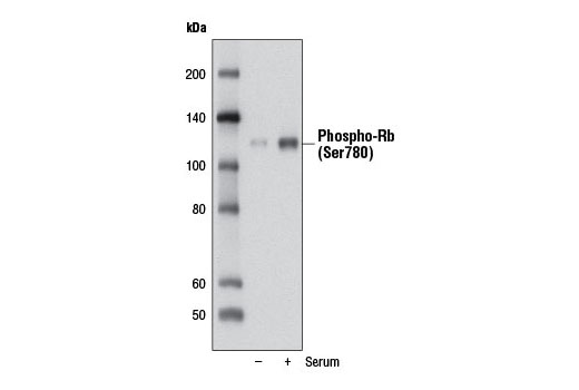
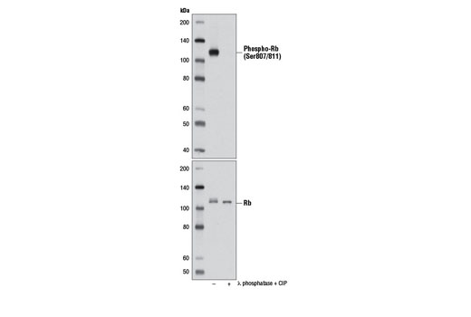
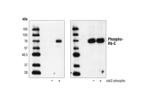
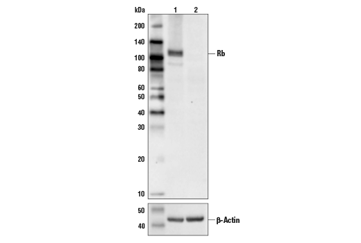
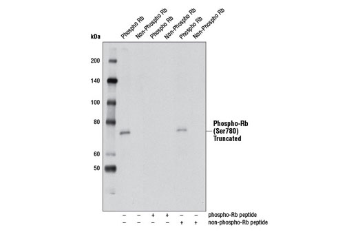
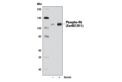
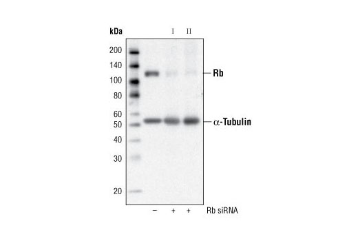
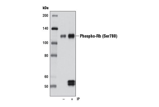
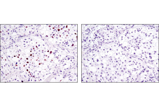
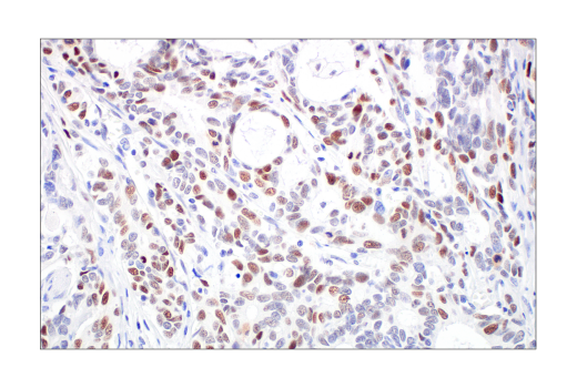
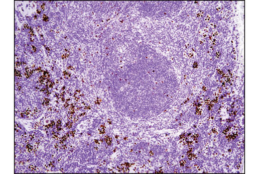
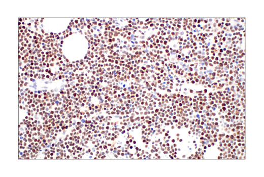
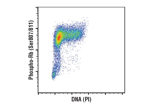
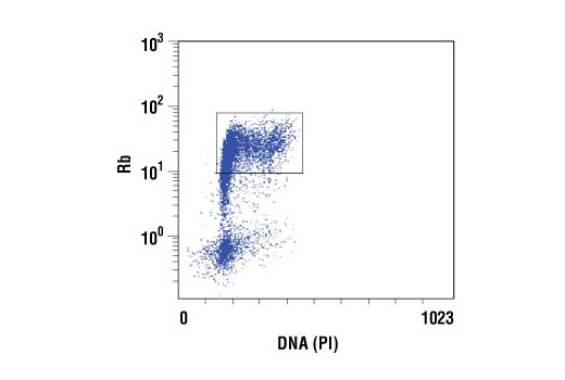
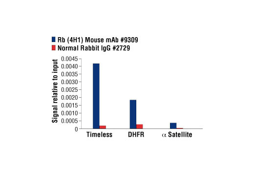
Phospho-Rb (Ser780) (D59B7) Rabbit mAb is produced by immunizing animals with a synthetic peptide corresponding to residues surrounding Ser780 of human Rb protein. Phospho-Rb (Ser807/811) (D20B12) XP® Rabbit mAb is produced by immunizing animals with a synthetic peptide corresponding to residues surrounding Ser807/811 of human Rb protein. Rb (4H1) monoclonal antibody is produced by immunizing animals with Rb-C Fusion Protein #6022, which contains residues 701-928 of human Rb .

Product Description

The PhosphoPlus® Rb (Ser780, Ser807/811) Antibody Kit provides reagents and protocols to investigate cell cycle progression within cells. The kit contains a total Rb antibody, two distinct phospho-Rb antibodies, and Rb control proteins along with secondary antibodies and reagents to perform up to 10 western blots.

Background

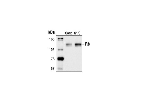
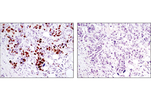
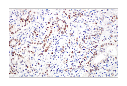
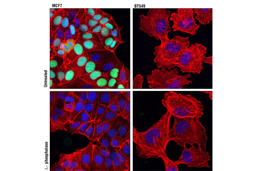
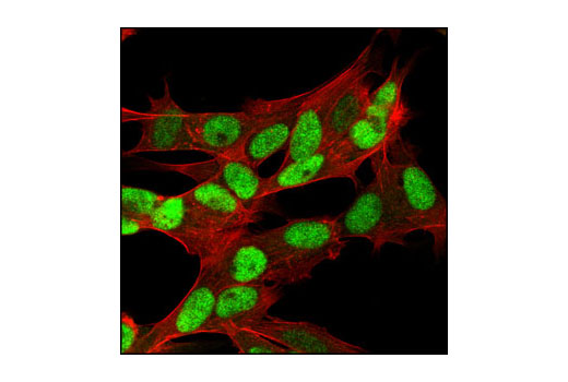
The retinoblastoma tumor suppressor protein Rb regulates cell proliferation by controlling progression through the restriction point within the G1-phase of the cell cycle (1). Rb has three functionally distinct binding domains and interacts with critical regulatory proteins including the E2F family of transcription factors, c-Abl tyrosine kinase, and proteins with a conserved LXCXE motif (2-4). Cell cycle-dependent phosphorylation by a CDK inhibits Rb target binding and allows cell cycle progression (5). Rb inactivation and subsequent cell cycle progression likely requires an initial phosphorylation by cyclin D-CDK4/6 followed by cyclin E-CDK2 phosphorylation (6). Specificity of different CDK/cyclin complexes has been observed in vitro (6-8) and cyclin D1 is required for Ser780 phosphorylation in vivo (9).

  1. Sherr, C.J. (1996) Science 274, 1672-7.
  2. Nevins, J.R. (1992) Science 258, 424-9.
  3. Welch, P.J. and Wang, J.Y. (1993) Cell 75, 779-90.
  4. Hu, Q.J. et al. (1990) EMBO J 9, 1147-55.
  5. Knudsen, E.S. and Wang, J.Y. (1997) Mol Cell Biol 17, 5771-83.
  6. Lundberg, A.S. and Weinberg, R.A. (1998) Mol Cell Biol 18, 753-61.
  7. Connell-Crowley, L. et al. (1997) Mol Biol Cell 8, 287-301.
  8. Kitagawa, M. et al. (1996) EMBO J 15, 7060-9.
  9. Geng, Y. et al. (2001) Proc Natl Acad Sci USA 98, 194-9.

Species Reactivity

Species reactivity is determined by testing in at least one approved application (e.g., western blot).

Cross-Reactivity Key

H: human M: mouse R: rat Hm: hamster Mk: monkey Vir: virus Mi: mink C: chicken Dm: D. melanogaster X: Xenopus Z: zebrafish B: bovine Dg: dog Pg: pig Sc: S. cerevisiae Ce: C. elegans Hr: horse GP: Guinea Pig Rab: rabbit All: all species expected

Trademarks and Patents

Cell Signaling Technology is a trademark of Cell Signaling Technology, Inc.
PhosphoPlus is a registered trademark of Cell Signaling Technology, Inc.
All other trademarks are the property of their respective owners. Visit cellsignal.com/trademarks for more information.

Limited Uses

Except as otherwise expressly agreed in a writing signed by a legally authorized representative of CST, the following terms apply to Products provided by CST, its affiliates or its distributors. Any Customer's terms and conditions that are in addition to, or different from, those contained herein, unless separately accepted in writing by a legally authorized representative of CST, are rejected and are of no force or effect.

Products are labeled with For Research Use Only or a similar labeling statement and have not been approved, cleared, or licensed by the FDA or other regulatory foreign or domestic entity, for any purpose. Customer shall not use any Product for any diagnostic or therapeutic purpose, or otherwise in any manner that conflicts with its labeling statement. Products sold or licensed by CST are provided for Customer as the end-user and solely for research and development uses. Any use of Product for diagnostic, prophylactic or therapeutic purposes, or any purchase of Product for resale (alone or as a component) or other commercial purpose, requires a separate license from CST. Customer shall (a) not sell, license, loan, donate or otherwise transfer or make available any Product to any third party, whether alone or in combination with other materials, or use the Products to manufacture any commercial products, (b) not copy, modify, reverse engineer, decompile, disassemble or otherwise attempt to discover the underlying structure or technology of the Products, or use the Products for the purpose of developing any products or services that would compete with CST products or services, (c) not alter or remove from the Products any trademarks, trade names, logos, patent or copyright notices or markings, (d) use the Products solely in accordance with CST Product Terms of Sale and any applicable documentation, and (e) comply with any license, terms of service or similar agreement with respect to any third party products or services used by Customer in connection with the Products.

Revision 1
#9306

PhosphoPlus® Rb (Ser780, Ser807/811) Antibody Kit

PhosphoPlus® Rb (Ser780, Ser807/811) Antibody Kit: Image 1 Expand Image
Confocal immunofluorescent analysis of fixed frozen mouse small intestine labeled with Phospho-Rb (Ser807/811) (D20B12) XP® Rabbit mAb (left, green) and co-labeled with DyLight 554 Phalloidin #13054 (right, red) and ProLong® Gold Antifade Reagent with DAPI #8961 (right, blue).
PhosphoPlus® Rb (Ser780, Ser807/811) Antibody Kit: Image 2 Expand Image
Confocal immunofluorescent analysis of fixed frozen mouse testis labeled with Phospho-Rb (Ser807/811) (D20B12) XP® Rabbit mAb (left, green) and co-labeled with DyLight 554 Phalloidin #13054 (right, red) and ProLong® Gold Antifade Reagent with DAPI #8961 (right, blue).
PhosphoPlus® Rb (Ser780, Ser807/811) Antibody Kit: Image 3 Expand Image
Confocal immunofluorescent analysis of fixed frozen mouse thymus labeled with Phospho-Rb (Ser807/811) (D20B12) XP® Rabbit mAb (left, green) and co-labeled with DyLight 554 Phalloidin #13054 (right, red) and ProLong® Gold Antifade Reagent with DAPI #8961 (right, blue).
PhosphoPlus® Rb (Ser780, Ser807/811) Antibody Kit: Image 4 Expand Image
Simple Western™ analysis of lysates (0.1 mg/mL) from MCF-7 cells using Phospho-Rb (Ser807/811) (D20B12) XP® Rabbit mAb #8516. The virtual lane view (left) shows the target band (as indicated) at 1:10 dilution of primary antibody. The corresponding electropherogram view (right) plots chemiluminescence by molecular weight along the capillary at 1:10 dilution of primary antibody. This experiment was performed under reducing conditions on the Jess™ Simple Western instrument from ProteinSimple, a BioTechne brand, using the 12-230 kDa separation module.
PhosphoPlus® Rb (Ser780, Ser807/811) Antibody Kit: Image 5 Expand Image
After the primary antibody is bound to the target protein, a complex with HRP-linked secondary antibody is formed. The LumiGLO® is added and emits light during enzyme catalyzed decomposition.
PhosphoPlus® Rb (Ser780, Ser807/811) Antibody Kit: Image 6 Expand Image
After the primary antibody is bound to the target protein, a complex with HRP-linked secondary antibody is formed. The LumiGLO® is added and emits light during enzyme catalyzed decomposition.
PhosphoPlus® Rb (Ser780, Ser807/811) Antibody Kit: Image 7 Expand Image
After the primary antibody is bound to the target protein, a complex with HRP-linked secondary antibody is formed. The LumiGLO* is added and emits light during enzyme catalyzed decomposition.
PhosphoPlus® Rb (Ser780, Ser807/811) Antibody Kit: Image 8 Expand Image
Western blot analysis of extracts from WI-38 cells, serum-starved for 3 days (-) or serum-starved for 3 days followed by treatment with 10% serum for 2 days (+), using Phospho-Rb (Ser780) (D59B7) Rabbit mAb.
PhosphoPlus® Rb (Ser780, Ser807/811) Antibody Kit: Image 9 Expand Image
Western blot analysis of extracts from MCF7 cells, untreated (-) or treated with calf intestinal phosphatase (CIP) and λ phosphatase (+), using Phospho-Rb (Ser807/811) (D20B12) XP® Rabbit mAb (upper) or Rb (4H1) Mouse mAb #9309 (lower).
PhosphoPlus® Rb (Ser780, Ser807/811) Antibody Kit: Image 10 Expand Image
Western blot analysis of Rb-C Fusion Protein #6022 (amino acids 701-928 of Rb fused to MBP) before (-) and after (+) in vitro phosphorylation by cdc2/cyclin B Protein Kinase (New England Biolabs #P6020), using Phospho-Rb (Ser795) Antibody #9301 (left) or the control antibody (right).
PhosphoPlus® Rb (Ser780, Ser807/811) Antibody Kit: Image 11 Expand Image
Western blot analysis of extracts from HeLa cells (lane 1) or Rb knock-out cells (lane 2) using Rb (4H1) Mouse mAb #9309 (upper), and β-actin (D6A8) Rabbit mAb #8457 (lower). The absence of signal in the Rb knock-out HeLa cells confirms specificity of the antibody for Rb.
PhosphoPlus® Rb (Ser780, Ser807/811) Antibody Kit: Image 12 Expand Image
Western blot analysis of phosphorylated or nonphosphorylated recombinant, truncated Rb, without or with Rb blocking peptide, using Phospho-Rb (Ser780) (D59B7) Rabbit mAb.
PhosphoPlus® Rb (Ser780, Ser807/811) Antibody Kit: Image 13 Expand Image
Western blot analysis of extracts from WI-38 cells, serum-starved for 3 days (-) or serum-starved for 3 days followed by treatment with 10% serum for 2 days (+), using Phospho-Rb (Ser807/811) (D20B12) XP® Rabbit mAb.
PhosphoPlus® Rb (Ser780, Ser807/811) Antibody Kit: Image 14 Expand Image
Western blot analysis of extracts from HeLa cells, transfected with 100 nM SignalSilence® Control siRNA (Fluorescein Conjugate) #6201 (-), SignalSilence® Rb siRNA I #6451 (+) or SignalSilence® Rb siRNA II (+), using Rb (4H1) Mouse mAb #9309 and α-Tubulin (11H10) Rabbit mAb #2125. The Rb (4H1) Mouse mAb confirms silencing of Rb expression, while the α-Tubulin (11H10) Rabbit mAb is used to control for loading and specificity of Rb siRNA.
PhosphoPlus® Rb (Ser780, Ser807/811) Antibody Kit: Image 15 Expand Image
Immunoprecipitation of phospho-Rb (Ser780) from WI-38 cell extracts using Phospho-Rb (Ser780) (D59B7) Rabbit mAb (lane 2). Western blot was performed using the same antibody. Lane 1 is 10% input.
PhosphoPlus® Rb (Ser780, Ser807/811) Antibody Kit: Image 16 Expand Image
Immunoprecipitation of phospho-Rb (Ser807/811) from Cos cell extracts using Rabbit (DA1E) mAb IgG XP® Isotype Control #3900 (lane 2) or Phospho-Rb (Ser807/811) (D20B12) XP® Rabbit mAb (lane 3). Lane 1 is 10% input. Western blot analysis was performed using Phospho-Rb (Ser807/811) (D20B12) XP® Rabbit mAb.
PhosphoPlus® Rb (Ser780, Ser807/811) Antibody Kit: Image 17 Expand Image
Western blot analysis of extracts from COS-7 cells, untreated or hydroxyurea-treated (G1/S), using Rb (4H1) Mouse mAb.
PhosphoPlus® Rb (Ser780, Ser807/811) Antibody Kit: Image 18 Expand Image
Immunohistochemical analysis of paraffin-embedded human breast carcinoma, showing nuclear localization, using Rb (4H1) Mouse mAb.
PhosphoPlus® Rb (Ser780, Ser807/811) Antibody Kit: Image 19 Expand Image
Immunohistochemical analysis of paraffin-embedded human colon carcinoma using Phospho-Rb (Ser807/811) (D20B12) XP® Rabbit mAb.
PhosphoPlus® Rb (Ser780, Ser807/811) Antibody Kit: Image 20 Expand Image
Immunohistochemical analysis of paraffin-embedded human lung carcinoma, using Rb (4H1) Mouse mAb.
PhosphoPlus® Rb (Ser780, Ser807/811) Antibody Kit: Image 21 Expand Image
Immunohistochemical analysis of paraffin-embedded human lung carcinoma, control (left) or λ phosphatase-treated (right), using Phospho-Rb (Ser807/811) (D20B12) XP® Rabbit mAb.
PhosphoPlus® Rb (Ser780, Ser807/811) Antibody Kit: Image 22 Expand Image
Immunohistochemical analysis of paraffin-embedded human esophageal carcinoma using Rb (4H1) Mouse mAb.
PhosphoPlus® Rb (Ser780, Ser807/811) Antibody Kit: Image 23 Expand Image
Immunohistochemical analysis of paraffin-embedded mouse spleen using Phospho-Rb (Ser807/811) (D20B12) XP® Rabbit mAb.
PhosphoPlus® Rb (Ser780, Ser807/811) Antibody Kit: Image 24 Expand Image
Immunohistochemical analysis of paraffin-embedded human T cell lymphoblastic lymphoma using Rb (4H1) Mouse mAb.
PhosphoPlus® Rb (Ser780, Ser807/811) Antibody Kit: Image 25 Expand Image
Immunohistochemical analysis of paraffin-embedded human ovarian serous adenocarcinoma using Phospho-Rb (Ser807/811) (D20B12) XP® Rabbit mAb in the presence of control peptide (left) or antigen-specific peptide (right).
PhosphoPlus® Rb (Ser780, Ser807/811) Antibody Kit: Image 26 Expand Image
Immunohistochemical analysis of paraffin-embedded human gastric adenocarcinoma using Rb (4H1) Mouse mAb.
PhosphoPlus® Rb (Ser780, Ser807/811) Antibody Kit: Image 27 Expand Image
Confocal immunofluorescent analysis of MCF7 (left) and BT-549 (right) cells, untreated (upper) or λ phosphatase-treated (lower) using Phospho-Rb (Ser807/Ser811) (D20B12) XP® Rabbit mAb (green). Actin filaments were labeled with DY-554 phalloidin (red). Blue pseudocolor = DRAQ5® #4084 (fluorescent DNA dye).
PhosphoPlus® Rb (Ser780, Ser807/811) Antibody Kit: Image 28 Expand Image
Confocal immunofluorescent image of SH-SY5Y cells, using RB (4H1) Mouse mAb (green). Actin filaments have been labeled with Alexa Fluor® 555 phalloidin (red).
PhosphoPlus® Rb (Ser780, Ser807/811) Antibody Kit: Image 29 Expand Image
Flow cytometric analysis of Jurkat cells using Phospho-Rb (Ser807/811) (D20B12) XP® Rabbit mAb and Propidium Iodide (PI/RNase Staining Solution #4087 to measure DNA content. Anti-rabbit IgG (H+L), F(ab')2 Fragment (Alexa Fluor® 488 Conjugate) #4412 was used as a secondary antibody.
PhosphoPlus® Rb (Ser780, Ser807/811) Antibody Kit: Image 30 Expand Image
Flow cytometric analysis of Jurkat cells, using Rb (4H1) Mouse mAb versus propidium iodide (DNA content). The box indicates Rb positive cells.
PhosphoPlus® Rb (Ser780, Ser807/811) Antibody Kit: Image 31 Expand Image
Chromatin immunoprecipitations were performed with cross-linked chromatin from Raji cells and either Rb (4H1) Mouse mAb or Normal Rabbit IgG #2729 using SimpleChIP® Enzymatic Chromatin IP Kit (Magnetic Beads) #9003. The enriched DNA was quantified by real-time PCR using using SimpleChIP® Human Timeless Intron 1 Primers #7001, human DHFR promoter primers, and SimpleChIP® Human α Satellite Repeat Primers #4486. The amount of immunoprecipitated DNA in each sample is represented as signal relative to the total amount of input chromatin, which is equivalent to one.