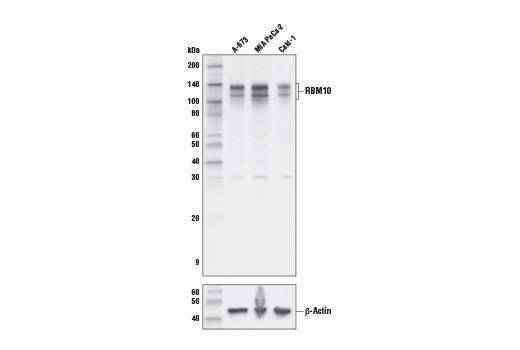
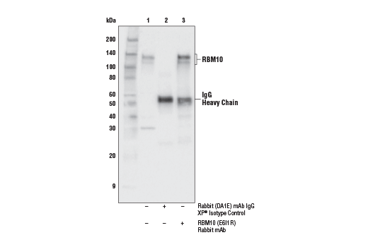
WB, IP
H Mk
Endogenous
110, 135
Rabbit IgG
#P98175
8241
Product Information
Product Usage Information
| Application | Dilution |
|---|---|
| Western Blotting | 1:1000 |
| Immunoprecipitation | 1:100 |
Storage
Specificity / Sensitivity
Species Reactivity:
Human, Monkey
Source / Purification
Monoclonal antibody is produced by immunizing animals with a synthetic peptide corresponding to residues surrounding Gly53 of human RBM10 protein.
Background
RBM10 is an RNA binding protein and a paralog to tumor suppressor RBM5 (1,2). Alternative splicing creates two highly expressed variants that differ by one exon. RBM10 controls alternative splicing and exon skipping of mRNAs, including Fas and Bcl-x (3,4). RBM10 has been shown to affect apoptosis via up regulation of TNF-α mRNA (5). In lung cancer, RBM10 mutations disrupt the splicing of NUMB, a regulator of Notch signaling (6,7). Fusion of the RBM10 gene with TFE3 has been found in renal cell carcinoma (8-9). Mutations of RBM10 causes TARP syndrome, an x-linked, lethal disorder characterized by various developmental defects (10-12).
- Timmer, T. et al. (1999) Genomics 60, 238-40.
- Bonnal, S. et al. (2008) Mol Cell 32, 81-95.
- Wang, Y. et al. (2013) EMBO Mol Med 5, 1431-42.
- Inoue, A. et al. (2014) FEBS Lett 588, 942-7.
- Wang, K. et al. (2012) J Cell Death 5, 1-19.
- Bechara, E.G. et al. (2013) Mol Cell 52, 720-33.
- Hernández, J. et al. (2016) RNA Biol 13, 466-72.
- Just, P.A. et al. (2016) Genes Chromosomes Cancer 55, 541-8.
- Argani, P. et al. (2017) Am J Surg Pathol 41, 655-62.
- Gorlin, R.J. et al. (1970) Am J Dis Child 119, 176-8.
- Johnston, J.J. et al. (2010) Am J Hum Genet 86, 743-8.
- Gripp, K.W. et al. (2011) Am J Med Genet A 155A, 2516-20.
Species Reactivity
Species reactivity is determined by testing in at least one approved application (e.g., western blot).
Western Blot Buffer
IMPORTANT: For western blots, incubate membrane with diluted primary antibody in 5% w/v BSA, 1X TBS, 0.1% Tween® 20 at 4°C with gentle shaking, overnight.
Applications Key
WB: Western Blotting IP: Immunoprecipitation
Cross-Reactivity Key
H: human M: mouse R: rat Hm: hamster Mk: monkey Vir: virus Mi: mink C: chicken Dm: D. melanogaster X: Xenopus Z: zebrafish B: bovine Dg: dog Pg: pig Sc: S. cerevisiae Ce: C. elegans Hr: horse GP: Guinea Pig Rab: rabbit All: all species expected
Trademarks and Patents
Limited Uses
Except as otherwise expressly agreed in a writing signed by a legally authorized representative of CST, the following terms apply to Products provided by CST, its affiliates or its distributors. Any Customer's terms and conditions that are in addition to, or different from, those contained herein, unless separately accepted in writing by a legally authorized representative of CST, are rejected and are of no force or effect.
Products are labeled with For Research Use Only or a similar labeling statement and have not been approved, cleared, or licensed by the FDA or other regulatory foreign or domestic entity, for any purpose. Customer shall not use any Product for any diagnostic or therapeutic purpose, or otherwise in any manner that conflicts with its labeling statement. Products sold or licensed by CST are provided for Customer as the end-user and solely for research and development uses. Any use of Product for diagnostic, prophylactic or therapeutic purposes, or any purchase of Product for resale (alone or as a component) or other commercial purpose, requires a separate license from CST. Customer shall (a) not sell, license, loan, donate or otherwise transfer or make available any Product to any third party, whether alone or in combination with other materials, or use the Products to manufacture any commercial products, (b) not copy, modify, reverse engineer, decompile, disassemble or otherwise attempt to discover the underlying structure or technology of the Products, or use the Products for the purpose of developing any products or services that would compete with CST products or services, (c) not alter or remove from the Products any trademarks, trade names, logos, patent or copyright notices or markings, (d) use the Products solely in accordance with CST Product Terms of Sale and any applicable documentation, and (e) comply with any license, terms of service or similar agreement with respect to any third party products or services used by Customer in connection with the Products.