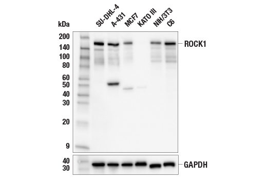
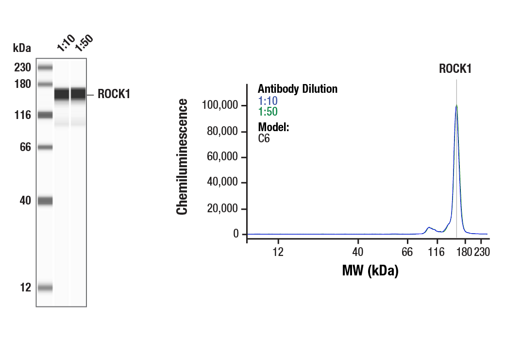
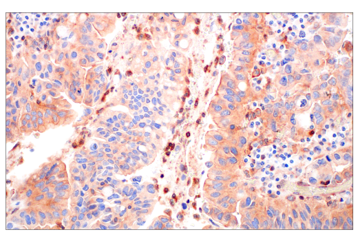
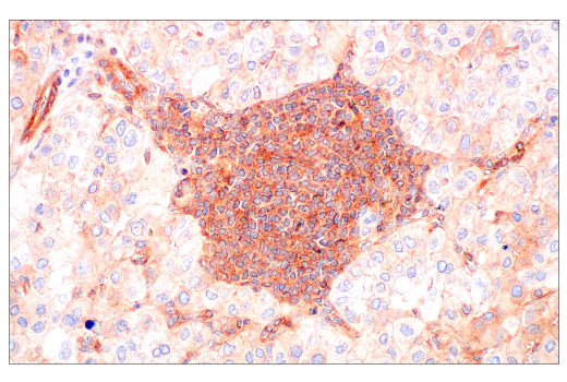
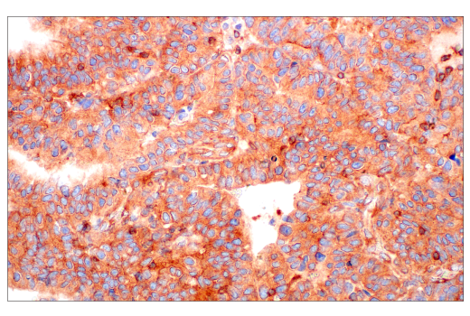
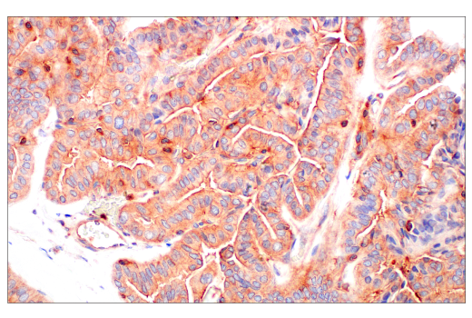
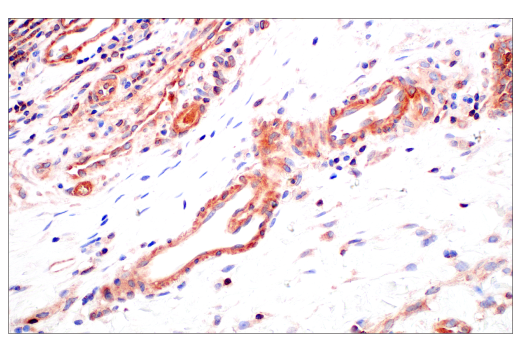
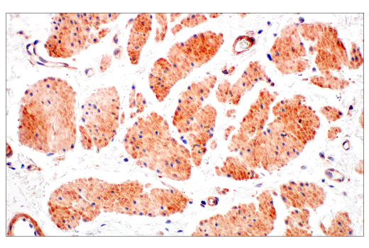
WB, W-S, IHC-P, IF-IC
H M R
Endogenous
160
Rabbit IgG
#Q13464
6093
Product Information
Product Usage Information
| Application | Dilution |
|---|---|
| Western Blotting | 1:1000 |
| Simple Western™ | 1:10 - 1:50 |
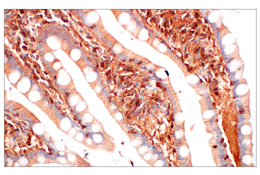
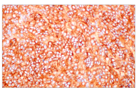
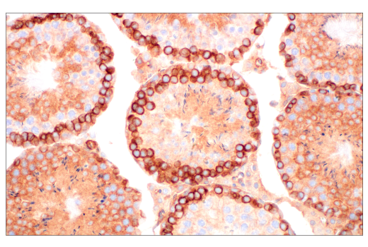
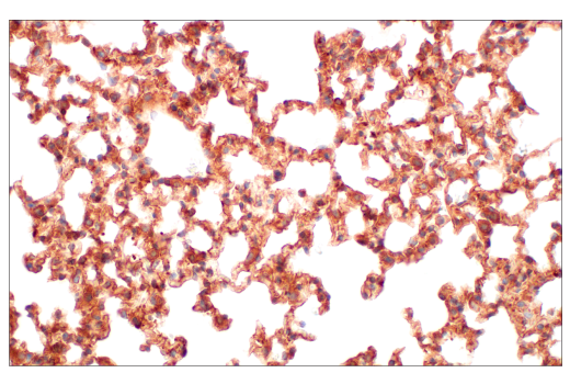
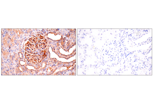
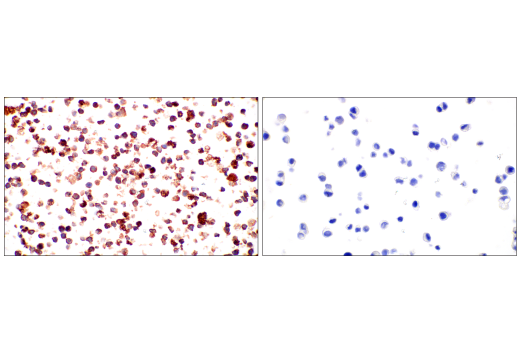
| Immunohistochemistry (Paraffin) | 1:400 - 1:1600 |
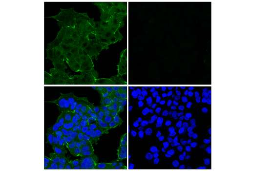
| Immunofluorescence (Immunocytochemistry) | 1:400 - 1:1600 |
Storage
Specificity / Sensitivity
Species Reactivity:
Human, Mouse, Rat
Source / Purification
Monoclonal antibody is produced by immunizing animals with recombinant protein specific to the central region of human ROCK1 protein.
Background
ROCK (Rho-associated kinase), a family of serine/threonine kinases, is an important downstream target of Rho-GTPase and plays an important role in Rho-mediated signaling. Two isoforms of ROCK have been identified: ROCK1 and ROCK2. ROCK is composed of N-terminal catalytic, coiled-coil, and C-terminal PH (pleckstrin homology) domains. The C-terminus of ROCK negatively regulates its kinase activity (1,2). ROCK1 is cleaved by caspase-3 at a conserved DETD1113/G sequence resulting in loss of its C-terminal inhibitory domain (3). ROCK2 is directly cleaved by granzyme B (grB). Cleavage activates ROCK and leads to phosphorylation of myosin light chain (MLC) and inhibition of myosin phosphatase (4). This phosphorylation may account for the mechanism by which Rho regulates cytokinesis, cell motility, cell membrane blebbing during apoptosis, and smooth muscle contraction (5-7).
- Nakagawa, O. et al. (1996) FEBS Lett. 392, 189-193.
- Lee, J.H. et al. (2004) J. Cell. Biol. 167, 327-337.
- Sebbagh, M. et al. (2001) Nat Cell Biol 3, 346-52.
- Sebbagh, M. et al. (2005) J Exp Med 201, 465-71.
- Amano, M. et al. (1996) J Biol Chem 271, 20246-9.
- Kureishi, Y. et al. (1997) J Biol Chem 272, 12257-60.
- Totsukawa, G. et al. (2000) J Cell Biol 150, 797-806.
Species Reactivity
Species reactivity is determined by testing in at least one approved application (e.g., western blot).
Western Blot Buffer
IMPORTANT: For western blots, incubate membrane with diluted primary antibody in 5% w/v nonfat dry milk, 1X TBS, 0.1% Tween® 20 at 4°C with gentle shaking, overnight.
Applications Key
WB: Western Blotting W-S: Simple Western™ IHC-P: Immunohistochemistry (Paraffin) IF-IC: Immunofluorescence (Immunocytochemistry)
Cross-Reactivity Key
H: human M: mouse R: rat Hm: hamster Mk: monkey Vir: virus Mi: mink C: chicken Dm: D. melanogaster X: Xenopus Z: zebrafish B: bovine Dg: dog Pg: pig Sc: S. cerevisiae Ce: C. elegans Hr: horse GP: Guinea Pig Rab: rabbit All: all species expected
Trademarks and Patents
Limited Uses
Except as otherwise expressly agreed in a writing signed by a legally authorized representative of CST, the following terms apply to Products provided by CST, its affiliates or its distributors. Any Customer's terms and conditions that are in addition to, or different from, those contained herein, unless separately accepted in writing by a legally authorized representative of CST, are rejected and are of no force or effect.
Products are labeled with For Research Use Only or a similar labeling statement and have not been approved, cleared, or licensed by the FDA or other regulatory foreign or domestic entity, for any purpose. Customer shall not use any Product for any diagnostic or therapeutic purpose, or otherwise in any manner that conflicts with its labeling statement. Products sold or licensed by CST are provided for Customer as the end-user and solely for research and development uses. Any use of Product for diagnostic, prophylactic or therapeutic purposes, or any purchase of Product for resale (alone or as a component) or other commercial purpose, requires a separate license from CST. Customer shall (a) not sell, license, loan, donate or otherwise transfer or make available any Product to any third party, whether alone or in combination with other materials, or use the Products to manufacture any commercial products, (b) not copy, modify, reverse engineer, decompile, disassemble or otherwise attempt to discover the underlying structure or technology of the Products, or use the Products for the purpose of developing any products or services that would compete with CST products or services, (c) not alter or remove from the Products any trademarks, trade names, logos, patent or copyright notices or markings, (d) use the Products solely in accordance with CST Product Terms of Sale and any applicable documentation, and (e) comply with any license, terms of service or similar agreement with respect to any third party products or services used by Customer in connection with the Products.