WB, W-S, IP
M
Endogenous
45
Rabbit IgG
#P11157
20135
Product Information
Product Usage Information
| Application | Dilution |
|---|---|
| Western Blotting | 1:1000 |
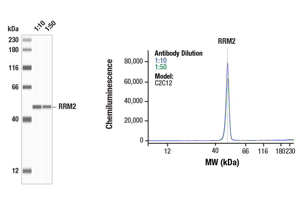
| Simple Western™ | 1:10 - 1:50 |
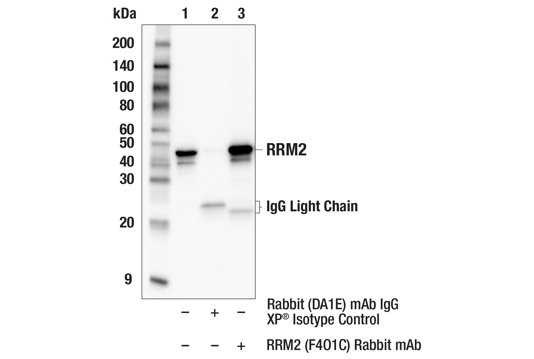
| Immunoprecipitation | 1:50 |
Storage
Specificity / Sensitivity
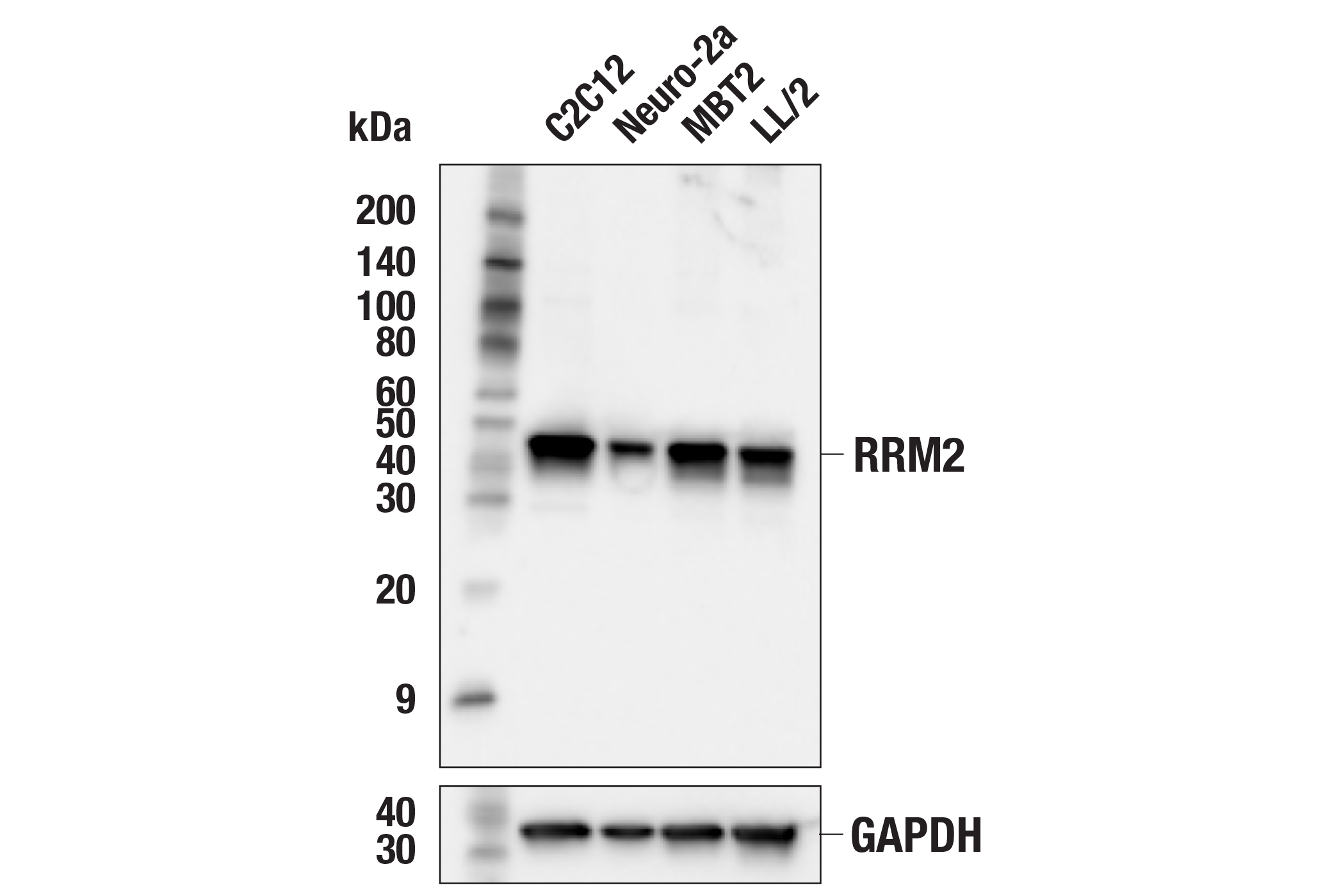
Species Reactivity:
Mouse
Source / Purification
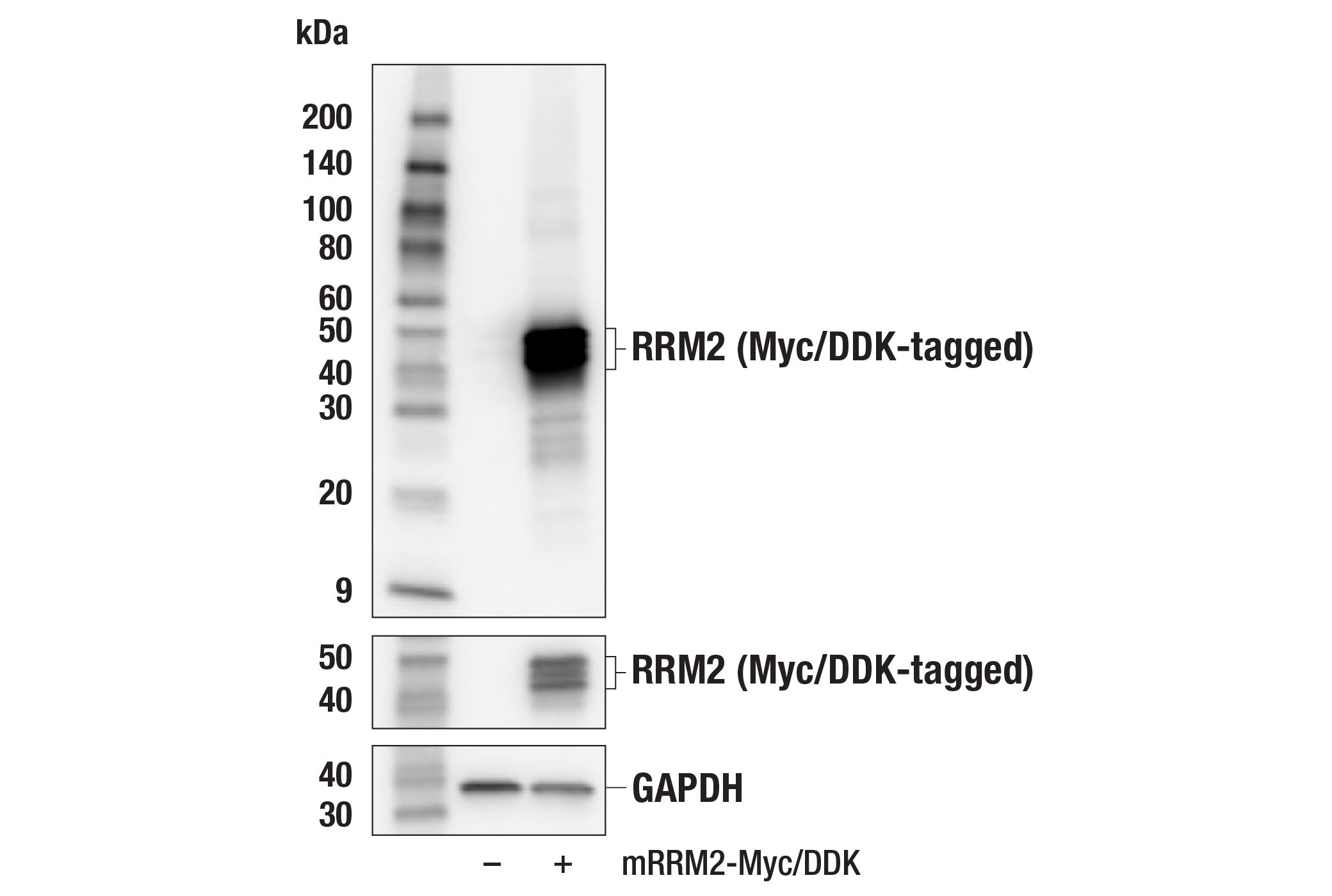
Monoclonal antibody is produced by immunizing animals with recombinant protein specific to the amino terminus of mouse RRM2 protein.
Background
Ribonucleotide reductase catalyzes the rate-limiting step in the synthesis of deoxynucleotide triphosphates (dNTPs). Ribonucleoside-diphosphate reductase subunit M2 (RRM2) is frequently overexpressed and associated with poor prognosis in multiple human cancers (1). RRM2/AKT/NF-κB signaling pathway is implicated in tumor invasiveness in gastric cancer (2). RRM2 is highly expressed in melanoma, and correlated with poor prognosis in BRAF-mutant melanoma. Knockdown of RRM2 stabilized the transient response of cells and patient-derived xenograft (PDX) model system to BRAF inhibition (3).
Cyclin-dependent kinase (CDK) mediated phosphorylation of RRM2 at Thr33 targets the protein for degradation, allowing cells to maintain balanced dNTP pools (4).
Species Reactivity
Species reactivity is determined by testing in at least one approved application (e.g., western blot).
Western Blot Buffer
IMPORTANT: For western blots, incubate membrane with diluted primary antibody in 5% w/v nonfat dry milk, 1X TBS, 0.1% Tween® 20 at 4°C with gentle shaking, overnight.
Applications Key
WB: Western Blotting W-S: Simple Western™ IP: Immunoprecipitation
Cross-Reactivity Key
H: human M: mouse R: rat Hm: hamster Mk: monkey Vir: virus Mi: mink C: chicken Dm: D. melanogaster X: Xenopus Z: zebrafish B: bovine Dg: dog Pg: pig Sc: S. cerevisiae Ce: C. elegans Hr: horse GP: Guinea Pig Rab: rabbit All: all species expected
Trademarks and Patents
Limited Uses
Except as otherwise expressly agreed in a writing signed by a legally authorized representative of CST, the following terms apply to Products provided by CST, its affiliates or its distributors. Any Customer's terms and conditions that are in addition to, or different from, those contained herein, unless separately accepted in writing by a legally authorized representative of CST, are rejected and are of no force or effect.
Products are labeled with For Research Use Only or a similar labeling statement and have not been approved, cleared, or licensed by the FDA or other regulatory foreign or domestic entity, for any purpose. Customer shall not use any Product for any diagnostic or therapeutic purpose, or otherwise in any manner that conflicts with its labeling statement. Products sold or licensed by CST are provided for Customer as the end-user and solely for research and development uses. Any use of Product for diagnostic, prophylactic or therapeutic purposes, or any purchase of Product for resale (alone or as a component) or other commercial purpose, requires a separate license from CST. Customer shall (a) not sell, license, loan, donate or otherwise transfer or make available any Product to any third party, whether alone or in combination with other materials, or use the Products to manufacture any commercial products, (b) not copy, modify, reverse engineer, decompile, disassemble or otherwise attempt to discover the underlying structure or technology of the Products, or use the Products for the purpose of developing any products or services that would compete with CST products or services, (c) not alter or remove from the Products any trademarks, trade names, logos, patent or copyright notices or markings, (d) use the Products solely in accordance with CST Product Terms of Sale and any applicable documentation, and (e) comply with any license, terms of service or similar agreement with respect to any third party products or services used by Customer in connection with the Products.