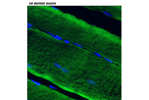
WB, IF-F
H M R
Endogenous
~560
Rabbit IgG
#P21817
6261
Product Information
Product Usage Information
| Application | Dilution |
|---|---|
| Western Blotting | 1:1000 |
| Immunofluorescence (Frozen) | 1:100 |
Storage
Specificity / Sensitivity
Species Reactivity:
Human, Mouse, Rat
Species predicted to react based on 100% sequence homology
The antigen sequence used to produce this antibody shares
100% sequence homology with the species listed here, but
reactivity has not been tested or confirmed to work by CST.
Use of this product with these species is not covered under
our
Product Performance Guarantee.
Pig
Source / Purification
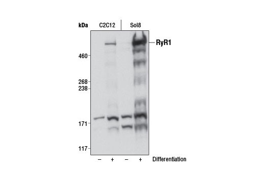
Monoclonal antibody is produced by immunizing animals with a synthetic peptide corresponding to residues surrounding Arg830 of human RyR1 protein.
Background
Ryanodine receptors (RyRs) are large (>500 kDa), intracellular calcium channels found in the sarcoplasmic/endoplasmic reticulum membrane and are responsible for the release of Ca2+ from intracellular stores in excitable cells, such as muscle and neurons. RyRs exist as three mammalian isoforms (RyR1-3), all of which form homotetramers regulated by phosphorylation and/or direct or indirect interaction with a variety of proteins (L-type calcium channels, PKA, FKBP12/12.6, CaMKII, calmodulin, calsequestrin, junctin, and triadin) and ions (Mg2+ and Ca2+). Regulation of the RyR channel by protein modulators occurs within the large cytoplasmic domain, whereas the carboxy-terminal portion of the protein forms the ion-binding and conducting pore (1,2). RyR1 and RyR2 are predominantly expressed in skeletal and cardiac muscle, respectively, where they localize exclusively to the sarcoplasmic reticulum (SR) and facilitate calcium-mediated communication between transverse-tubules and sarcoplasmic reticulum. Contraction of skeletal muscle is triggered by release of calcium ions from the SR following depolarization of T-tubules. Research studies have shown that defects in RyR1 are the cause of malignant hyperthermia susceptibility type 1 (MHS1), central core disease of muscle (CCD), multiminicore disease with external ophthalmoplegia, and congenital myopathy with fiber-type disproportion (CFTD), each of which is manifested by defects in muscle function, metabolism, and development (2). Investigators have shown that defects in RyR2 are the cause of familial arrhythmogenic right ventricular dysplasia type 2 (ARVD2) and catecholaminergic polymorphic ventricular tachycardia type 1 (CPVT1), both of which are implicated in sudden death syndromes as a result of electrical instability and degeneration of the ventricular myocardium or stress-induced ventricular tachycardia (2). Despite low levels of expression in skeletal and smooth muscle, RyR3 is the dominant isoform in neuronal cells (hippocampal neurons, thalamus, Purkinje cells) and has been implicated in synaptic plasticity, dendritic spine remodeling, and spatial memory formation (3). The role of RyR3 in neuronal function has been substantiated by mice lacking RyR3, which demonstrate normal motor function, but possess numerous behavioral and social defects (4).
Species Reactivity
Species reactivity is determined by testing in at least one approved application (e.g., western blot).
Western Blot Buffer
IMPORTANT: For western blots, incubate membrane with diluted primary antibody in 5% w/v BSA, 1X TBS, 0.1% Tween® 20 at 4°C with gentle shaking, overnight.
Applications Key
WB: Western Blotting IF-F: Immunofluorescence (Frozen)
Cross-Reactivity Key
H: human M: mouse R: rat Hm: hamster Mk: monkey Vir: virus Mi: mink C: chicken Dm: D. melanogaster X: Xenopus Z: zebrafish B: bovine Dg: dog Pg: pig Sc: S. cerevisiae Ce: C. elegans Hr: horse GP: Guinea Pig Rab: rabbit All: all species expected
Trademarks and Patents
Limited Uses
Except as otherwise expressly agreed in a writing signed by a legally authorized representative of CST, the following terms apply to Products provided by CST, its affiliates or its distributors. Any Customer's terms and conditions that are in addition to, or different from, those contained herein, unless separately accepted in writing by a legally authorized representative of CST, are rejected and are of no force or effect.
Products are labeled with For Research Use Only or a similar labeling statement and have not been approved, cleared, or licensed by the FDA or other regulatory foreign or domestic entity, for any purpose. Customer shall not use any Product for any diagnostic or therapeutic purpose, or otherwise in any manner that conflicts with its labeling statement. Products sold or licensed by CST are provided for Customer as the end-user and solely for research and development uses. Any use of Product for diagnostic, prophylactic or therapeutic purposes, or any purchase of Product for resale (alone or as a component) or other commercial purpose, requires a separate license from CST. Customer shall (a) not sell, license, loan, donate or otherwise transfer or make available any Product to any third party, whether alone or in combination with other materials, or use the Products to manufacture any commercial products, (b) not copy, modify, reverse engineer, decompile, disassemble or otherwise attempt to discover the underlying structure or technology of the Products, or use the Products for the purpose of developing any products or services that would compete with CST products or services, (c) not alter or remove from the Products any trademarks, trade names, logos, patent or copyright notices or markings, (d) use the Products solely in accordance with CST Product Terms of Sale and any applicable documentation, and (e) comply with any license, terms of service or similar agreement with respect to any third party products or services used by Customer in connection with the Products.