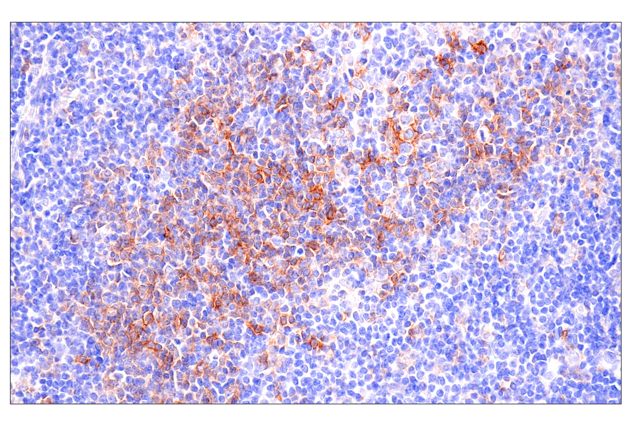
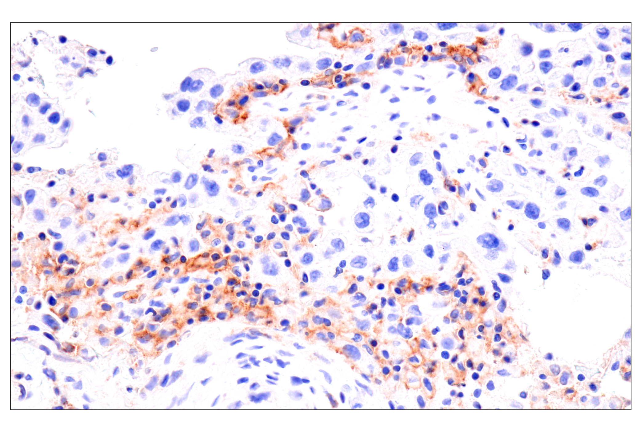
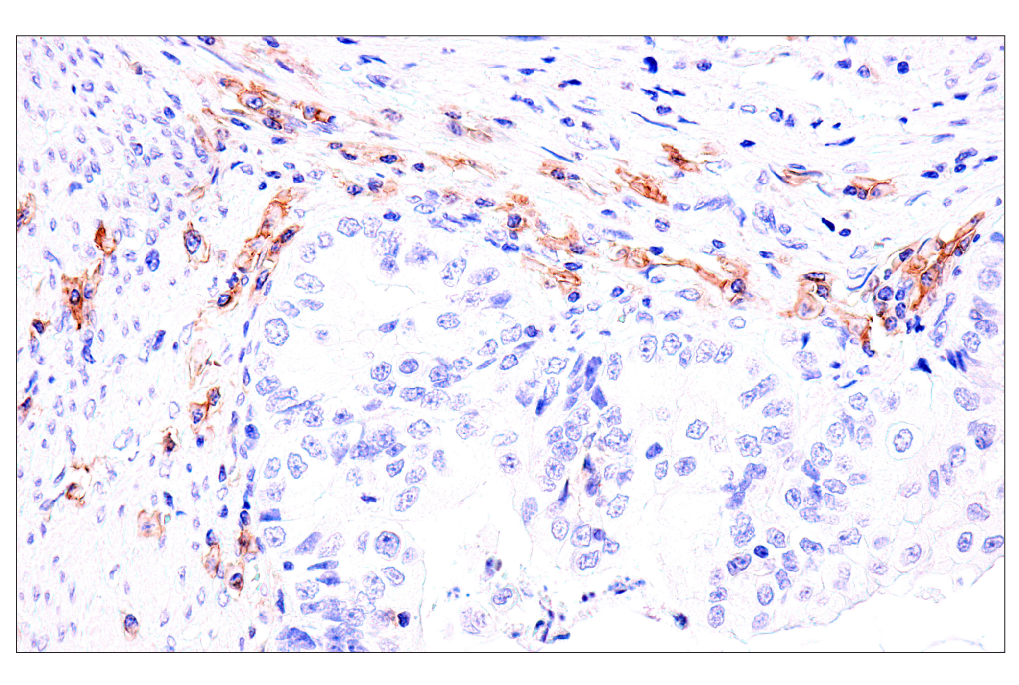
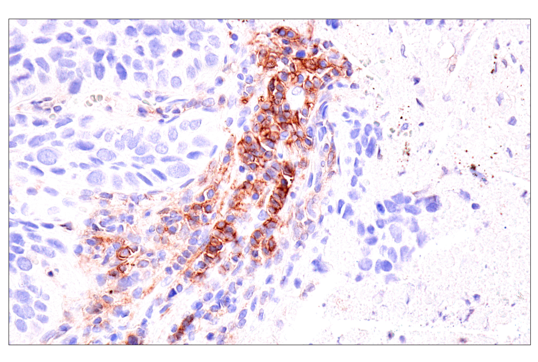
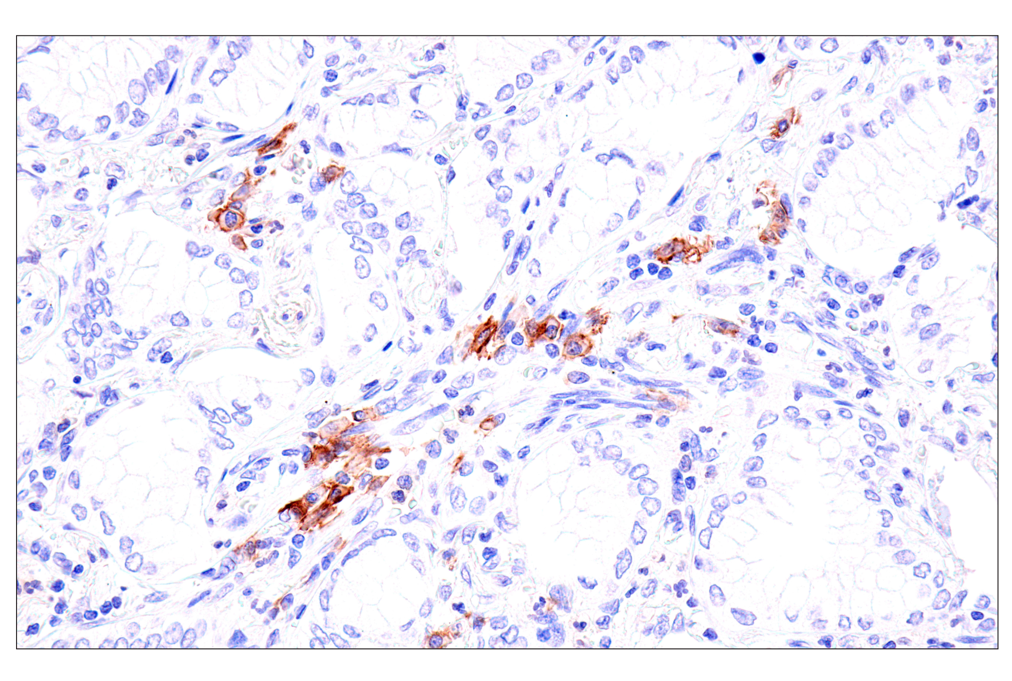
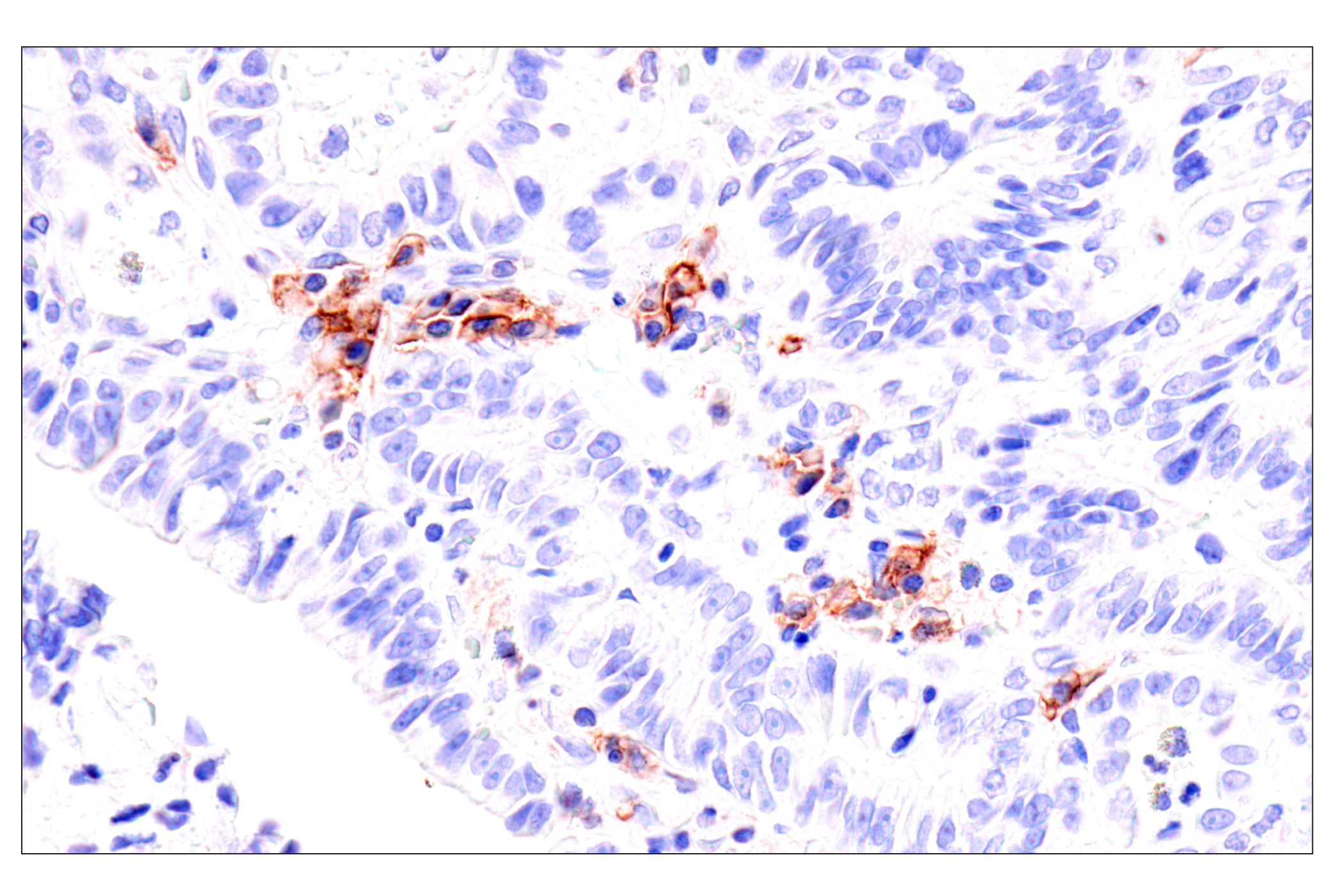
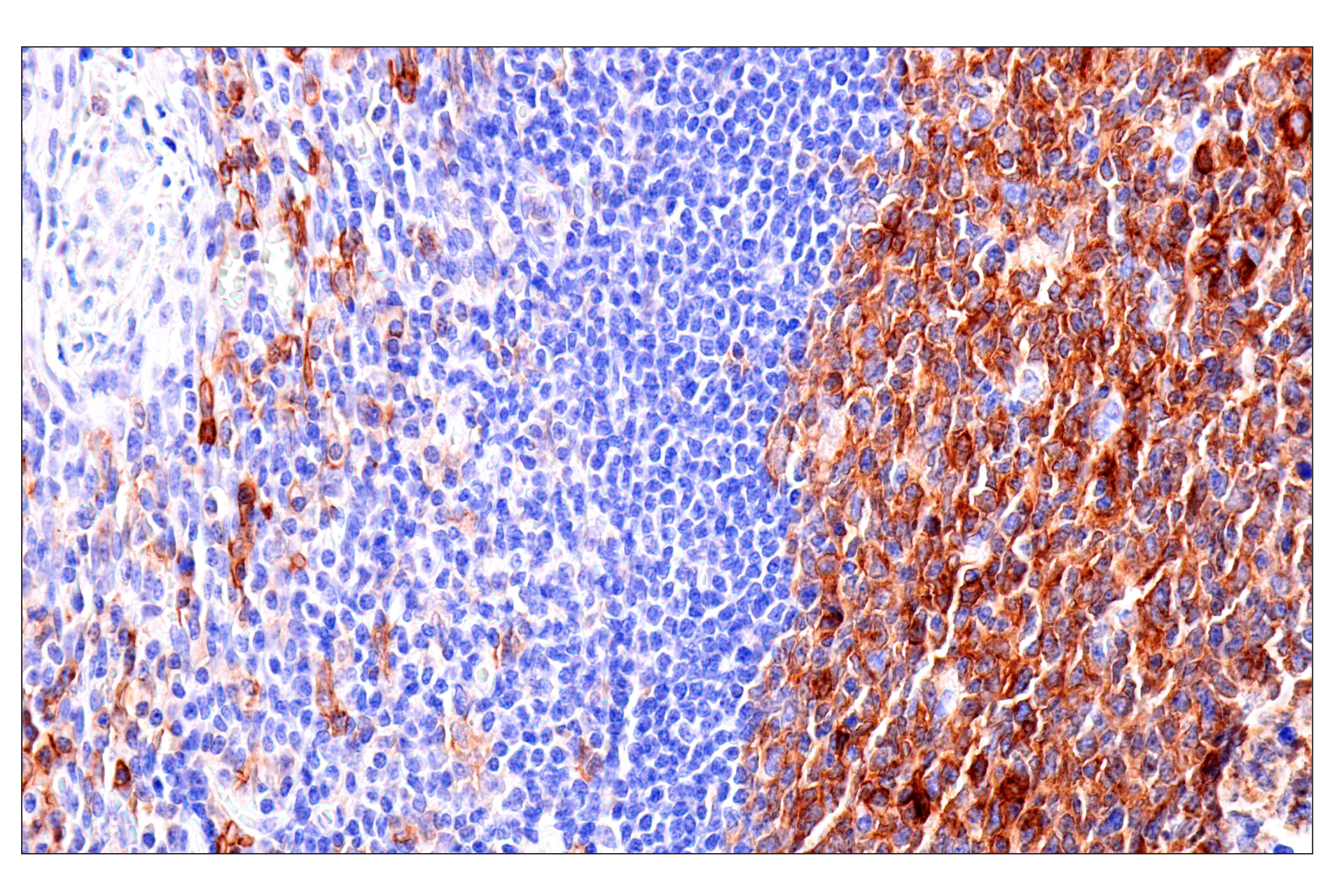
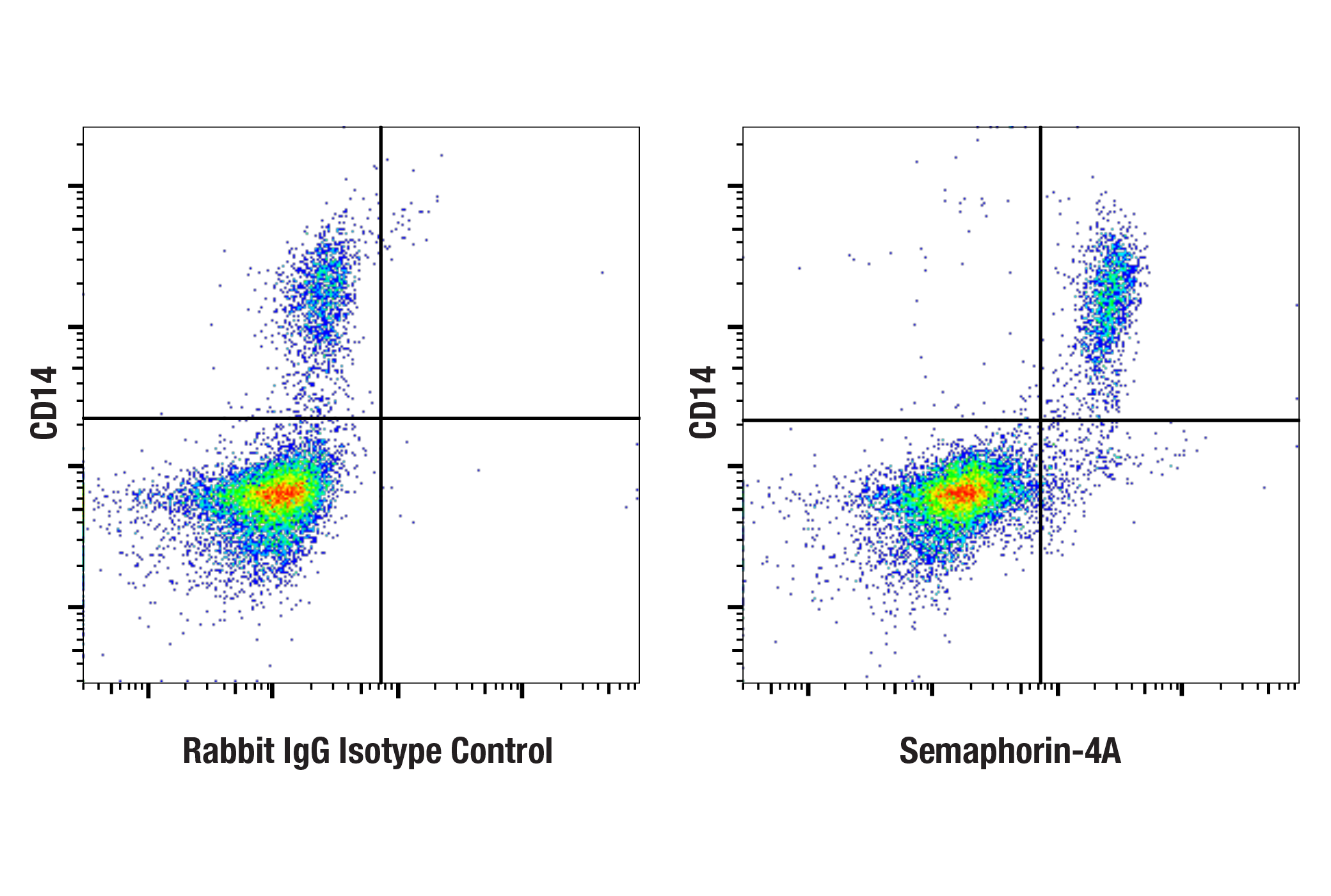
WB, IHC-P, FC-FP
H
Endogenous
90
Rabbit IgG
#Q9H3S1
64218
Product Information
Product Usage Information
| Application | Dilution |
|---|---|
| Western Blotting | 1:1000 |
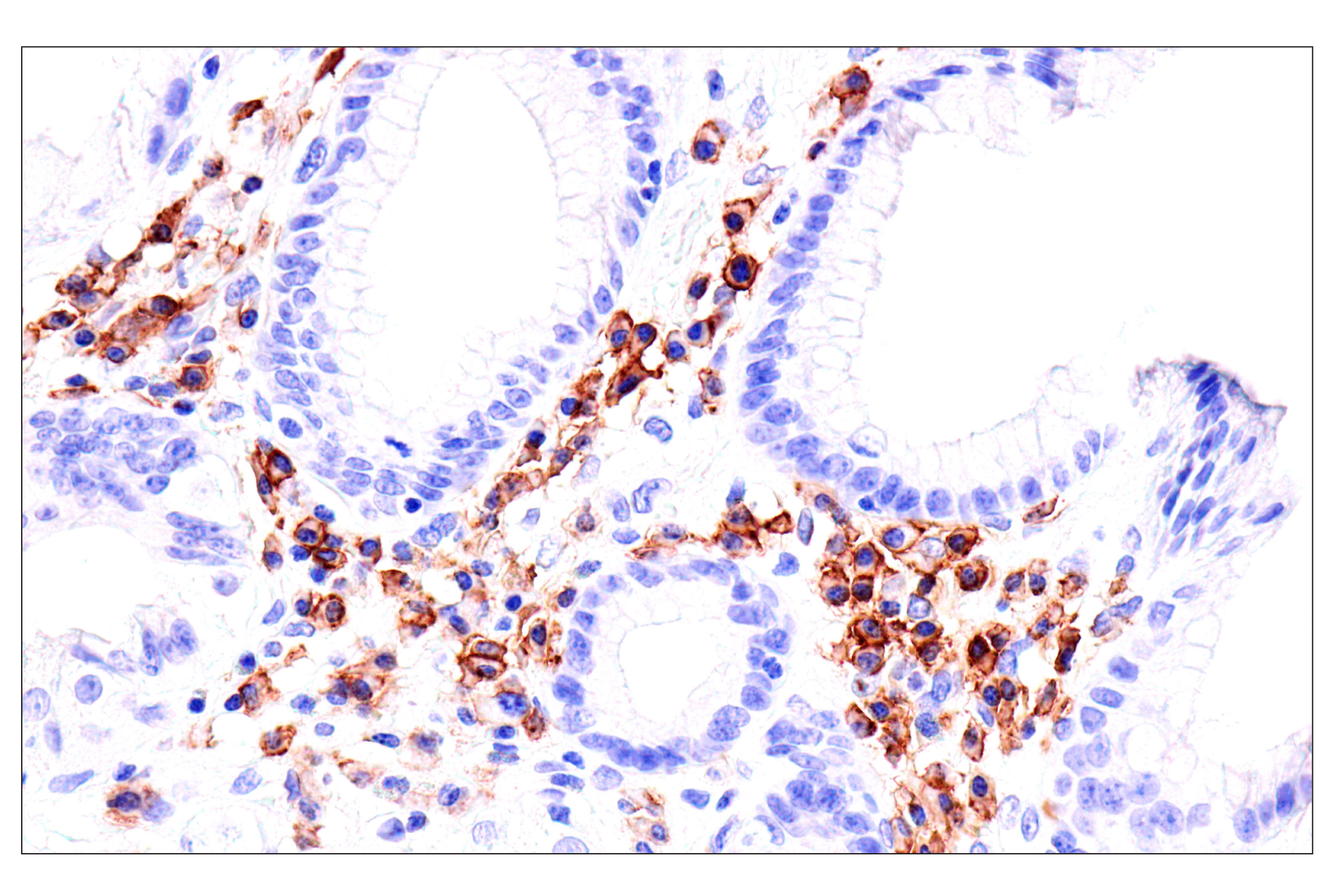
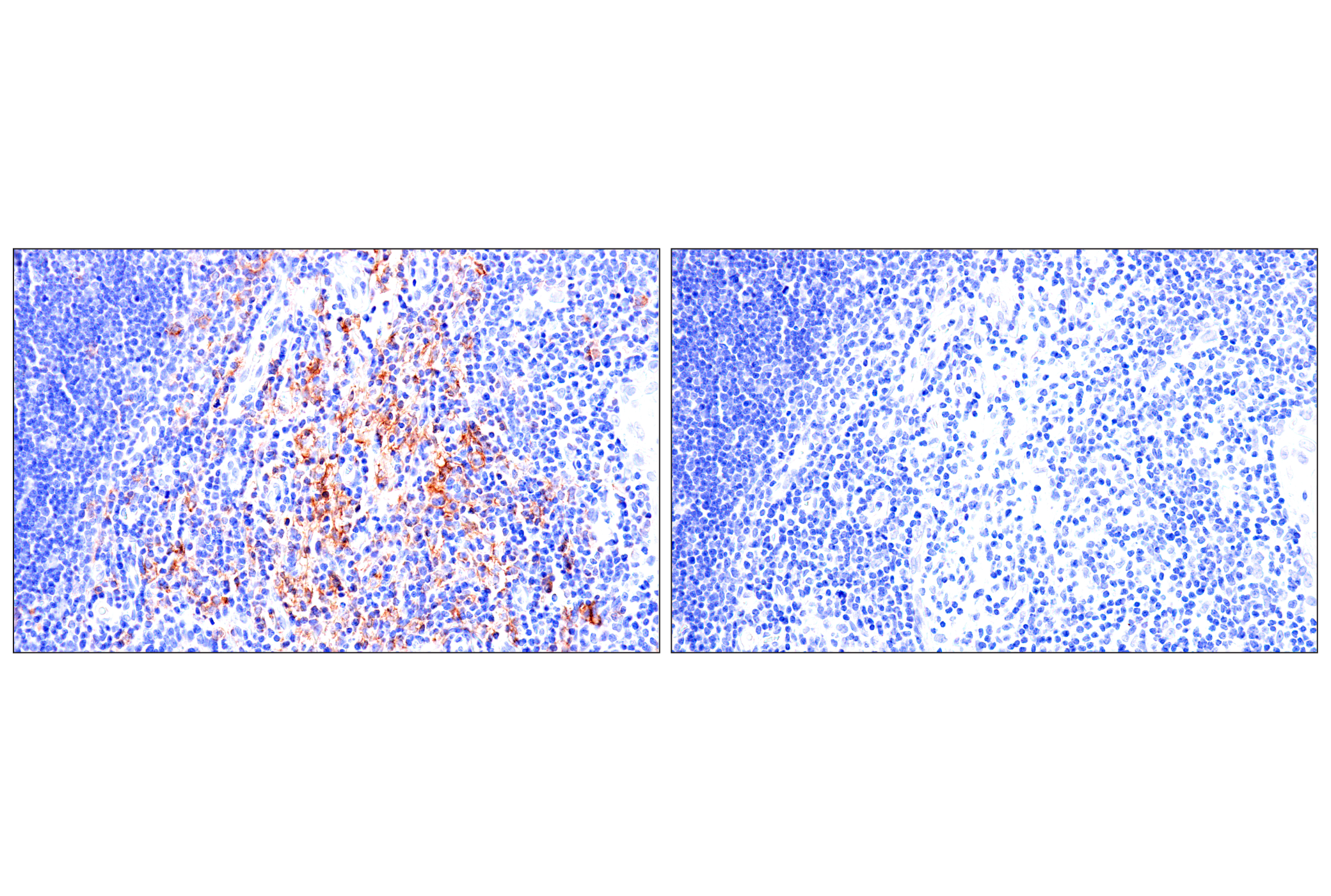
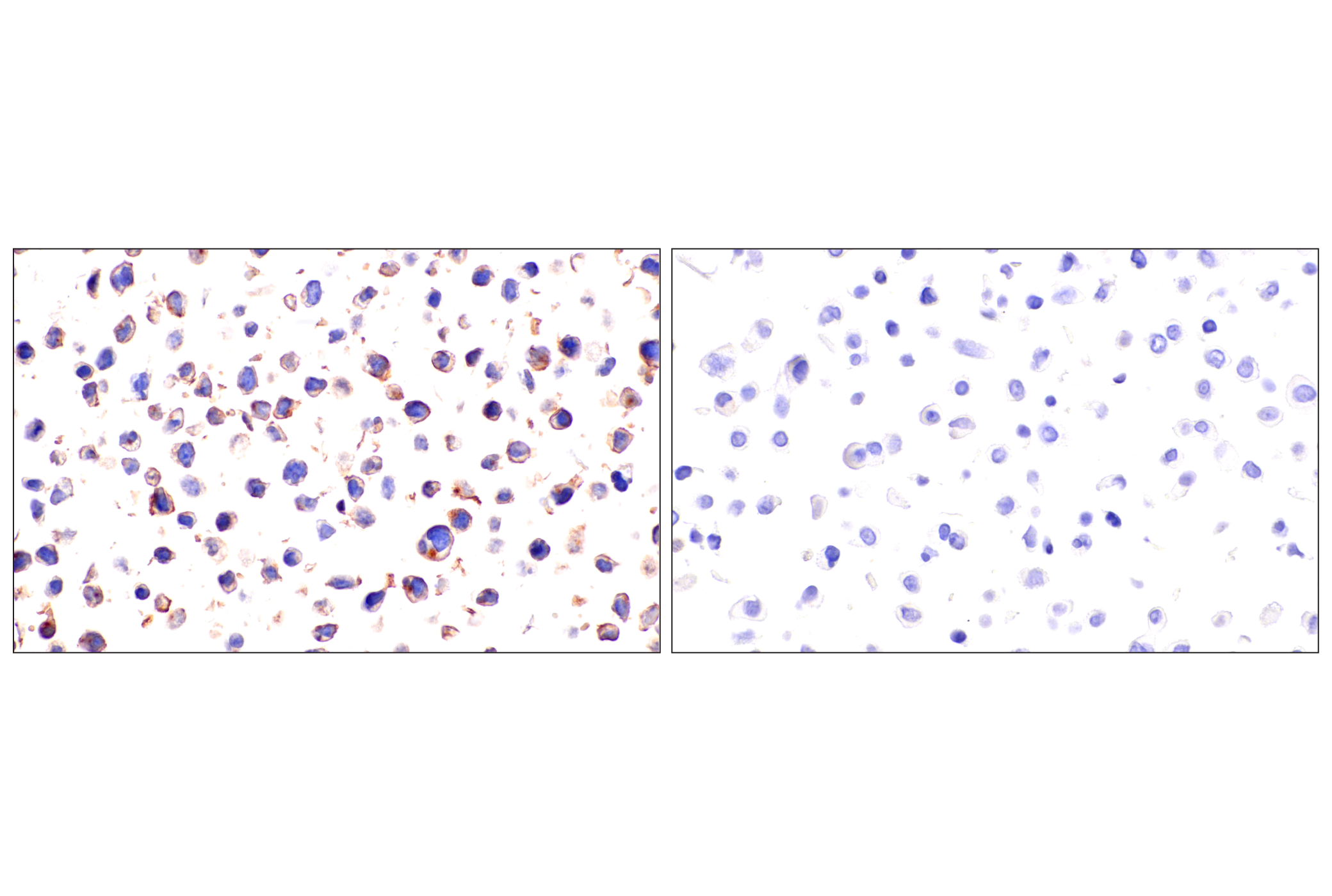
| Immunohistochemistry (Paraffin) | 1:500 - 1:2000 |
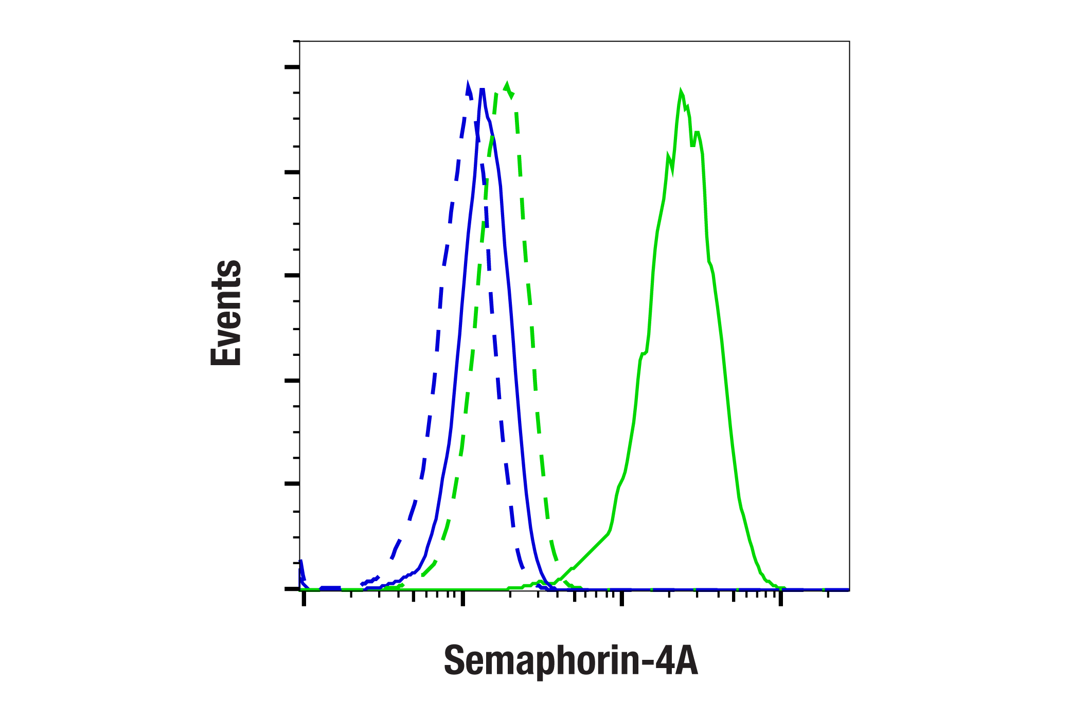
| Flow Cytometry (Fixed/Permeabilized) | 1:200 - 1:800 |
Storage
Specificity / Sensitivity
Species Reactivity:
Human
Source / Purification
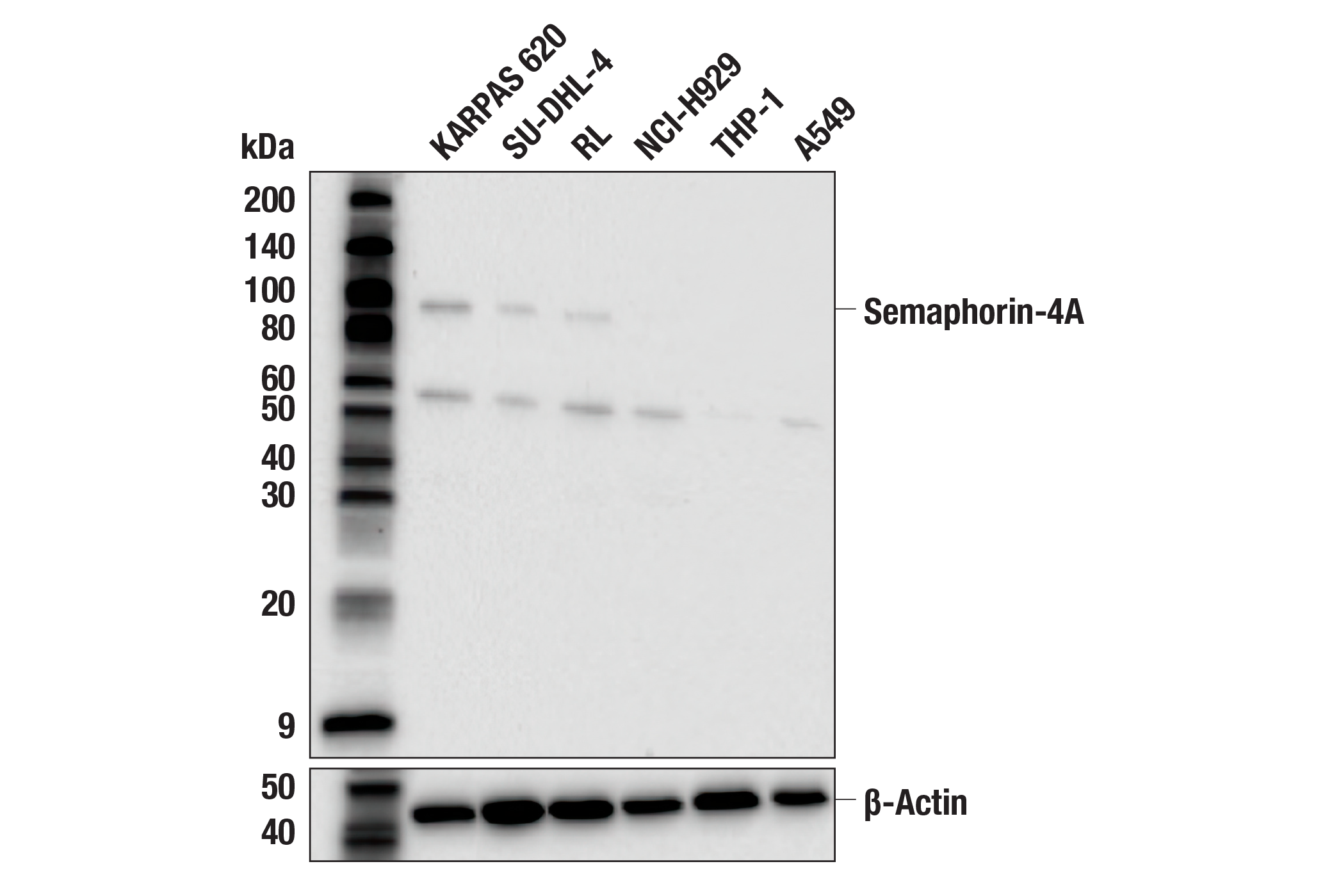
Monoclonal antibody is produced by immunizing animals with a synthetic peptide corresponding to residues surrounding Leu756 of human Semaphorin-4A protein.
Background
Semaphorin-4A, or Sema4A, is a membrane-type class IV semaphorin family member glycoprotein and is expressed in dendritic cells, B cells, T cells, and endothelial cells (1). The protein structure consists of an N-terminus signal peptide, a sema domain, a C2 type Ig domain, a transmembrane region, and a cytoplasmic tail (2). Semaphorin-4A exerts diverse biological effects through several different receptor-mediated pathways and plays important roles in immunity, angiogenesis, neuronal migration, and tumor progression. Semaphorin-4A enhances T cell activation by interacting with TIM-2, and T cell-derived Semaphorin-4A plays a role in helper T cell (Th) differentiation (3,4). In regulatory T cells (Tregs), Semaphorin-4A plays an important role in enhancing stability through interactions with neuropilin-1 (Nrp1) (5). Semaphorin-4A interacts with PlexinD1, which is expressed in endothelial cells and negatively regulates angiogenesis (6). In presynaptic neurons, Semaphorin-4A promotes the development of inhibitory or excitatory synapses through interactions with PlexinB1 or PlexinB2, respectively (7). Semaphorin-4A has been identified as a therapeutic target to promote antitumor responses, and its expression in the tumor microenvironment (TME) has been suggested as a biomarker to predict response to immunotherapy (8).
- Ito, D. and Kumanogoh, A. (2016) Cell Adh Migr 10, 692-699.
- Nkyimbeng-Takwi, E. and Chapoval, S.P. (2011) Immunol Res 50, 10-21.
- Kumanogoh, A. et al. (2002) Nature 419, 629-33.
- Kumanogoh, A. et al. (2005) Immunity 22, 305-16.
- Delgoffe, G.M. et al. (2013) Nature 501, 252-6.
- Toyofuku, T. et al. (2007) EMBO J 26, 1373-84.
- McDermott, J.E. et al. (2018) Mol Cell Neurosci 92, 50-66.
- Naito, Y. et al. (2023) Sci Adv 9, eade0718.
Species Reactivity
Species reactivity is determined by testing in at least one approved application (e.g., western blot).
Western Blot Buffer
IMPORTANT: For western blots, incubate membrane with diluted primary antibody in 5% w/v BSA, 1X TBS, 0.1% Tween® 20 at 4°C with gentle shaking, overnight.
Applications Key
WB: Western Blotting IHC-P: Immunohistochemistry (Paraffin) FC-FP: Flow Cytometry (Fixed/Permeabilized)
Cross-Reactivity Key
H: human M: mouse R: rat Hm: hamster Mk: monkey Vir: virus Mi: mink C: chicken Dm: D. melanogaster X: Xenopus Z: zebrafish B: bovine Dg: dog Pg: pig Sc: S. cerevisiae Ce: C. elegans Hr: horse GP: Guinea Pig Rab: rabbit All: all species expected
Trademarks and Patents
Limited Uses
Except as otherwise expressly agreed in a writing signed by a legally authorized representative of CST, the following terms apply to Products provided by CST, its affiliates or its distributors. Any Customer's terms and conditions that are in addition to, or different from, those contained herein, unless separately accepted in writing by a legally authorized representative of CST, are rejected and are of no force or effect.
Products are labeled with For Research Use Only or a similar labeling statement and have not been approved, cleared, or licensed by the FDA or other regulatory foreign or domestic entity, for any purpose. Customer shall not use any Product for any diagnostic or therapeutic purpose, or otherwise in any manner that conflicts with its labeling statement. Products sold or licensed by CST are provided for Customer as the end-user and solely for research and development uses. Any use of Product for diagnostic, prophylactic or therapeutic purposes, or any purchase of Product for resale (alone or as a component) or other commercial purpose, requires a separate license from CST. Customer shall (a) not sell, license, loan, donate or otherwise transfer or make available any Product to any third party, whether alone or in combination with other materials, or use the Products to manufacture any commercial products, (b) not copy, modify, reverse engineer, decompile, disassemble or otherwise attempt to discover the underlying structure or technology of the Products, or use the Products for the purpose of developing any products or services that would compete with CST products or services, (c) not alter or remove from the Products any trademarks, trade names, logos, patent or copyright notices or markings, (d) use the Products solely in accordance with CST Product Terms of Sale and any applicable documentation, and (e) comply with any license, terms of service or similar agreement with respect to any third party products or services used by Customer in connection with the Products.