WB, IP, IF-F, IF-IC, FC-FP, FC-L
M
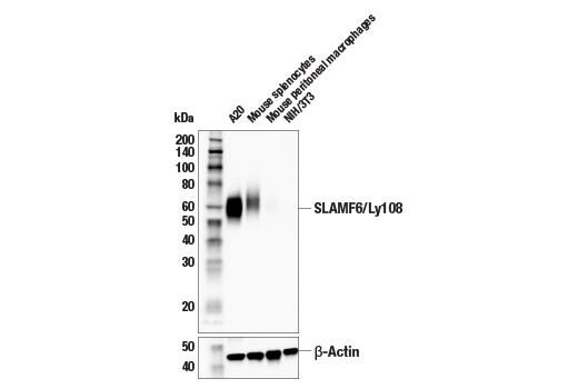
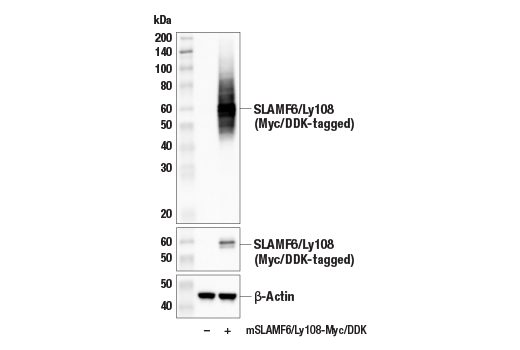
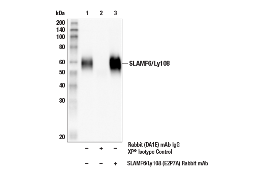
Endogenous
55-70
Rabbit IgG
#Q9ET39
30925
Product Information
Product Usage Information
| Application | Dilution |
|---|---|
| Western Blotting | 1:1000 |
| Immunoprecipitation | 1:100 |
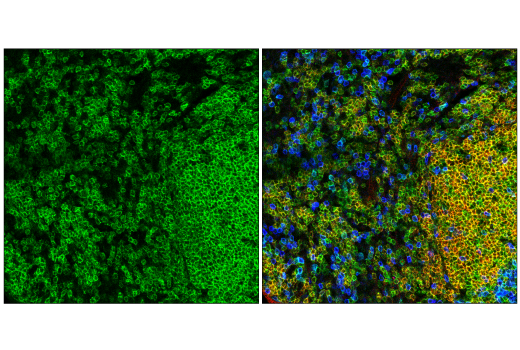
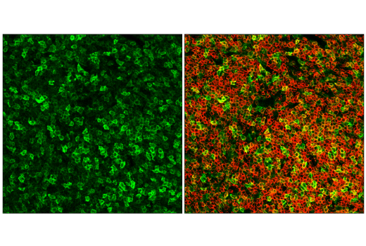
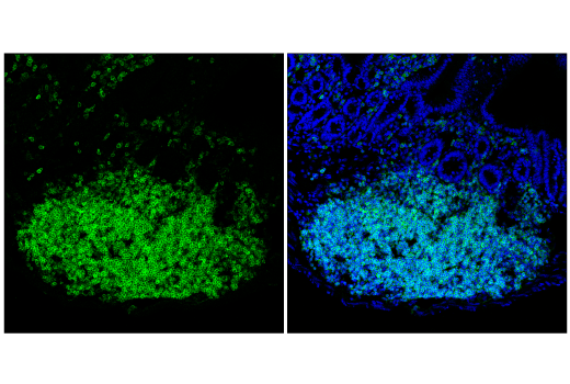
| Immunofluorescence (Frozen) | 1:200 - 1:800 |
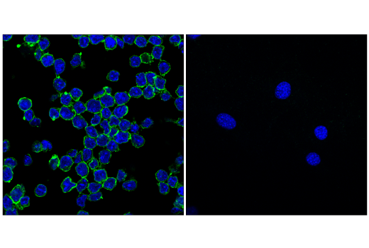
| Immunofluorescence (Immunocytochemistry) | 1:200 - 1:800 |
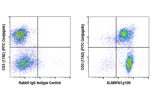
| Flow Cytometry (Fixed/Permeabilized) | 1:50 - 1:200 |
| Flow Cytometry (Live) | 1:50 - 1:200 |
Storage
Specificity / Sensitivity
Species Reactivity:
Mouse
Source / Purification
Monoclonal antibody is produced by immunizing animals with recombinant protein specific to the extracellular domain of mouse SLAMF6/Ly108 protein.
Background
SLAMF6 (CD352/NTB-A) is a type-I transmembrane glycoprotein belonging to the signaling lymphocytic activation molecule (SLAM) family of immunomodulatory receptors. Like other members of the SLAM receptor family, SLAMF6 contains Ig-like domains within its extracellular region and conserved tyrosine-based signaling motifs within its intracellular domain that, when phosphorylated, bind to the SAP and EAT-2 signaling adaptors (1). SLAMF6 is expressed on the surface of multiple types of immune cells, such as those of the B, T, and NK lineages. Its activation is triggered by homotypic interactions involving its extracellular domain (1-3). Indeed, research studies have shown that in T cells, SLAMF6 engagement facilitates activation and cytokine production (4). Similarly, homotypic ligand-mediated engagement of SLAMF6 on NK cells activates signaling cascades that drive proliferation, cytotoxicity, and cytokine production (1,5-7).
- Bottino, C. et al. (2001) J Exp Med 194, 235-46.
- Fraser, C.C. et al. (2002) Immunogenetics 53, 843-50.
- Flaig, R.M. et al. (2004) J Immunol 172, 6524-7.
- Valdez, P.A. et al. (2004) J Biol Chem 279, 18662-9.
- Stark, S. and Watzl, C. (2006) Int Immunol 18, 241-7.
- Falco, M. et al. (2004) Eur J Immunol 34, 1663-72.
- Eissmann, P. and Watzl, C. (2006) J Immunol 177, 3170-7.
Species Reactivity
Species reactivity is determined by testing in at least one approved application (e.g., western blot).
Western Blot Buffer
IMPORTANT: For western blots, incubate membrane with diluted primary antibody in 5% w/v BSA, 1X TBS, 0.1% Tween® 20 at 4°C with gentle shaking, overnight.
Applications Key
WB: Western Blotting IP: Immunoprecipitation IF-F: Immunofluorescence (Frozen) IF-IC: Immunofluorescence (Immunocytochemistry) FC-FP: Flow Cytometry (Fixed/Permeabilized) FC-L: Flow Cytometry (Live)
Cross-Reactivity Key
H: human M: mouse R: rat Hm: hamster Mk: monkey Vir: virus Mi: mink C: chicken Dm: D. melanogaster X: Xenopus Z: zebrafish B: bovine Dg: dog Pg: pig Sc: S. cerevisiae Ce: C. elegans Hr: horse GP: Guinea Pig Rab: rabbit All: all species expected
Trademarks and Patents
Limited Uses
Except as otherwise expressly agreed in a writing signed by a legally authorized representative of CST, the following terms apply to Products provided by CST, its affiliates or its distributors. Any Customer's terms and conditions that are in addition to, or different from, those contained herein, unless separately accepted in writing by a legally authorized representative of CST, are rejected and are of no force or effect.
Products are labeled with For Research Use Only or a similar labeling statement and have not been approved, cleared, or licensed by the FDA or other regulatory foreign or domestic entity, for any purpose. Customer shall not use any Product for any diagnostic or therapeutic purpose, or otherwise in any manner that conflicts with its labeling statement. Products sold or licensed by CST are provided for Customer as the end-user and solely for research and development uses. Any use of Product for diagnostic, prophylactic or therapeutic purposes, or any purchase of Product for resale (alone or as a component) or other commercial purpose, requires a separate license from CST. Customer shall (a) not sell, license, loan, donate or otherwise transfer or make available any Product to any third party, whether alone or in combination with other materials, or use the Products to manufacture any commercial products, (b) not copy, modify, reverse engineer, decompile, disassemble or otherwise attempt to discover the underlying structure or technology of the Products, or use the Products for the purpose of developing any products or services that would compete with CST products or services, (c) not alter or remove from the Products any trademarks, trade names, logos, patent or copyright notices or markings, (d) use the Products solely in accordance with CST Product Terms of Sale and any applicable documentation, and (e) comply with any license, terms of service or similar agreement with respect to any third party products or services used by Customer in connection with the Products.