| Product Includes | Product # | Quantity | Mol. Wt | Isotype/Source |
|---|---|---|---|---|
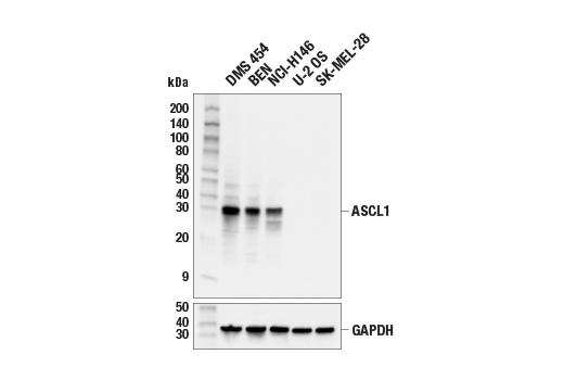
| ASCL1 (E5S4Q) XP® Rabbit mAb | 10585 | 20 µl | 30 kDa | Rabbit IgG |
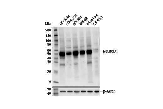
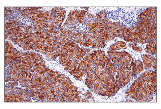
| NeuroD1 (D90G12) Rabbit mAb | 7019 | 20 µl | 49 kDa | Rabbit IgG |
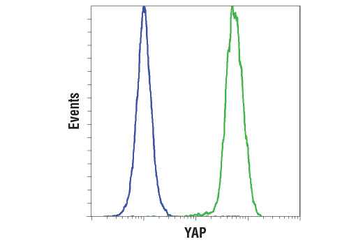
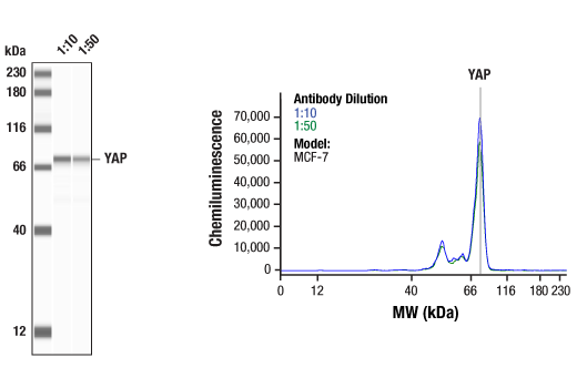
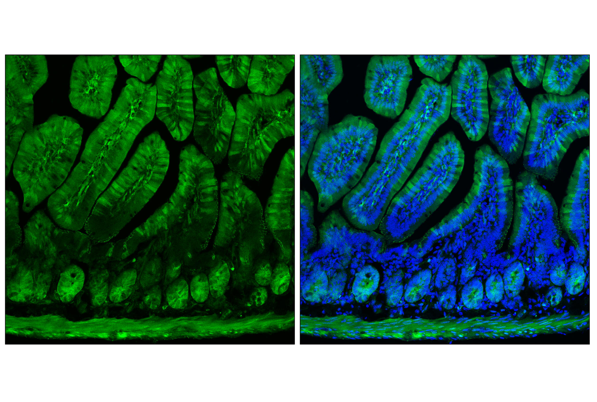
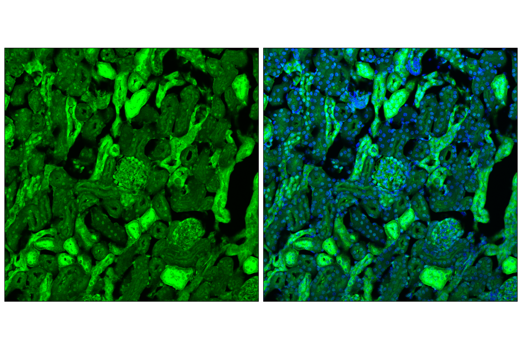
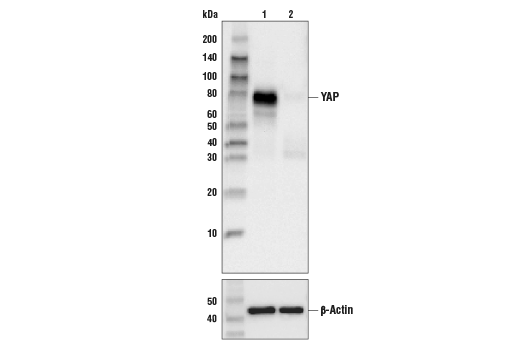
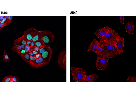
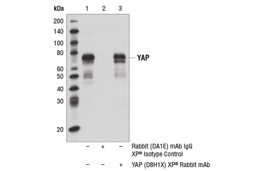
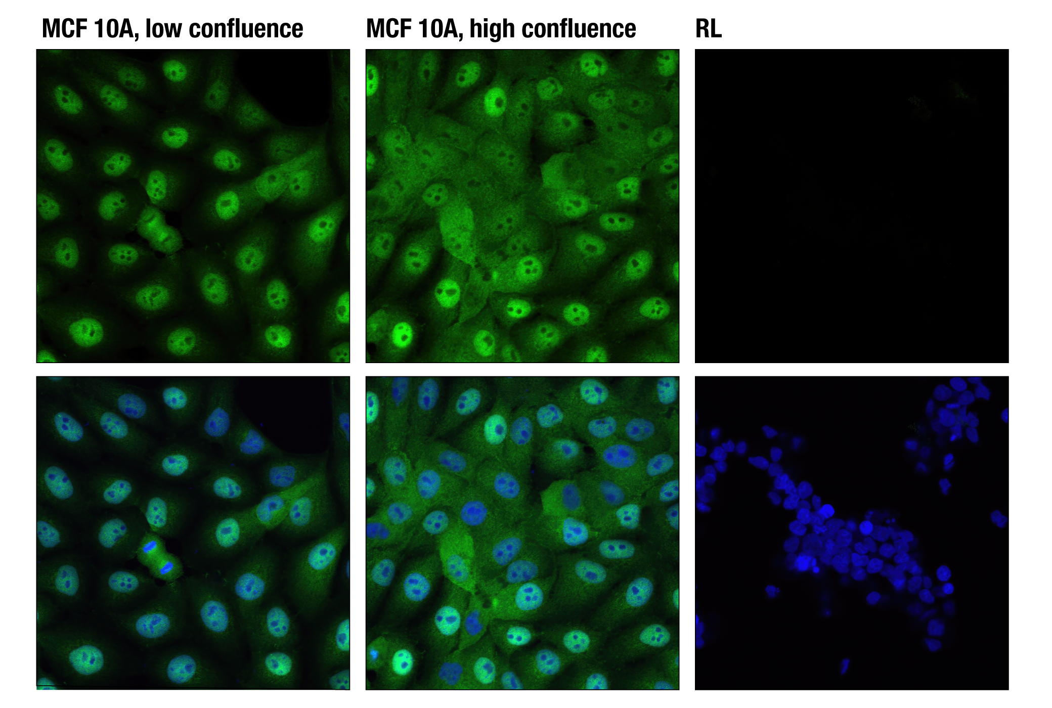
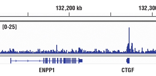
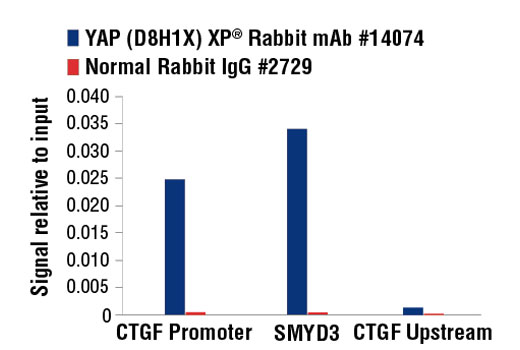
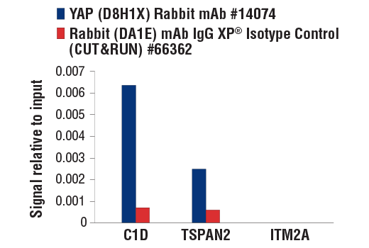
| YAP (D8H1X) XP® Rabbit mAb | 14074 | 20 µl | 65-78 kDa | Rabbit IgG |
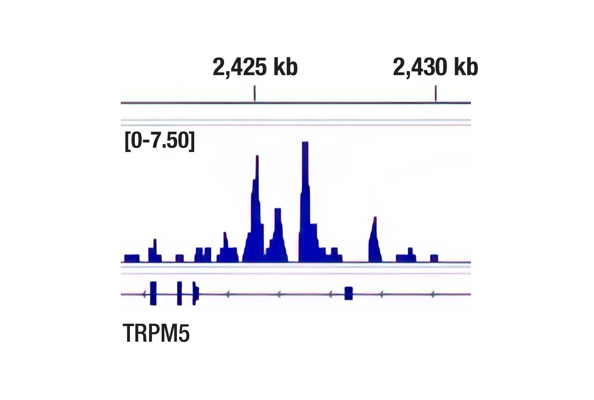
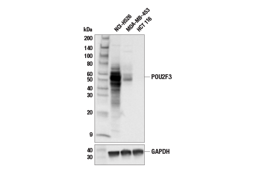
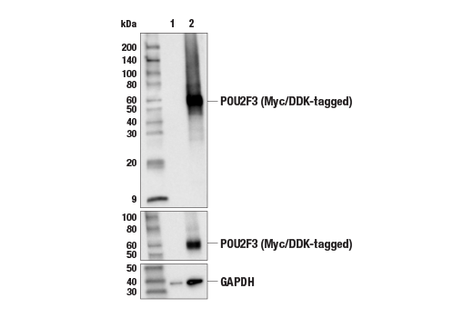
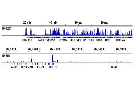
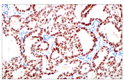
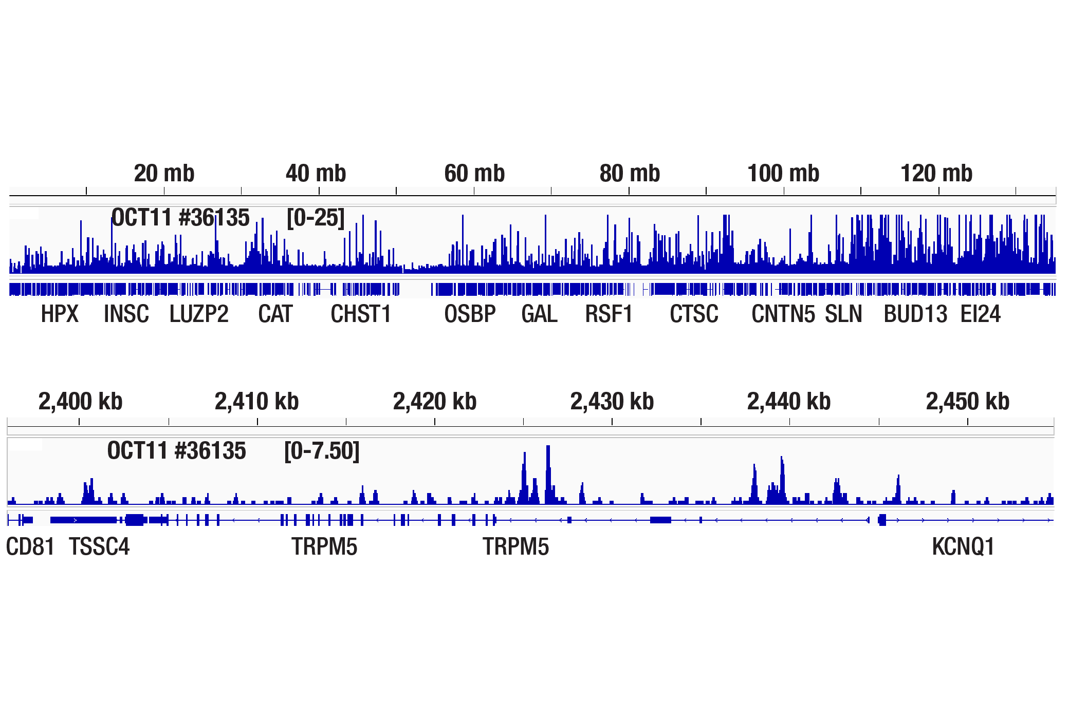
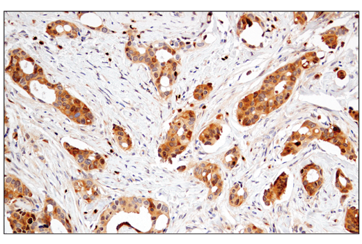
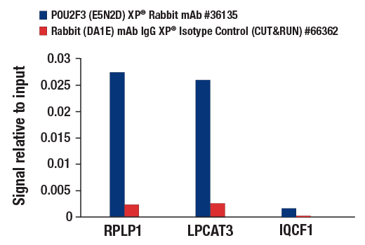
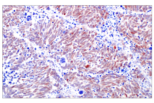
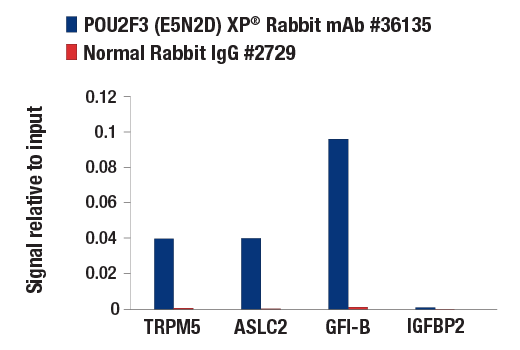
| POU2F3 (E5N2D) XP® Rabbit mAb | 36135 | 20 µl | 45-60 kDa | Rabbit IgG |
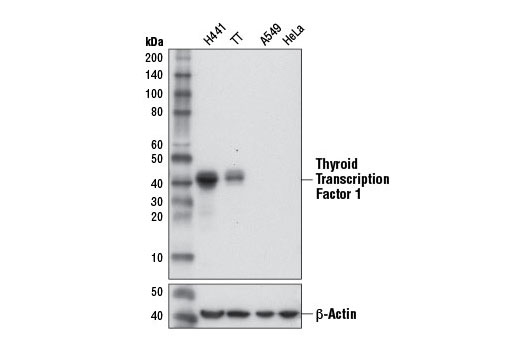
| Thyroid Transcription Factor 1 (TTF-1) (D2E8) Rabbit mAb | 12373 | 20 µl | 39, 42 kDa | Rabbit |
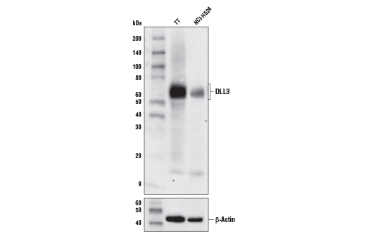
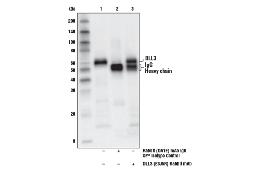
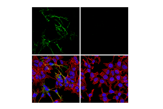
| DLL3 (E3J5R) Rabbit mAb | 71804 | 20 µl | 65 kDa | Rabbit IgG |
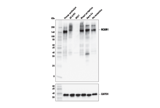
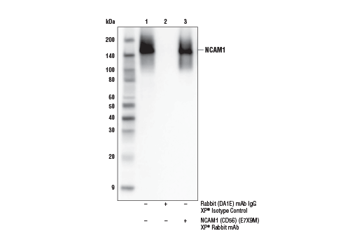
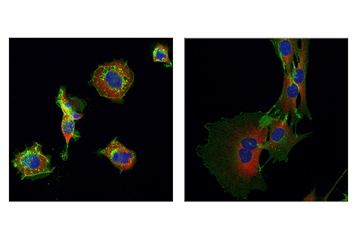
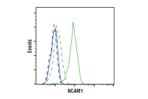
| NCAM1 (CD56) (E7X9M) XP® Rabbit mAb | 99746 | 20 µl | 120 to 220 kDa | Rabbit IgG |
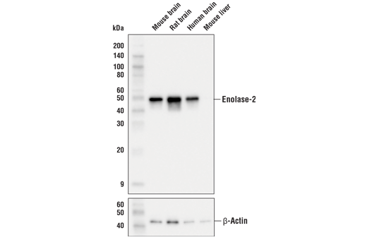
| Enolase-2 (E2H9X) XP® Rabbit mAb | 24330 | 20 µl | 47 kDa | Rabbit IgG |
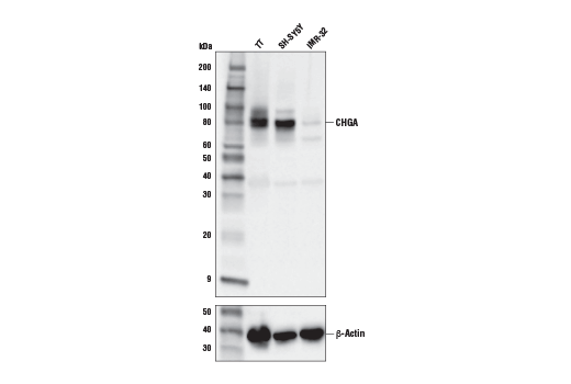
| CHGA (E8X7R) Rabbit mAb | 85798 | 20 µl | 80 kDa | Rabbit IgG |
| Anti-rabbit IgG, HRP-linked Antibody | 7074 | 100 µl | Goat |
Please visit cellsignal.com for individual component applications, species cross-reactivity, dilutions, protocols, and additional product information.
Description
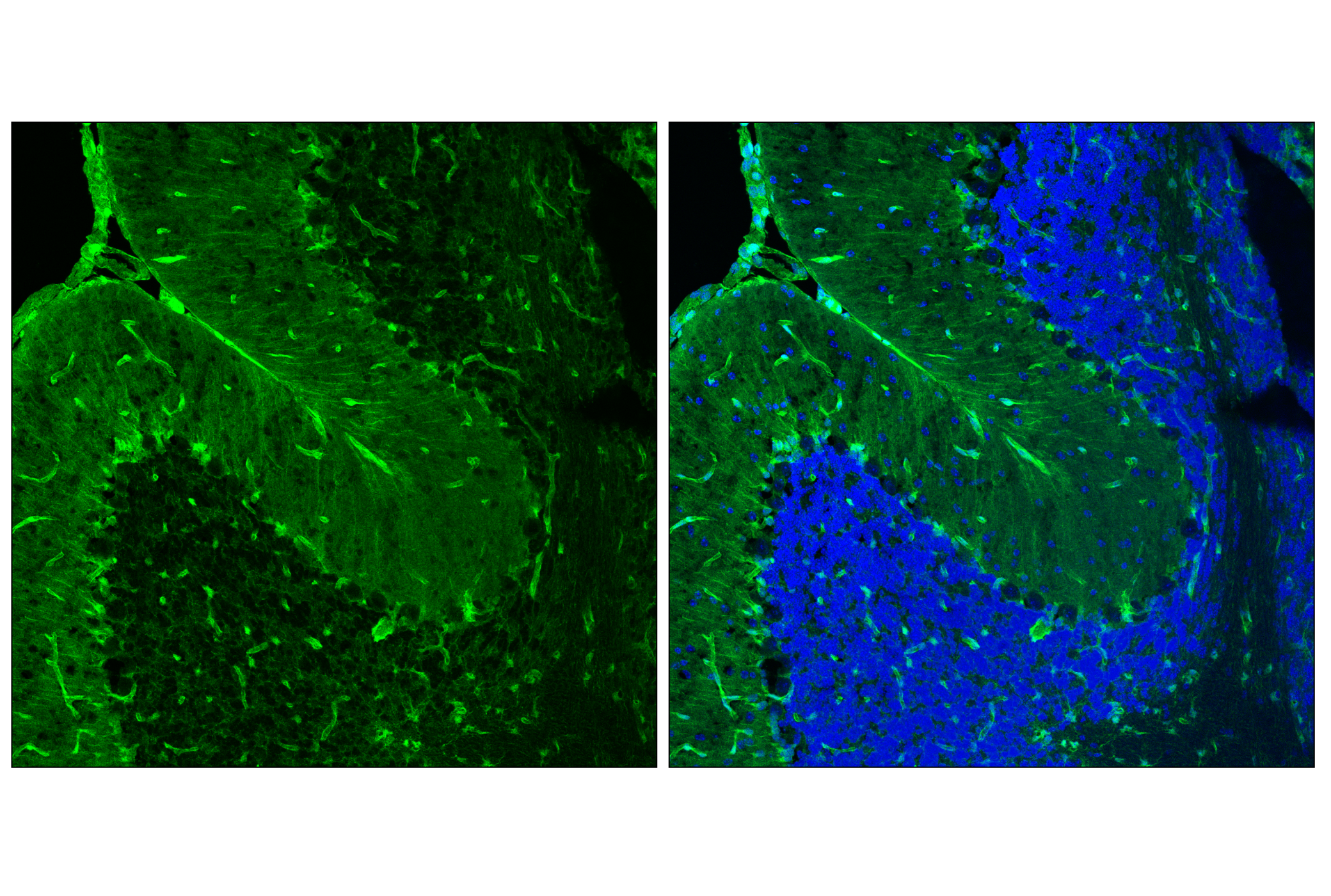
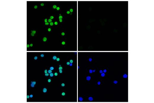
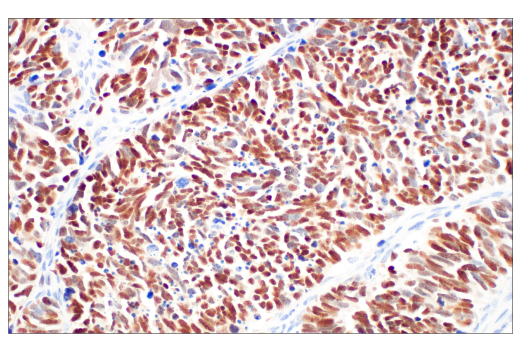
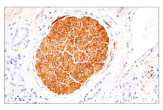
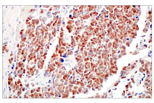
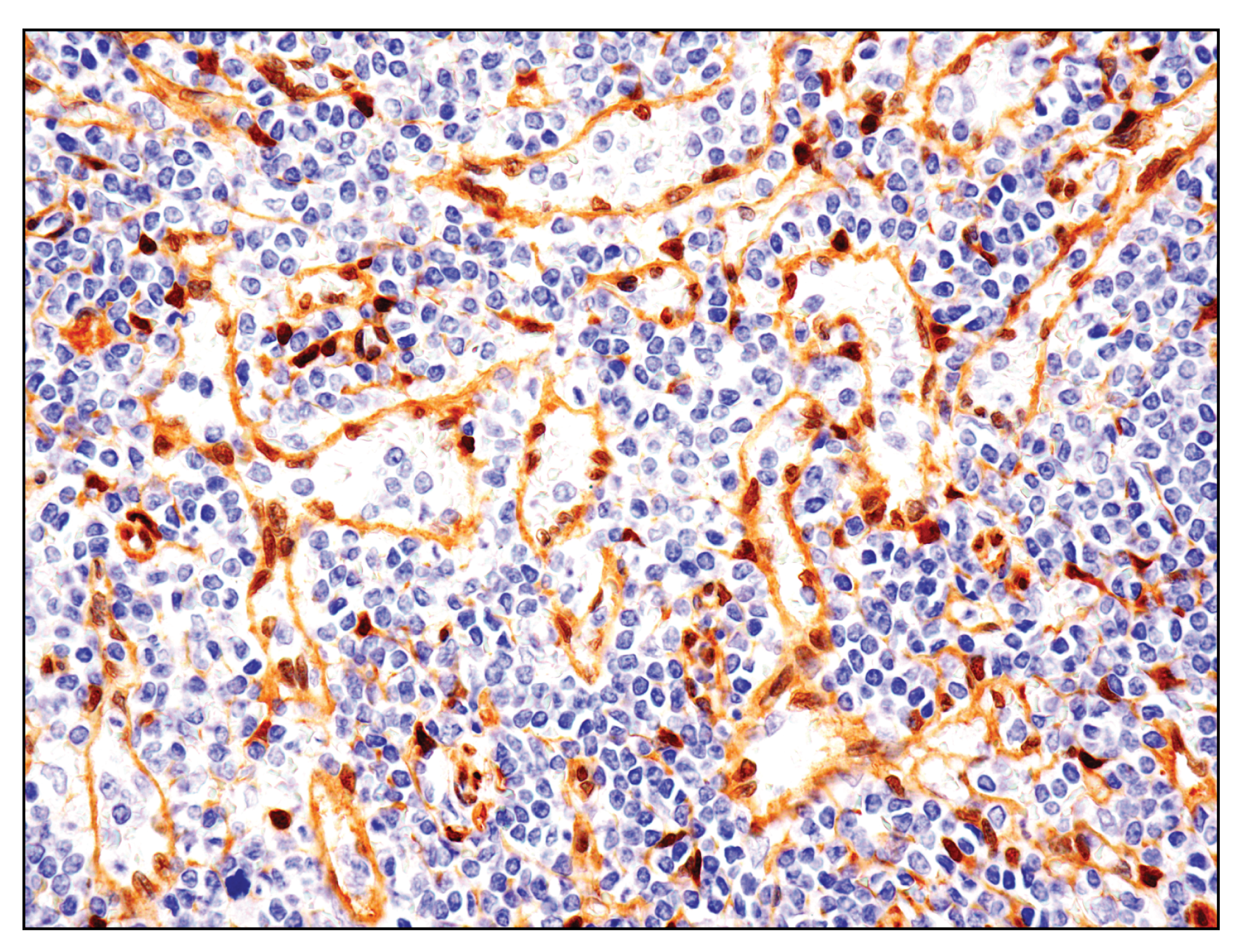
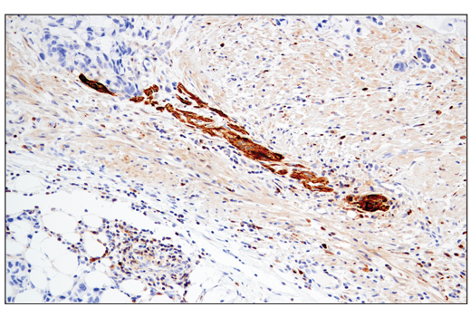
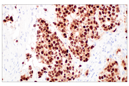
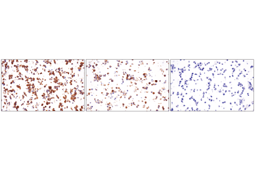
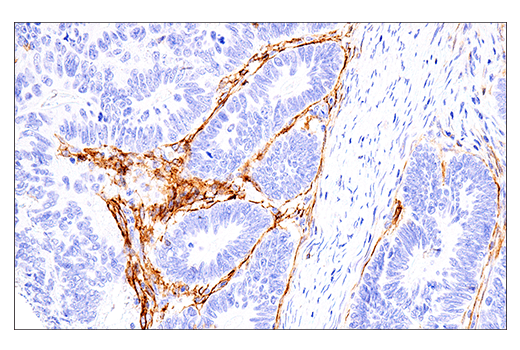
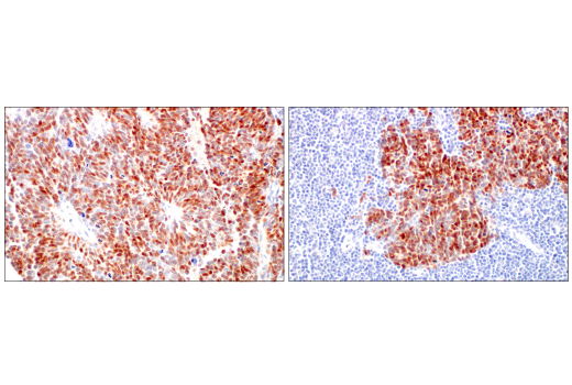
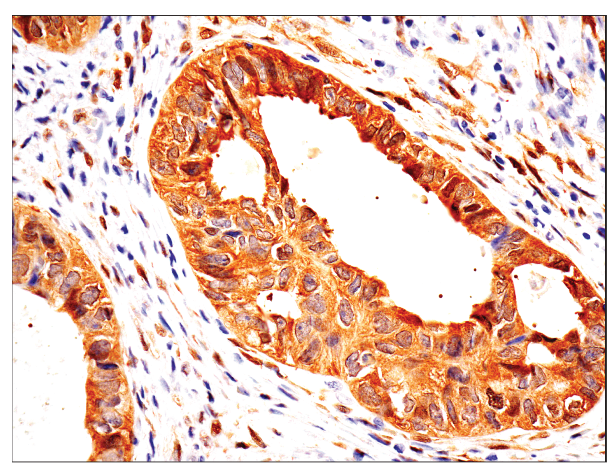
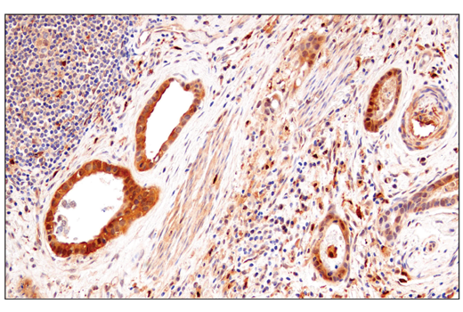
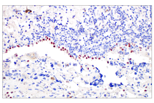
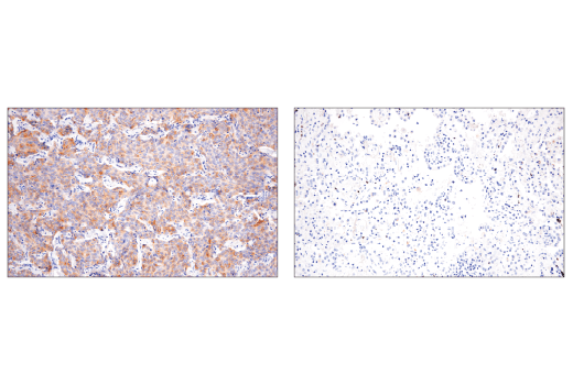
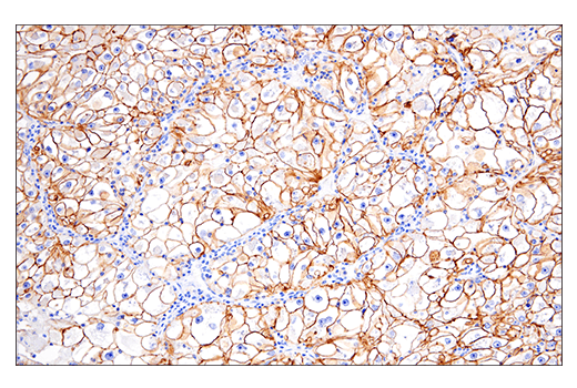
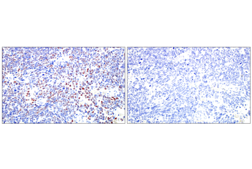
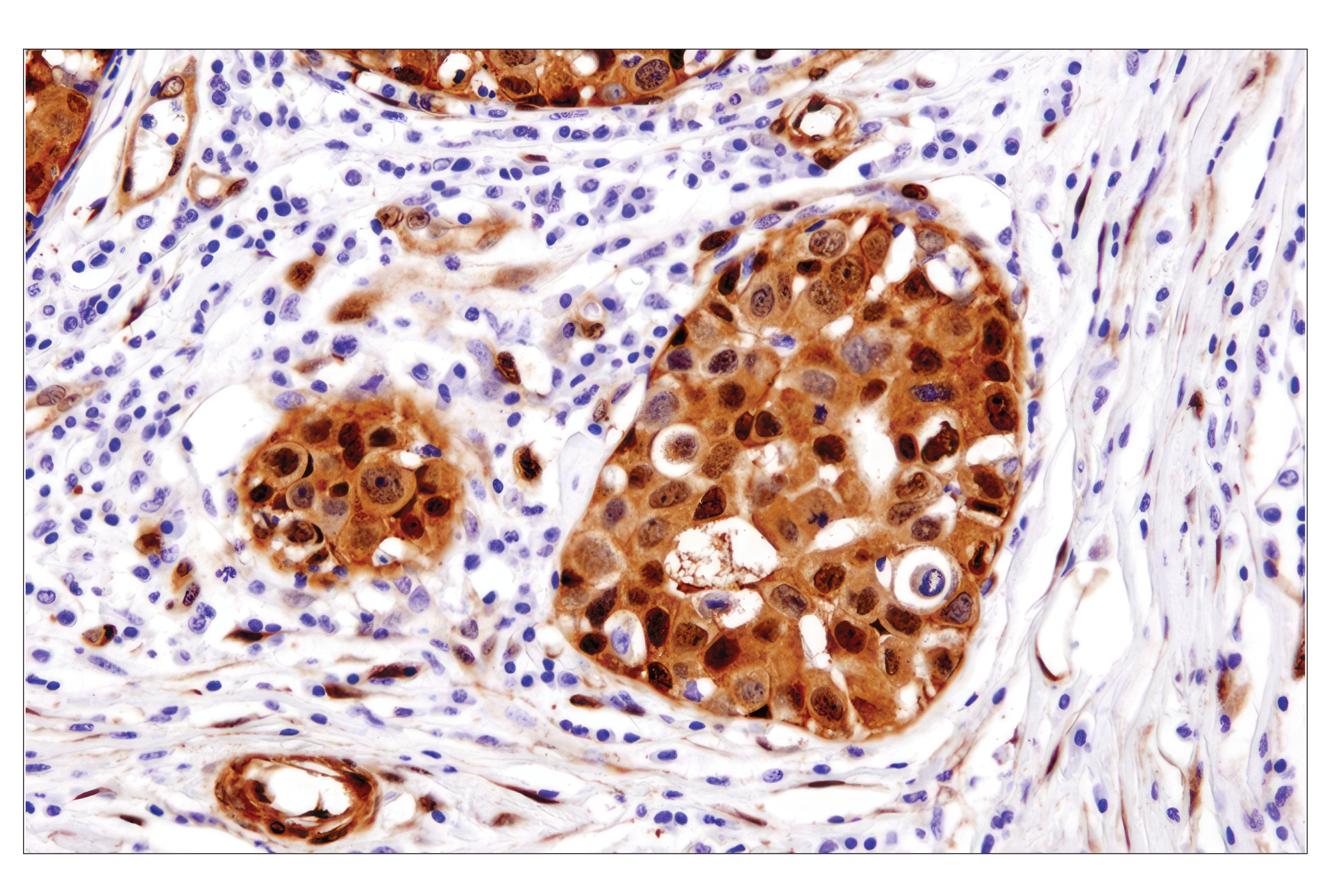
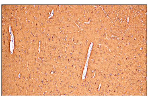
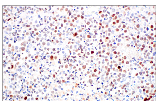
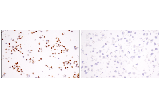
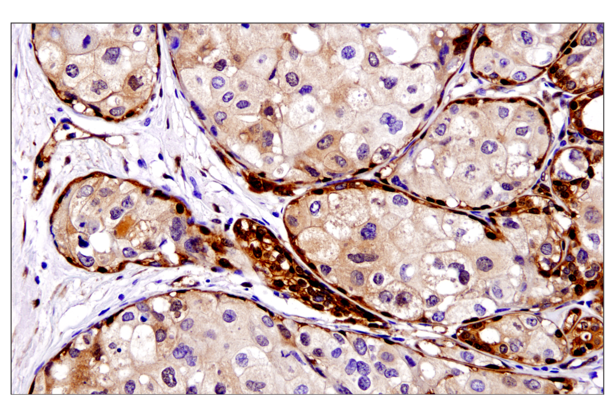
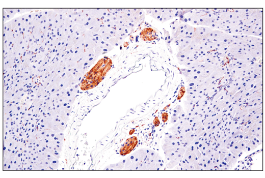
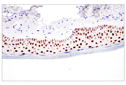
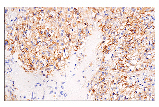
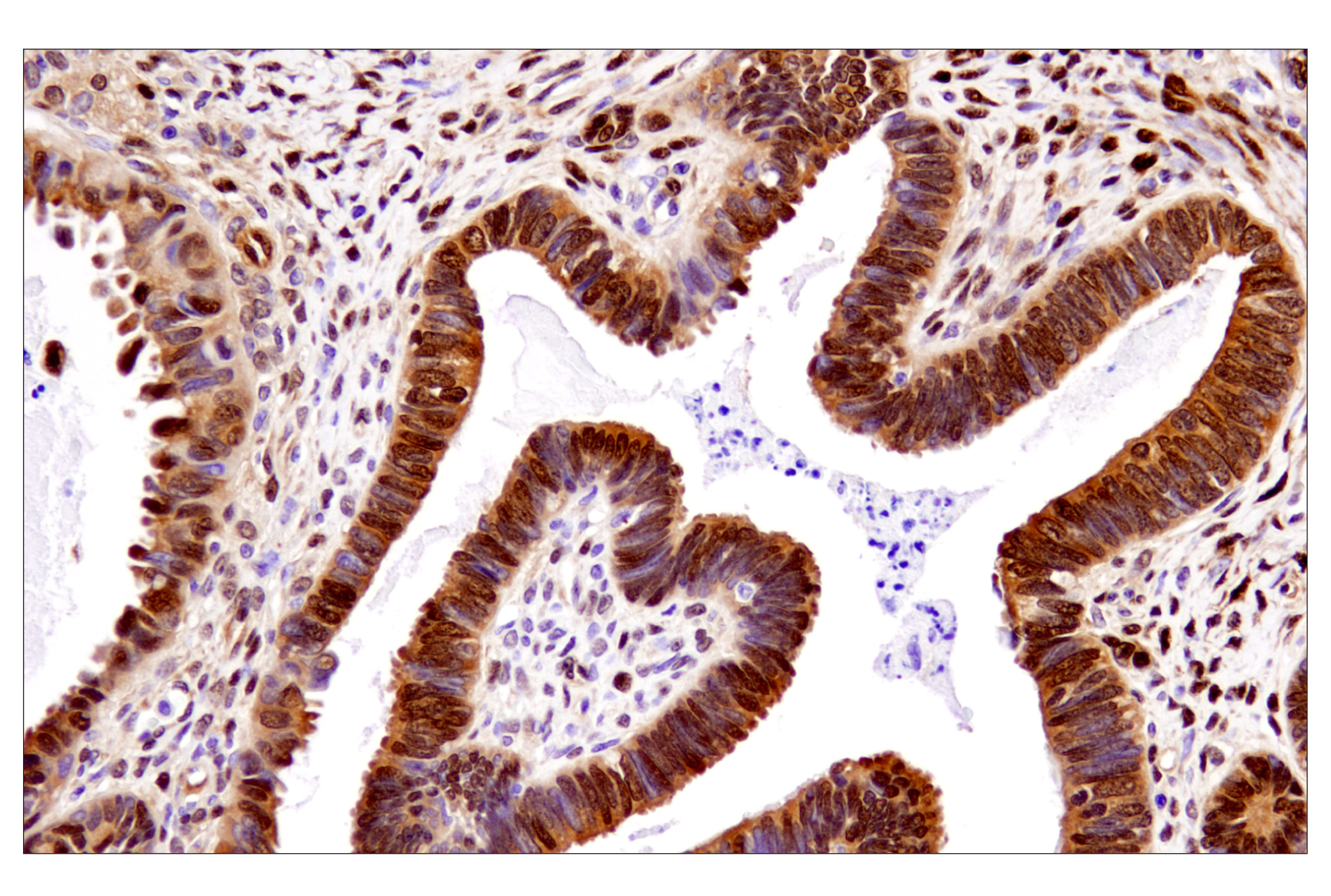
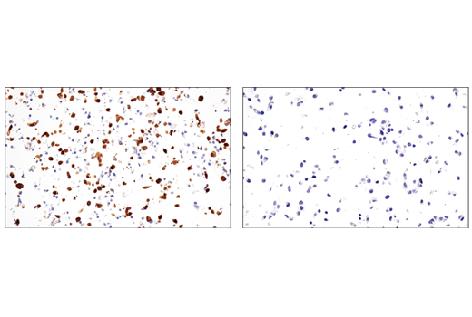
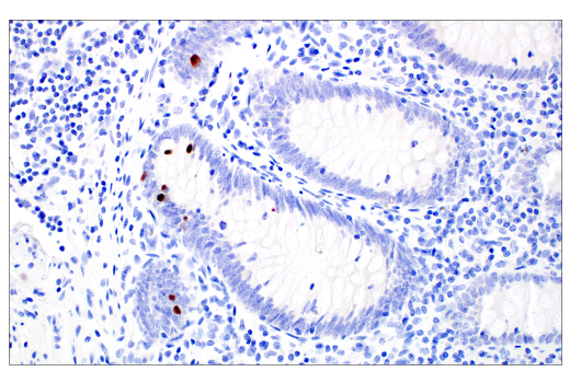
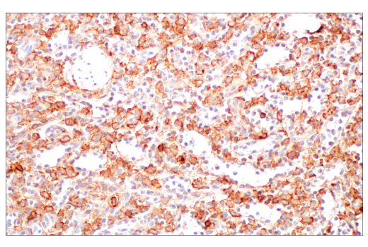
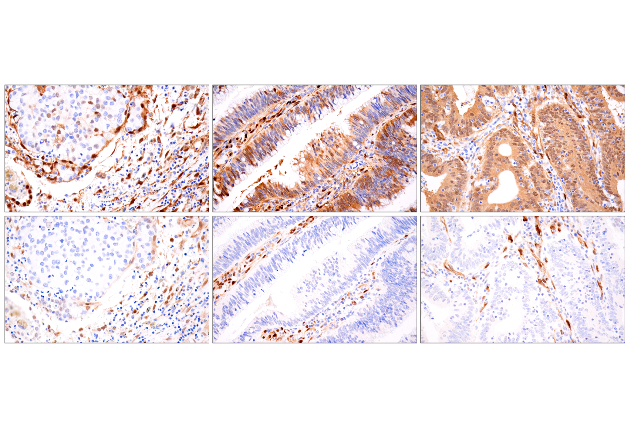
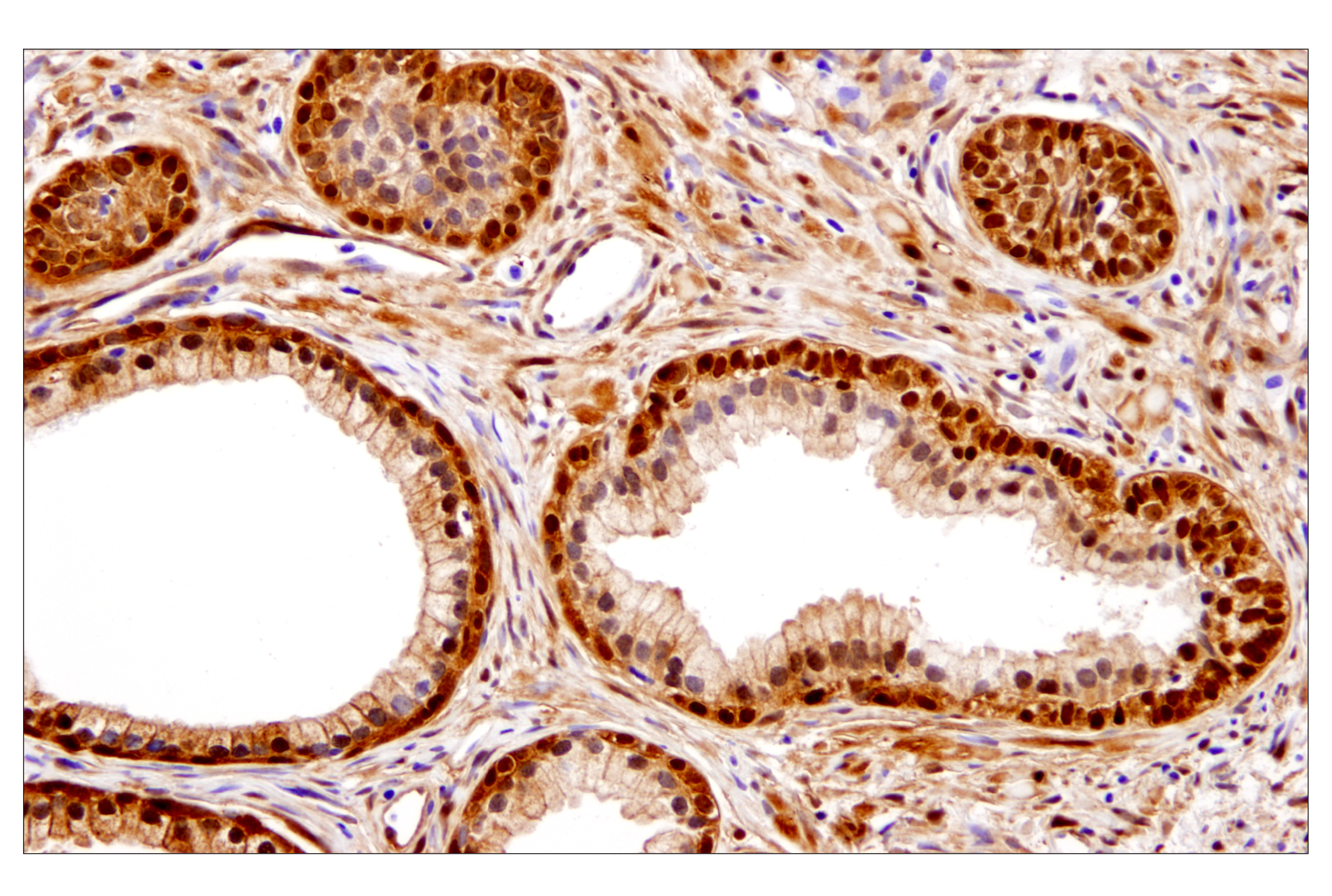
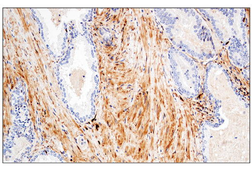
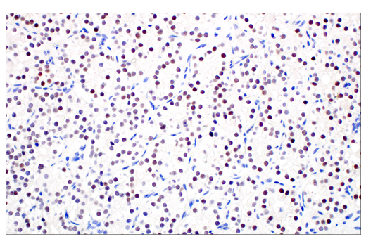
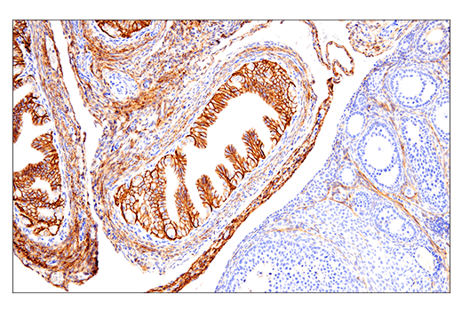
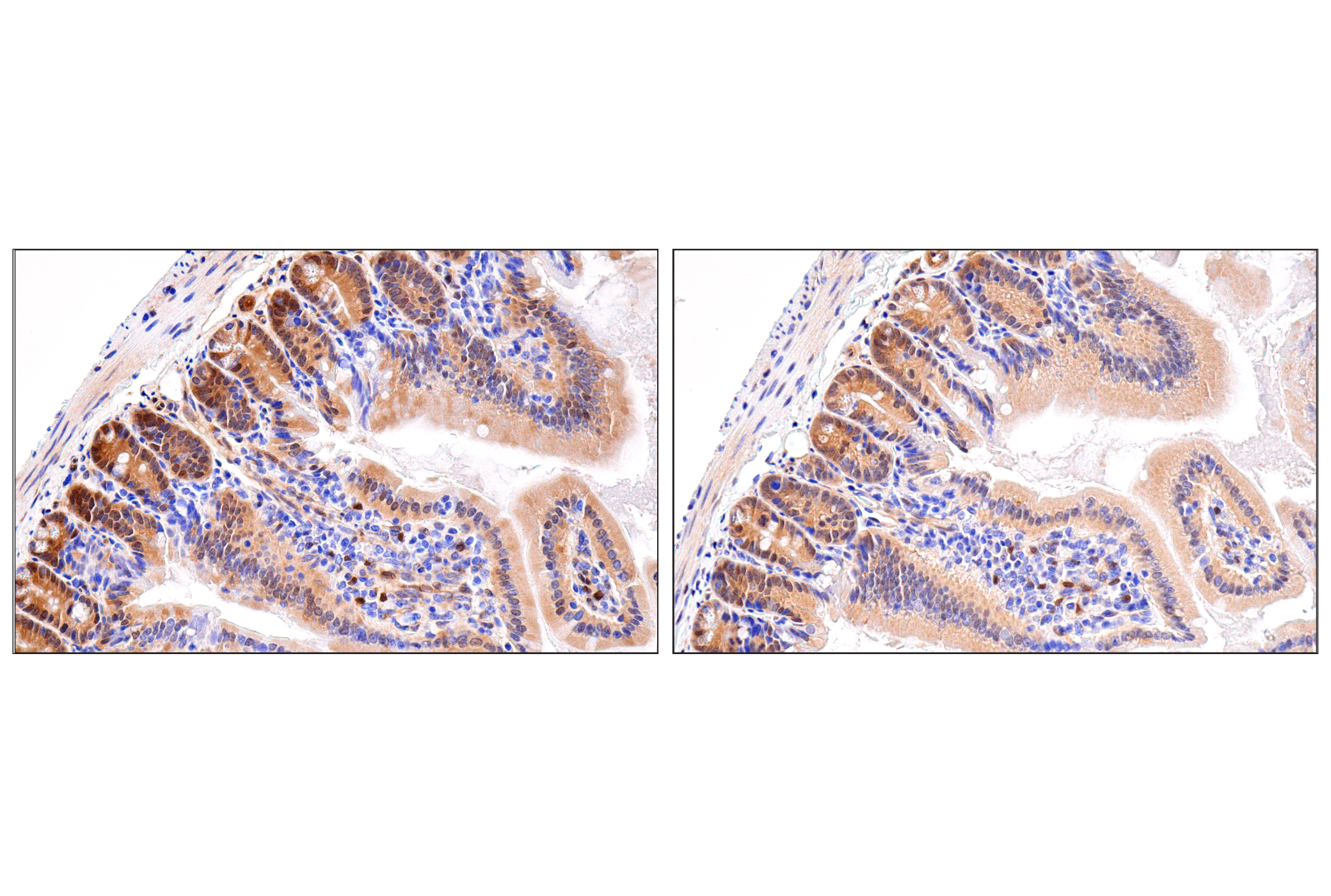
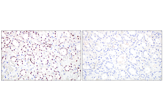
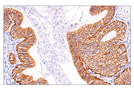
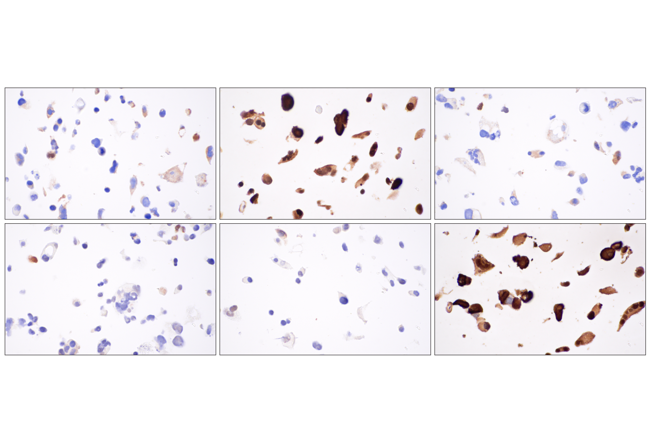
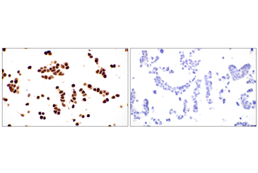
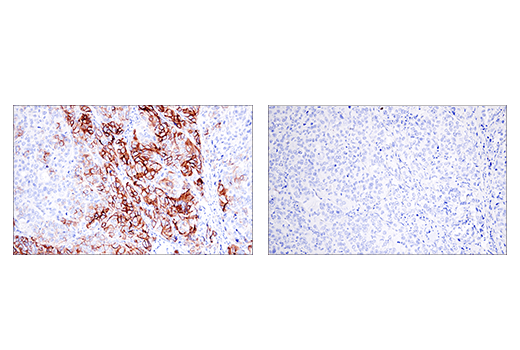
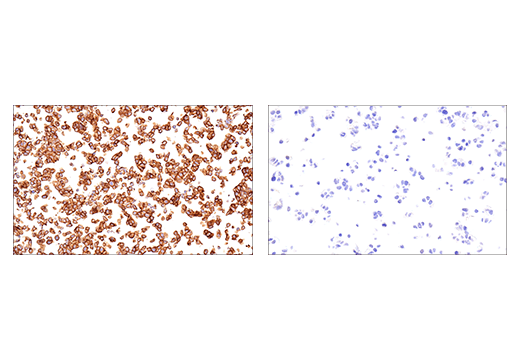
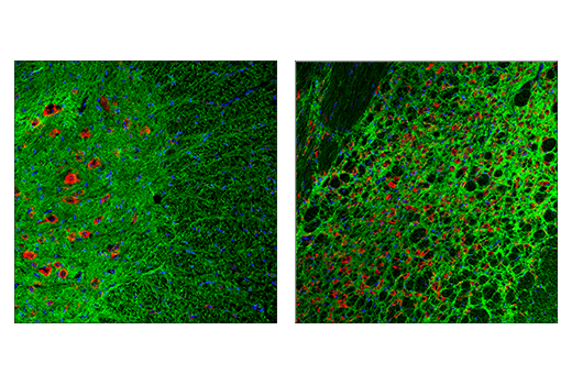
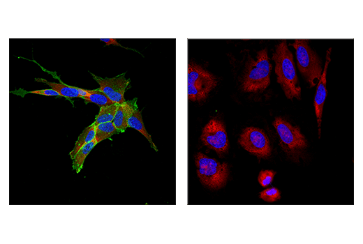
The Small Cell Lung Cancer Biomarker Antibody Sampler Kit provides a means of detecting common biomarkers studied in small cell lung cancer (SCLC). The kit includes enough antibodies to perform two western blot experiments with each primary antibody.
Storage
Background
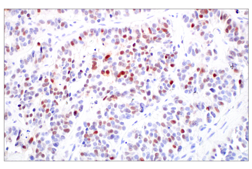
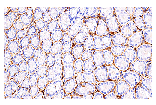
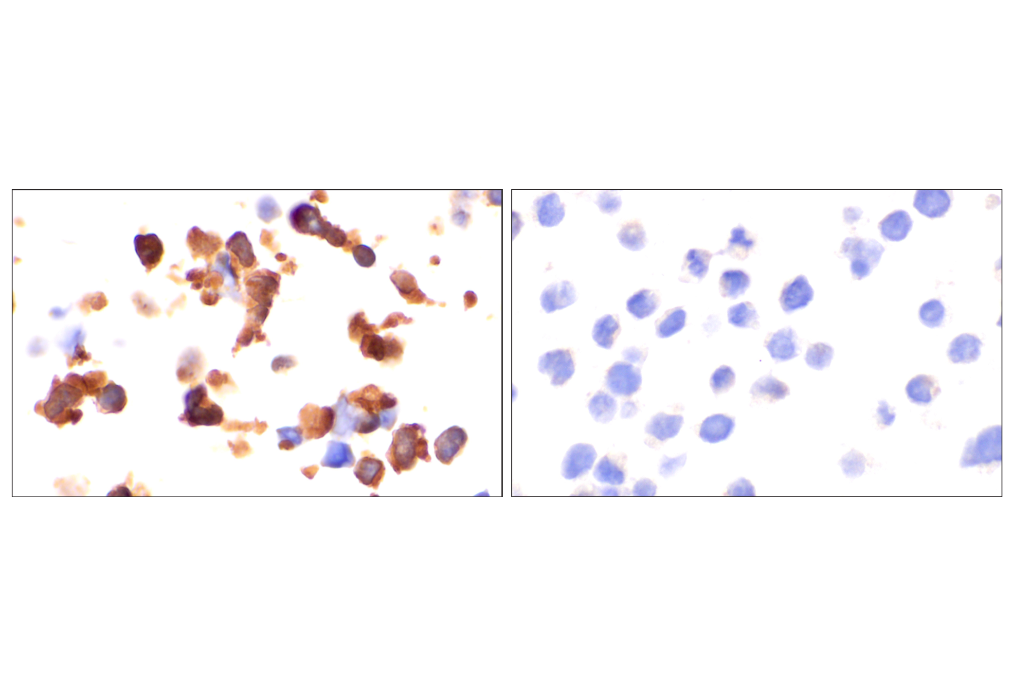
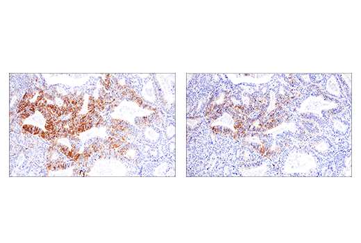
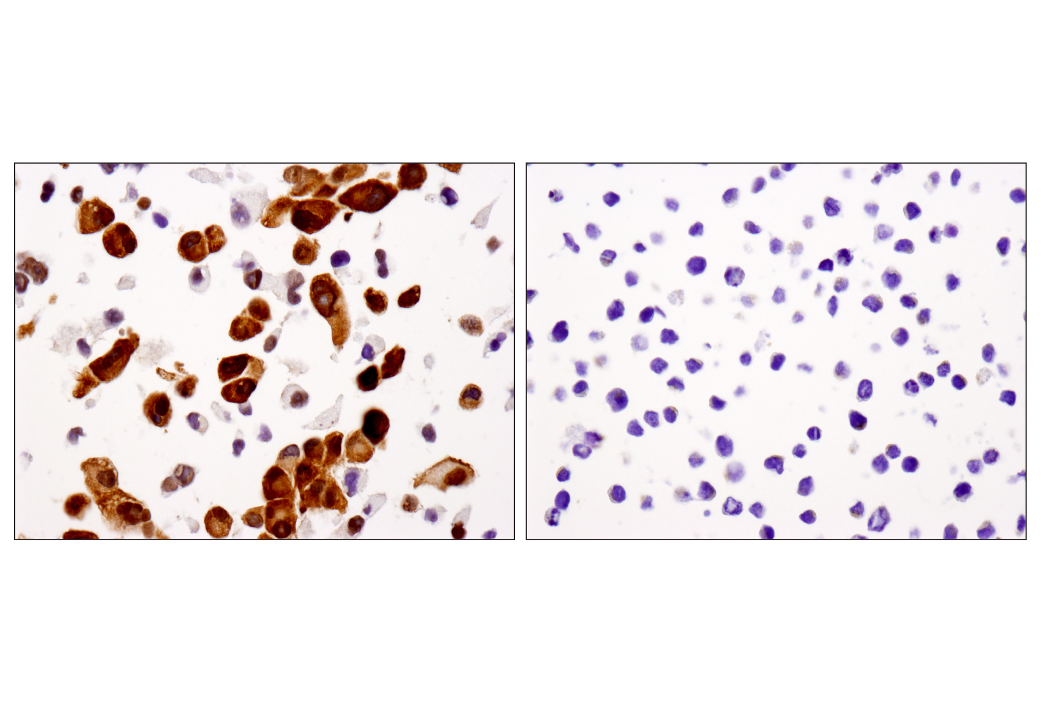
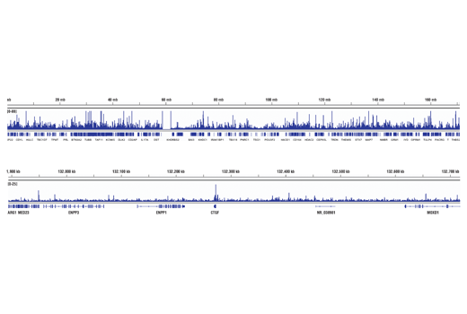
Lung cancer is the leading cause of cancer-related mortality worldwide (1). It is generally divided into two broad histological classifications: small cell lung cancer (SCLC) and non-small cell lung cancer (NSCLC). SCLC is particularly aggressive and has been further subdivided by biological heterogeneity. Subtypes of SCLC have recently been described based on expression distinct transcriptional regulators (2,3). These subtypes were labeled as SCLC-A expressing achaete-scute homolog 1 (ASCL1), SCLC-N expressing neurogenic differentiation factor 1 (NeuroD1), SCLC-Y expressing yes-associated protein 1 (YAP), and SCLC-P expressing POU class 2 homeobox 3 (POU2F3). ASCL1 and NeuroD1 drive a neuroendocrine phenotype through regulation of distinct genes. DLL3, an inhibitor of NOTCH signaling, is upregulated by ASCL1 (4). NCAM1 (neural cell adhesion molecule, CD56) is an adhesion glycoprotein that mediates neuronal attachment, neurite extension, and is a marker for the neuroendocrine phenotype (5). Thyroid transcription factor 1 (TTF-1), a member of the NKX homeobox transcription factor family, is expressed in malignant tumors of the thyroid and lung, and it is commonly used as a marker for both primary and malignant lung cancers (6-8). Enolase-2 is a glycolytic enzyme that is involved in the conversion of 2-phosphoglycerate to phosphoenolpyruvate (9). Research studies have shown elevated levels of neuro-specific enolase-2 in neuroblastoma and SCLC (10,11). Chromogranin A (CHGA) is a member of the chromogranin/secretogranin family of neuroendocrine secretory proteins. It is expressed in the secretory vesicles of neurons and endocrine cells (1,2). CHGA is also useful as a serological and immunohistological marker for the presence of neuroendocrine tumors from various tissue sources (12,13). POU2F3 and YAP drive non-neuroendocrine phenotypes. POU2F3 is normally selectively expressed in chemosensory tuft cells, and SCLC expressing POU2F3 resemble that cell type (14). YAP is widely recognized as a key mediator of the Hippo growth signaling pathway (15). Expression of these key biomarkers in SCLC are thought to help predict therapeutic treatment (16).
- Sung, H. et al. (2021) CA Cancer J Clin 71, 209-249.
- Baine, M.K. et al. (2020) J Thorac Oncol 15, 1823-1835.
- Rudin, C.M. et al. (2019) Nat Rev Cancer 19, 289-297.
- Borromeo, M.D. et al. (2016) Cell Rep 16, 1259-1272.
- Seidenfaden, R. et al. (2003) Mol Cell Biol 23, 5908-18.
- Whithaus, K. et al. (2012) Arch Pathol Lab Med 136, 155-62.
- Yoshida, A. et al. (2011) Lung Cancer 72, 309-15.
- Moldvay, J. et al. (2004) Pathol Oncol Res 10, 85-8.
- Van Obberghen, E. et al. (1988) J Neurosci Res 19, 450-6.
- Stern, P. et al. (2007) Tumour Biol 28, 84-92.
- O'Shea, P. et al. (1995) Ir J Med Sci 164, 31-6.
- Weisbrod, A.B. et al. (2013) Horm Cancer 4, 165-75.
- Annaratone, L. et al. (2014) Endocr Pathol 25, 219-28.
- Rudin, C.M. et al. (2019) Nat Rev Cancer 19, 289-297.
- Zhao, B. et al. (2010) Genes Dev 24, 862-74.
- Wang, W.Z. et al. (2022) Semin Cancer Biol 00095-5, doi: 10.1016/j.semcancer.2022.04.001.
Background References
Trademarks and Patents
Limited Uses
Except as otherwise expressly agreed in a writing signed by a legally authorized representative of CST, the following terms apply to Products provided by CST, its affiliates or its distributors. Any Customer's terms and conditions that are in addition to, or different from, those contained herein, unless separately accepted in writing by a legally authorized representative of CST, are rejected and are of no force or effect.
Products are labeled with For Research Use Only or a similar labeling statement and have not been approved, cleared, or licensed by the FDA or other regulatory foreign or domestic entity, for any purpose. Customer shall not use any Product for any diagnostic or therapeutic purpose, or otherwise in any manner that conflicts with its labeling statement. Products sold or licensed by CST are provided for Customer as the end-user and solely for research and development uses. Any use of Product for diagnostic, prophylactic or therapeutic purposes, or any purchase of Product for resale (alone or as a component) or other commercial purpose, requires a separate license from CST. Customer shall (a) not sell, license, loan, donate or otherwise transfer or make available any Product to any third party, whether alone or in combination with other materials, or use the Products to manufacture any commercial products, (b) not copy, modify, reverse engineer, decompile, disassemble or otherwise attempt to discover the underlying structure or technology of the Products, or use the Products for the purpose of developing any products or services that would compete with CST products or services, (c) not alter or remove from the Products any trademarks, trade names, logos, patent or copyright notices or markings, (d) use the Products solely in accordance with CST Product Terms of Sale and any applicable documentation, and (e) comply with any license, terms of service or similar agreement with respect to any third party products or services used by Customer in connection with the Products.