| Product Includes | Product # | Quantity | Mol. Wt | Isotype/Source |
|---|---|---|---|---|
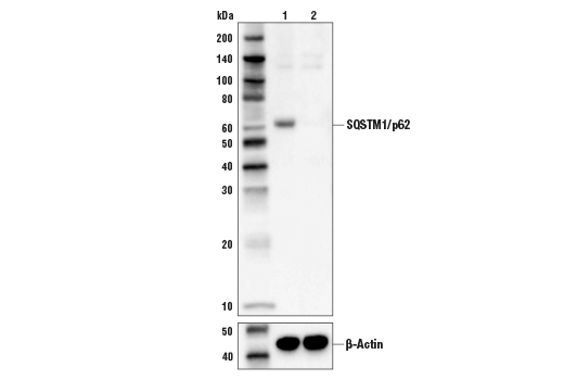
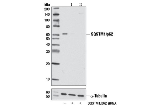
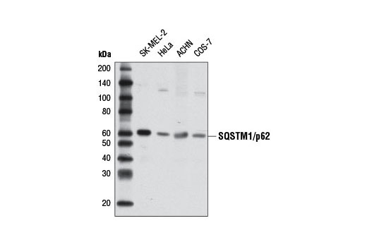
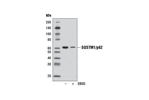
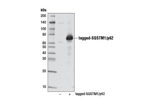
| SQSTM1/p62 (D5E2) Rabbit mAb | 8025 | 20 µl | 62 kDa | Rabbit IgG |
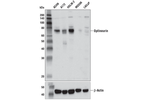
| Optineurin (D2L8S) Rabbit mAb | 58981 | 20 µl | 75 kDa | Rabbit IgG |
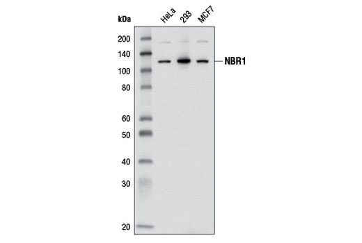
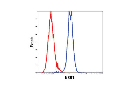
| NBR1 (D2E6) Rabbit mAb | 9891 | 20 µl | 120 kDa | Rabbit IgG |
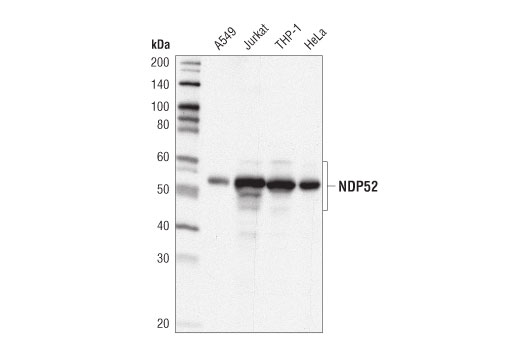
| NDP52 (D1E4A) Rabbit mAb | 60732 | 20 µl | 52, 60 kDa | Rabbit IgG |
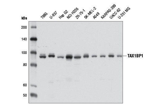
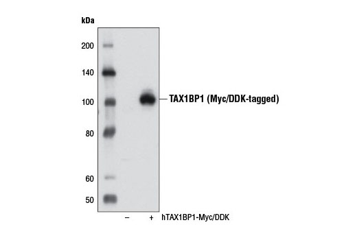
| TAX1BP1 (D1D5) Rabbit mAb | 5105 | 20 µl | 92 kDa | Rabbit IgG |
| Anti-rabbit IgG, HRP-linked Antibody | 7074 | 100 µl | Goat |
Please visit cellsignal.com for individual component applications, species cross-reactivity, dilutions, protocols, and additional product information.
Description
The SQSTM1/p62-like Receptor Antibody Sampler Kit provides an economical means of detecting members of the SQSTM1/p62-like Receptor (SLR) family. The kit includes enough antibody to perform two western blot experiments with each primary antibody.
Storage
Background
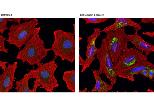
Autophagy is a catabolic process for the autophagosome-lysosomal degradation of bulk cytoplasmic contents (1,2). Selective autophagy targets the degradation of distinct sets of substrates and organelles and can occur through the utilization of a number of autophagy cargo receptors (3-5). Autophagy cargo receptors contain an LC3-interacting region (LIR) required for interaction with Atg8/LC3 family members targeted to the autophagosome. SQSTM1/p62-like receptors (SLRs) are a family of autophagy cargo receptors that contain domains for binding to ubiquitin. This family includes prototypical member SQSTM1/p62, NBR1, NDP52, Optineurin, and TAX1BP1. Targets of SLRs include ubiquitylated protein aggregates (aggrephagy), organelles such as mitochondria (mitoophagy) and peroxisomes (pexophagy), and intracellular bacteria (xenophagy).
Upon binding of cargo to these receptors, the complex is delivered to the autophagosome where both the cargo and receptor are degraded through the autophagic process. While some redundancy may exist among SLR family members, they can have unique activities. Many SLRs can have additional roles as scaffolding proteins for various signaling pathways. For example, SQSTM1/p62 interacts with KEAP1, a cytoplasmic inhibitor of NRF2, a key transcription factor involved in cellular responses to oxidative stress (6). Thus, accumulation of SQSTM1/p62 can lead to an increase in NRF2 activity.
- Reggiori, F. and Klionsky, D.J. (2002) Eukaryot Cell 1, 11-21.
- Codogno, P. and Meijer, A.J. (2005) Cell Death Differ 12 Suppl 2, 1509-18.
- Birgisdottir, Å.B. et al. (2013) J Cell Sci 126, 3237-47.
- Xu, Z. et al. (2015) Acta Biochim Biophys Sin (Shanghai) 47, 571-80.
- Mancias, J.D. and Kimmelman, A.C. (2016) J Mol Biol 428, 1659-80.
- Komatsu, M. et al. (2010) Nat Cell Biol 12, 213-23.
Background References
Trademarks and Patents
Limited Uses
Except as otherwise expressly agreed in a writing signed by a legally authorized representative of CST, the following terms apply to Products provided by CST, its affiliates or its distributors. Any Customer's terms and conditions that are in addition to, or different from, those contained herein, unless separately accepted in writing by a legally authorized representative of CST, are rejected and are of no force or effect.
Products are labeled with For Research Use Only or a similar labeling statement and have not been approved, cleared, or licensed by the FDA or other regulatory foreign or domestic entity, for any purpose. Customer shall not use any Product for any diagnostic or therapeutic purpose, or otherwise in any manner that conflicts with its labeling statement. Products sold or licensed by CST are provided for Customer as the end-user and solely for research and development uses. Any use of Product for diagnostic, prophylactic or therapeutic purposes, or any purchase of Product for resale (alone or as a component) or other commercial purpose, requires a separate license from CST. Customer shall (a) not sell, license, loan, donate or otherwise transfer or make available any Product to any third party, whether alone or in combination with other materials, or use the Products to manufacture any commercial products, (b) not copy, modify, reverse engineer, decompile, disassemble or otherwise attempt to discover the underlying structure or technology of the Products, or use the Products for the purpose of developing any products or services that would compete with CST products or services, (c) not alter or remove from the Products any trademarks, trade names, logos, patent or copyright notices or markings, (d) use the Products solely in accordance with CST Product Terms of Sale and any applicable documentation, and (e) comply with any license, terms of service or similar agreement with respect to any third party products or services used by Customer in connection with the Products.