Revision 1
#86791
Store at -20C
Suppressive Myeloid Cell Phenotyping IHC Antibody Sampler Kit
1 Kit
(9 x 20 microliters)
877-616-CELL (2355)
877-678-TECH (8324)
3 Trask Lane | Danvers | Massachusetts | 01923 | USA
For Research Use Only. Not for Use in Diagnostic Procedures.
| Product Includes | Product # | Quantity | Mol. Wt | Isotype/Source |
|---|---|---|---|---|
| CD14 (D7A2T) Rabbit Monoclonal Antibody (IHC Formulated) | 75181 | 20 µl | Rabbit IgG | |
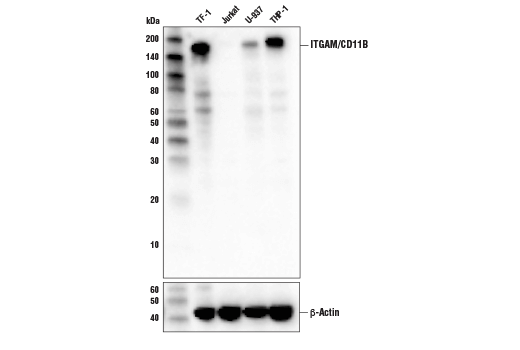
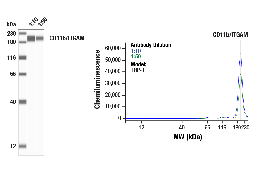
| CD11b/ITGAM (D6X1N) Rabbit Monoclonal Antibody | 49420 | 20 µl | 170 kDa | Rabbit IgG |
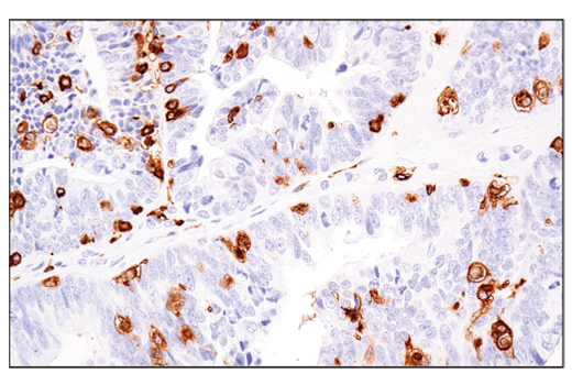
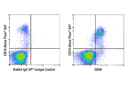
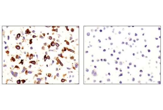
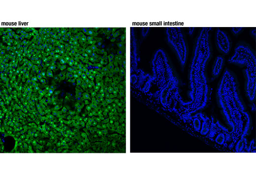
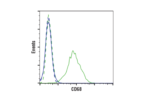
| CD68 (D4B9C) Rabbit Monoclonal Antibody | 76437 | 20 µl | Rabbit IgG | |
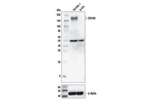
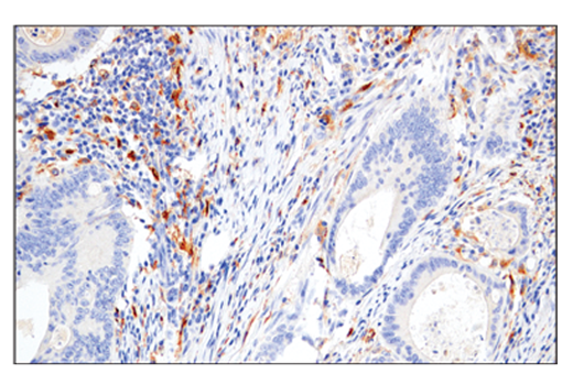
| CD163 (D6U1J) Rabbit Monoclonal Antibody | 93498 | 20 µl | 160, 170 kDa | Rabbit IgG |
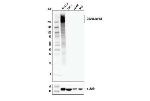
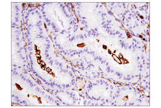
| CD206/MRC1 (E2L9N) Rabbit Monoclonal Antibody | 91992 | 20 µl | 190-250 kDa | Rabbit IgG |
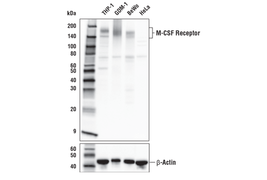
| CSF-1R/M-CSF-R (E4T8Z) Rabbit Monoclonal Antibody | 28917 | 20 µl | 140-200 kDa | Rabbit IgG |
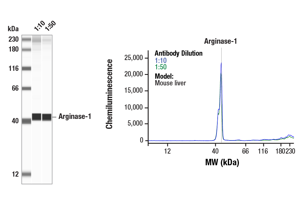
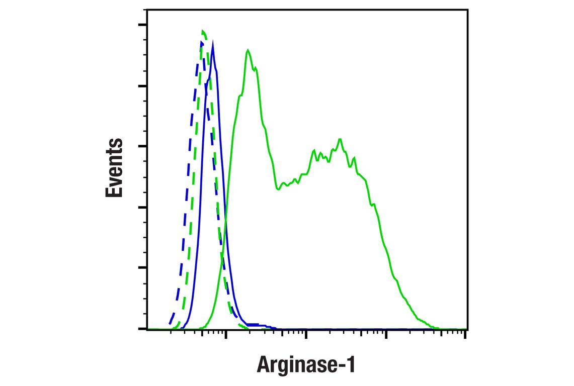
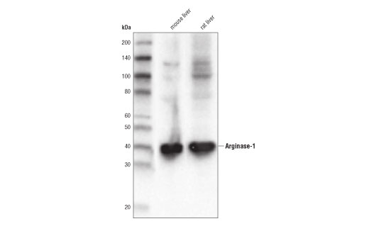
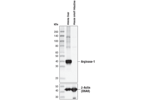
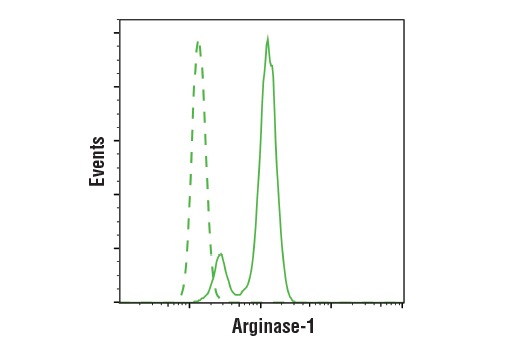
| Arginase-1 (D4E3M) Rabbit Monoclonal Antibody | 93668 | 20 µl | 40 kDa | Rabbit IgG |
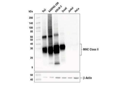
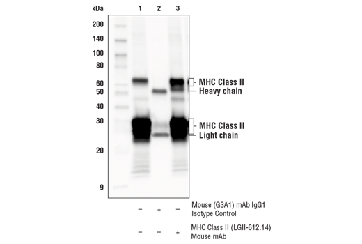
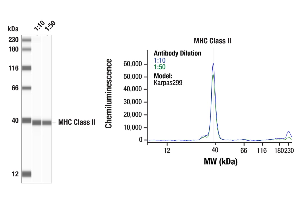
| MHC Class II (LGII-612.14) Mouse Monoclonal Antibody | 68258 | 20 µl | 25-35, 50-65 kDa | Mouse IgG1 |
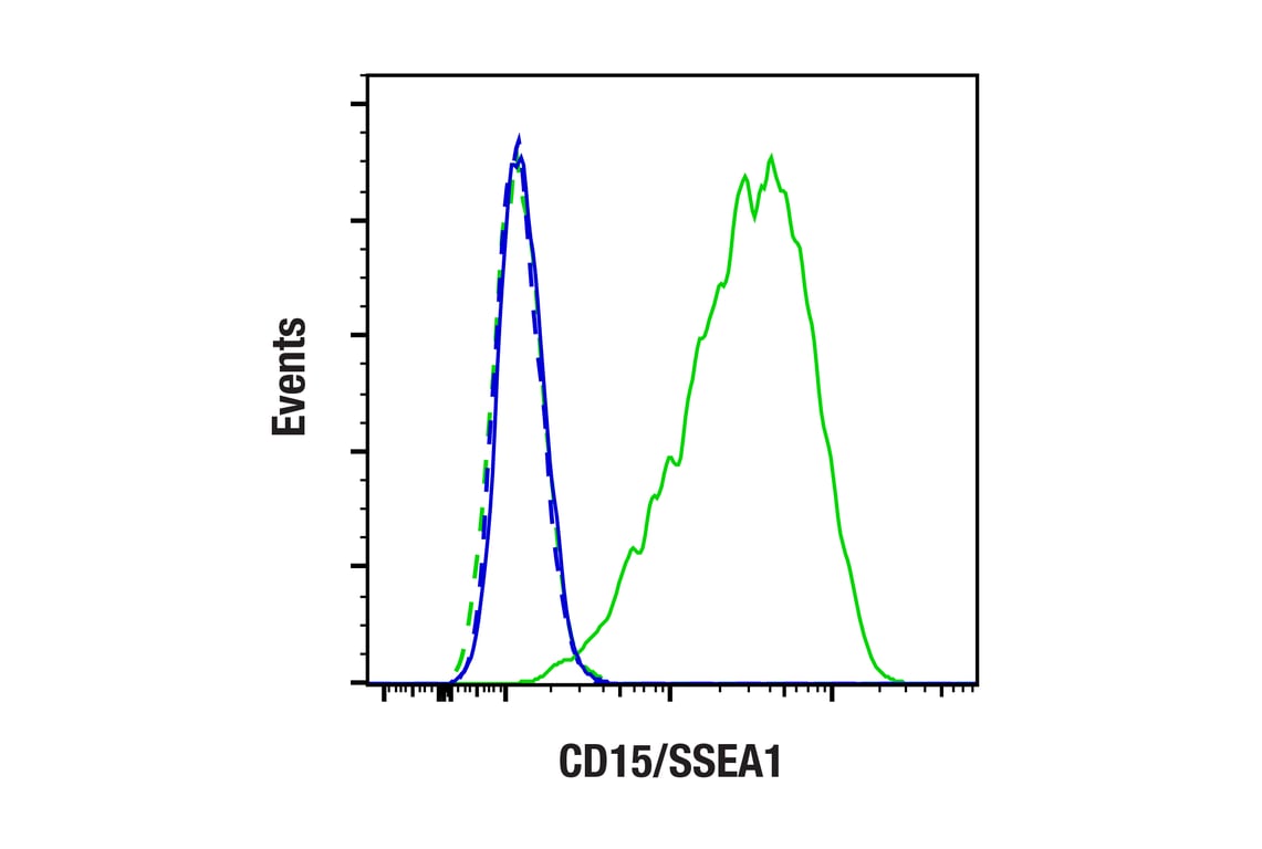
| CD15/SSEA1 (MC480) Mouse Monoclonal Antibody | 4744 | 20 µl | N/A kDa | Mouse IgM |
Please visit cellsignal.com for individual component applications, species cross-reactivity, dilutions, protocols, and additional product information.
Description
Storage
Background
CD163 is a transmembrane scavenger receptor expressed on the macrophage surface. It has 9 B-type SRCR extracellular domains mediating serum haptoglobin clearing/endocytosis, pathogen binding and signal transduction, and calcium binding (18,19). The mannose receptor (CD206/MR/CLEC13D/MMR/MRC1/Macrophage mannose receptor 1) is an endocytic receptor expressed by populations of dendritic cells, macrophages, and nonvascular endothelium (20). CD206/MRC1 receptor functions include a role in antigen cross-presentation, clearance of endogenous proteins, pathogen detection and trafficking through lymphatic vessels (21-24). Macrophage-colony stimulating factor (M-CSF, CSF-1) receptor is an integral membrane tyrosine kinase encoded by the c-fms proto-oncogene. M-CSF receptor is expressed in monocytes (macrophages and their progenitors) and drives growth and development of this blood cell lineage (25,26). CD163, CD206, and M-CSF receptors are used as surface markers of M2 type macrophages, including M2 type tumor associated macrophages (TAMs), which facilitate cancer progression by secreting cytokines to promote angiogenesis, immunosuppression, and metastasis (20,27,28). Arginase-1 catalyzes the final step of the urea cycle converting L-arginine to L-ornithine and urea (29). Myeloid-derived suppressor cells express high levels of arginase-1, increasing the catabolism of L-arginine resulting in L-arginine depletion in the inflammatory microenvironment of cancer. The reduced availability of L-arginine suppresses T cell proliferation and function and thus contributes to tumor progression (30,31).
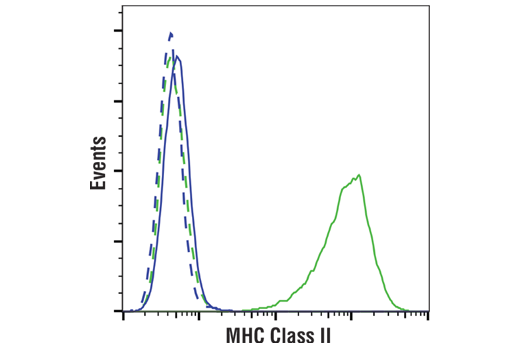
Major histocompatibility complex class II (MHC class II) molecules are heterodimeric, transmembrane glycoproteins expressed on the surface of antigen-presenting cells such as macrophages, dendritic cells, and B cells. Expression can also be induced through interferon-γ signaling (32). Prior to being displayed on the cell membrane, MHC class II molecules are loaded with exogenous peptide antigens approximately 15-24 amino acids in length that were derived from endocytosed extracellular proteins digested in the lysosome. Antigen-presentation through MHC class II is required for T cell activation during the immune response to extracellular pathogens (33). High expression of MHC class II on myeloid cell lineages is used as a surface marker of M1 type macrophages, including M1 type TAMs, which can assist in tumor eradication by secreting cytokines to activate anti-tumor immune responses, and inhibit angiogenesis and metastasis (27,28).
Background References
- Wright, S.D. et al. (1991) J Exp Med 173, 1281-6.
- Schumann, R.R. et al. (1994) Med Microbiol Immunol 183, 279-97.
- Ziegler-Heitbrock, H.W. and Ulevitch, R.J. (1993) Immunol Today 14, 121-5.
- Solovjov, D.A. et al. (2005) J Biol Chem 280, 1336-45.
- Murray, P.J. and Wynn, T.A. (2011) Nat Rev Immunol 11, 723-37.
- Kawai, K. et al. (2005) J Allergy Clin Immunol 116, 192-7.
- Payne, D. Nurs Times 92, 18.
- Merad, M. et al. (2013) Annu Rev Immunol 31, 563-604.
- Rabinowitz, S.S. and Gordon, S. (1991) J Exp Med 174, 827-36.
- Ramprasad, M.P. et al. (1995) Proc Natl Acad Sci U S A 92, 9580-4.
- Gottfried, E. et al. (2008) Scand J Immunol 67, 453-63.
- Oriol, R. et al. (1986) Vox Sang 51, 161-71.
- Hanjan, S.N. et al. (1982) Clin Immunol Immunopathol 23, 172-88.
- Civin, C.I. et al. (1981) Blood 57, 842-5.
- Hakomori, S. et al. (1984) J Biol Chem 259, 4672-80.
- Itzkowitz, S.H. et al. (1986) Cancer Res 46, 2627-32.
- Streit, A. et al. (1996) J Neurochem 66, 834-44.
- Graversen, J.H. and Moestrup, S.K. (2015) Membranes (Basel) 5, 228-52.
- Etzerodt, A. and Moestrup, S.K. (2013) Antioxid Redox Signal 18, 2352-63.
- Martinez-Pomares, L. (2012) J Leukoc Biol 92, 1177-86.
- Burgdorf, S. et al. (2006) J Immunol 176, 6770-6.
- Lee, S.J. et al. (2002) Science 295, 1898-901.
- Milone, M.C. and Fitzgerald-Bocarsly, P. (1998) J Immunol 161, 2391-9.
- Marttila-Ichihara, F. et al. (2008) Blood 112, 64-72.
- Stanley, E.R. et al. (1978) Nature 274, 168-70.
- Bourette, R.P. and Rohrschneider, L.R. (2000) Growth Factors 17, 155-66.
- Komohara, Y. et al. (2014) Cancer Sci 105, 1-8.
- Mills, C.D. et al. (2000) J Immunol 164, 6166-73.
- Wu, G. and Morris, S.M. (1998) Biochem J 336 ( Pt 1), 1-17.
- Gabrilovich, D.I. and Nagaraj, S. (2009) Nat Rev Immunol 9, 162-74.
- Raber, P. et al. (2012) Immunol Invest 41, 614-34.
- Ting, J.P. and Trowsdale, J. (2002) Cell 109 Suppl, S21-33.
- Cresswell, P. (1994) Annu Rev Immunol 12, 259-93.
Trademarks and Patents
Cell Signaling Technology is a trademark of Cell Signaling Technology, Inc.
Alexa Fluor is a registered trademark of Life Technologies Corporation.
All other trademarks are the property of their respective owners. Visit cellsignal.com/trademarks for more information.
Limited Uses
Except as otherwise expressly agreed in a writing signed by a legally authorized representative of CST, the following terms apply to Products provided by CST, its affiliates or its distributors. Any Customer's terms and conditions that are in addition to, or different from, those contained herein, unless separately accepted in writing by a legally authorized representative of CST, are rejected and are of no force or effect.
Products are labeled with For Research Use Only or a similar labeling statement and have not been approved, cleared, or licensed by the FDA or other regulatory foreign or domestic entity, for any purpose. Customer shall not use any Product for any diagnostic or therapeutic purpose, or otherwise in any manner that conflicts with its labeling statement. Products sold or licensed by CST are provided for Customer as the end-user and solely for research and development uses. Any use of Product for diagnostic, prophylactic or therapeutic purposes, or any purchase of Product for resale (alone or as a component) or other commercial purpose, requires a separate license from CST. Customer shall (a) not sell, license, loan, donate or otherwise transfer or make available any Product to any third party, whether alone or in combination with other materials, or use the Products to manufacture any commercial products, (b) not copy, modify, reverse engineer, decompile, disassemble or otherwise attempt to discover the underlying structure or technology of the Products, or use the Products for the purpose of developing any products or services that would compete with CST products or services, (c) not alter or remove from the Products any trademarks, trade names, logos, patent or copyright notices or markings, (d) use the Products solely in accordance with CST Product Terms of Sale and any applicable documentation, and (e) comply with any license, terms of service or similar agreement with respect to any third party products or services used by Customer in connection with the Products.
Revision 1
Revision 1
Revision 1
Revision 1
Revision 1

Revision 1
Revision 1
Revision 1
Revision 1
Revision 1
Revision 1
Revision 1
Revision 1
Revision 1
Revision 1
Revision 1
Revision 1
Revision 1
Revision 1
Revision 1
Revision 1
Revision 1

Revision 1
Revision 1
Revision 1

Revision 1
Revision 1