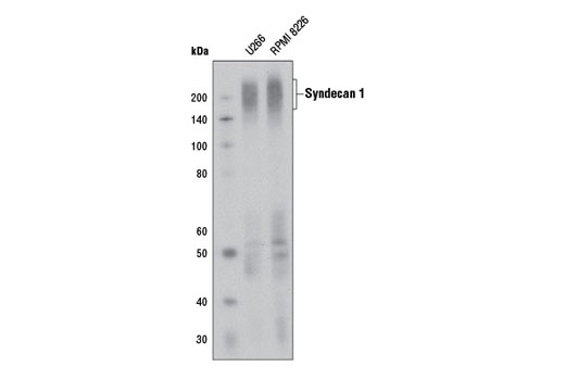
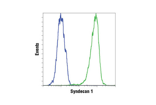
WB, IP, FC-FP
H
Endogenous
180-250, 70
Rabbit IgG
#P18827
6382
Product Information
Product Usage Information
| Application | Dilution |
|---|---|
| Western Blotting | 1:1000 |
| Immunoprecipitation | 1:50 |
| Flow Cytometry (Fixed/Permeabilized) | 1:200 - 1:800 |
Storage
Specificity / Sensitivity
Species Reactivity:
Human
Source / Purification
Monoclonal antibody is produced by immunizing animals with a synthetic peptide corresponding to residues surrounding Ala294 of human syndecan 1 protein.
Background
Syndecans are a family of type 1 transmembrane heparan sulfate proteoglycans comprising four members in mammals (SDC1-4) (1) encoded by four syndecan genes. Syndecans are involved in embryonic development, tumorigenesis, and angiogenesis (2). The extracellular domain harbors attachment sites for heparan sulfate and chondroitin sulfate chains, facilitating interaction with an array of proteins, including a plethora of growth factors. In addition, the hydrophobic C-terminal intracellular domain can interact with proteins containing a PDZ domain (2). These interactions place syndecans as important integrators of membrane signaling (3). Syndecans undergo proteolytic cleavage causing the release of their extracellular domain (shedding), converting the membrane-bound proteins into soluble molecular effectors (4).
Syndecan 1 (SDC1) is a specific marker for plasmacytic differentiation in hematologic disorders (5-7). This cell surface proteoglycan is also expressed in normal epithelial cells and tissues as well as various types of cancer tissues (8-11). The extracellular shed form of syndecan 1 remains soluble or accumulates in the extracellular matrix where it binds growth factors, cytokines and other extracellular matrix proteins (12,13). This binding activates signaling of bound growth factors or cytokines, which results in enhanced tumor growth, dissemination, angiogenesis, and osteolysis (14-17). As a result, the level of syndecan1 protein and its shed form may serve as prognostic factors for a list of malignancies (6,18,19). Syndecan 1 has recently been found to be a critical mediator of macropinocytosis in pancreatic cancer (20).
- Couchman, J.R. (2003) Nat Rev Mol Cell Biol 4, 926-37.
- Multhaupt, H.A. et al. (2009) J Physiol Pharmacol 60 Suppl 4, 31-8.
- Zimmermann, P. and David, G. (1999) FASEB J 13 Suppl, S91-S100.
- Manon-Jensen, T. et al. (2010) FEBS J 277, 3876-89.
- Chilosi, M. et al. (1999) Mod Pathol 12, 1101-6.
- Seidel, C. et al. (2000) Blood 95, 388-92.
- O'Connell, F.P. et al. (2004) Am J Clin Pathol 121, 254-63.
- Inki, P. and Jalkanen, M. (1996) Ann Med 28, 63-7.
- Matsumoto, A. et al. (1997) Int J Cancer 74, 482-91.
- Conejo, J.R. et al. (2000) Int J Cancer 88, 12-20.
- Zellweger, T. et al. (2003) Prostate 55, 20-9.
- Bayer-Garner, I.B. et al. (2001) Mod Pathol 14, 1052-8.
- Ramani, V.C. et al. (2013) FEBS J 280, 2294-306.
- Derksen, P.W. et al. (2002) Blood 99, 1405-10.
- You, W.K. and McDonald, D.M. (2008) BMB Rep 41, 833-9.
- Ramani, V.C. et al. (2011) J Biol Chem 286, 6490-9.
- Aragão, A.Z. et al. (2012) PLoS One 7, e43521.
- Joensuu, H. et al. (2002) Cancer Res 62, 5210-7.
- Lee, S.H. et al. (2013) Int J Clin Oncol (Epub ahead of print).
- Yao, W. et al. (2019) Nature 568, 410-414.
Species Reactivity
Species reactivity is determined by testing in at least one approved application (e.g., western blot).
Western Blot Buffer
IMPORTANT: For western blots, incubate membrane with diluted primary antibody in 5% w/v BSA, 1X TBS, 0.1% Tween® 20 at 4°C with gentle shaking, overnight.
Applications Key
WB: Western Blotting IP: Immunoprecipitation FC-FP: Flow Cytometry (Fixed/Permeabilized)
Cross-Reactivity Key
H: human M: mouse R: rat Hm: hamster Mk: monkey Vir: virus Mi: mink C: chicken Dm: D. melanogaster X: Xenopus Z: zebrafish B: bovine Dg: dog Pg: pig Sc: S. cerevisiae Ce: C. elegans Hr: horse GP: Guinea Pig Rab: rabbit All: all species expected
Trademarks and Patents
Limited Uses
Except as otherwise expressly agreed in a writing signed by a legally authorized representative of CST, the following terms apply to Products provided by CST, its affiliates or its distributors. Any Customer's terms and conditions that are in addition to, or different from, those contained herein, unless separately accepted in writing by a legally authorized representative of CST, are rejected and are of no force or effect.
Products are labeled with For Research Use Only or a similar labeling statement and have not been approved, cleared, or licensed by the FDA or other regulatory foreign or domestic entity, for any purpose. Customer shall not use any Product for any diagnostic or therapeutic purpose, or otherwise in any manner that conflicts with its labeling statement. Products sold or licensed by CST are provided for Customer as the end-user and solely for research and development uses. Any use of Product for diagnostic, prophylactic or therapeutic purposes, or any purchase of Product for resale (alone or as a component) or other commercial purpose, requires a separate license from CST. Customer shall (a) not sell, license, loan, donate or otherwise transfer or make available any Product to any third party, whether alone or in combination with other materials, or use the Products to manufacture any commercial products, (b) not copy, modify, reverse engineer, decompile, disassemble or otherwise attempt to discover the underlying structure or technology of the Products, or use the Products for the purpose of developing any products or services that would compete with CST products or services, (c) not alter or remove from the Products any trademarks, trade names, logos, patent or copyright notices or markings, (d) use the Products solely in accordance with CST Product Terms of Sale and any applicable documentation, and (e) comply with any license, terms of service or similar agreement with respect to any third party products or services used by Customer in connection with the Products.