WB, IF-F, IF-IC
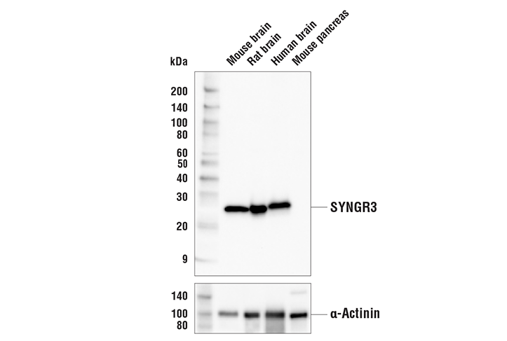
H M R
Endogenous
26
Rabbit IgG
#O43761
9143
Product Information
Product Usage Information
| Application | Dilution |
|---|---|
| Western Blotting | 1:1000 |
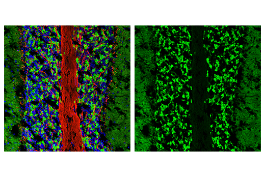
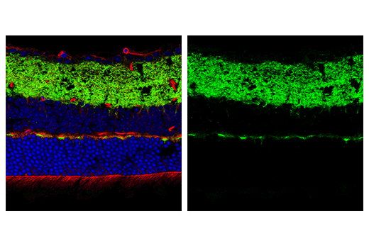
| Immunofluorescence (Frozen) | 1:400 - 1:800 |
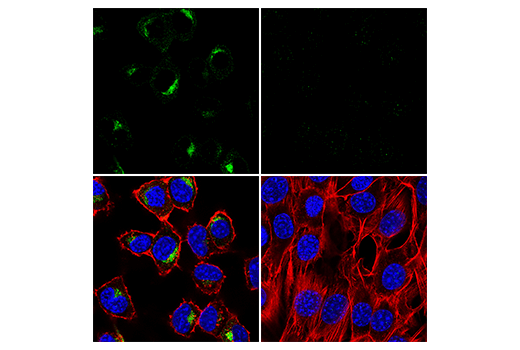
| Immunofluorescence (Immunocytochemistry) | 1:800 - 1:1600 |
Storage
For a carrier free (BSA and azide free) version of this product see product #29745.
Specificity / Sensitivity
Species Reactivity:
Human, Mouse, Rat
Source / Purification
Monoclonal antibody is produced by immunizing animals with a synthetic peptide corresponding to residues near the carboxy terminus of human SYNGR3 protein.
Background
Synaptogyrin, or SYNGR, are a family of tyrosine-phosphorylated proteins, including neuronal SYNGR1 and SYNGR3 that are found in synaptic vesicles and contribute to the proper synapse function. Synaptogyrin-2 (SYNGR2) expresses ubiquitously and it is not only associated with synaptic vesicles, but also plays an important role in exocytosis processes (1,3). In addition, it has been shown that SYNGRs modulate calcium currents in excitable cells during potassium chloride-dependent exocytosis (3). SYNGR3 and SYNGR1 specifically localize in synaptic vesicles. SYNGR1 modulates synaptic vesicle function similar to SYNGR3 (2,3). SYNGR1 and SYNGR3 contribute to the neurotransmitter release in neurons by interactions with the GABA and VGLUT transporters in primary neurons and in C. elegans (4-6). SYNGRs are associated with disease including Schizophrenia (7,8) and Alzheimer's disease (9,10).
- Kedra, D. et al. (1998) Hum Genet 103, 131-41.
- Janz, R. et al. (1999) Neuron 24, 687-700.
- Sugita, S. et al. (1999) J Biol Chem 274, 18893-901.
- Belizaire, R. et al. (2004) J Comp Neurol 470, 266-81.
- Bragina, L. et al. (2010) Neuroscience 165, 934-43.
- Abraham, C. et al. (2011) Neuroscience 190, 75-88.
- Verma, R. et al. (2005) J Hum Genet 50, 635-40.
- Cheng, M.C. and Chen, C.H. (2007) J Psychiatr Res 41, 1027-31.
- Liu, C. et al. (2016) J Biol Chem 291, 8173-88.
- McInnes, J. et al. (2018) Neuron 97, 823-835.e8.
Species Reactivity
Species reactivity is determined by testing in at least one approved application (e.g., western blot).
Western Blot Buffer
IMPORTANT: For western blots, incubate membrane with diluted primary antibody in 5% w/v BSA, 1X TBS, 0.1% Tween® 20 at 4°C with gentle shaking, overnight.
Applications Key
WB: Western Blotting IF-F: Immunofluorescence (Frozen) IF-IC: Immunofluorescence (Immunocytochemistry)
Cross-Reactivity Key
H: human M: mouse R: rat Hm: hamster Mk: monkey Vir: virus Mi: mink C: chicken Dm: D. melanogaster X: Xenopus Z: zebrafish B: bovine Dg: dog Pg: pig Sc: S. cerevisiae Ce: C. elegans Hr: horse GP: Guinea Pig Rab: rabbit All: all species expected
Trademarks and Patents
Limited Uses
Except as otherwise expressly agreed in a writing signed by a legally authorized representative of CST, the following terms apply to Products provided by CST, its affiliates or its distributors. Any Customer's terms and conditions that are in addition to, or different from, those contained herein, unless separately accepted in writing by a legally authorized representative of CST, are rejected and are of no force or effect.
Products are labeled with For Research Use Only or a similar labeling statement and have not been approved, cleared, or licensed by the FDA or other regulatory foreign or domestic entity, for any purpose. Customer shall not use any Product for any diagnostic or therapeutic purpose, or otherwise in any manner that conflicts with its labeling statement. Products sold or licensed by CST are provided for Customer as the end-user and solely for research and development uses. Any use of Product for diagnostic, prophylactic or therapeutic purposes, or any purchase of Product for resale (alone or as a component) or other commercial purpose, requires a separate license from CST. Customer shall (a) not sell, license, loan, donate or otherwise transfer or make available any Product to any third party, whether alone or in combination with other materials, or use the Products to manufacture any commercial products, (b) not copy, modify, reverse engineer, decompile, disassemble or otherwise attempt to discover the underlying structure or technology of the Products, or use the Products for the purpose of developing any products or services that would compete with CST products or services, (c) not alter or remove from the Products any trademarks, trade names, logos, patent or copyright notices or markings, (d) use the Products solely in accordance with CST Product Terms of Sale and any applicable documentation, and (e) comply with any license, terms of service or similar agreement with respect to any third party products or services used by Customer in connection with the Products.