| Product Includes | Product # | Quantity | Mol. Wt | Isotype/Source |
|---|---|---|---|---|
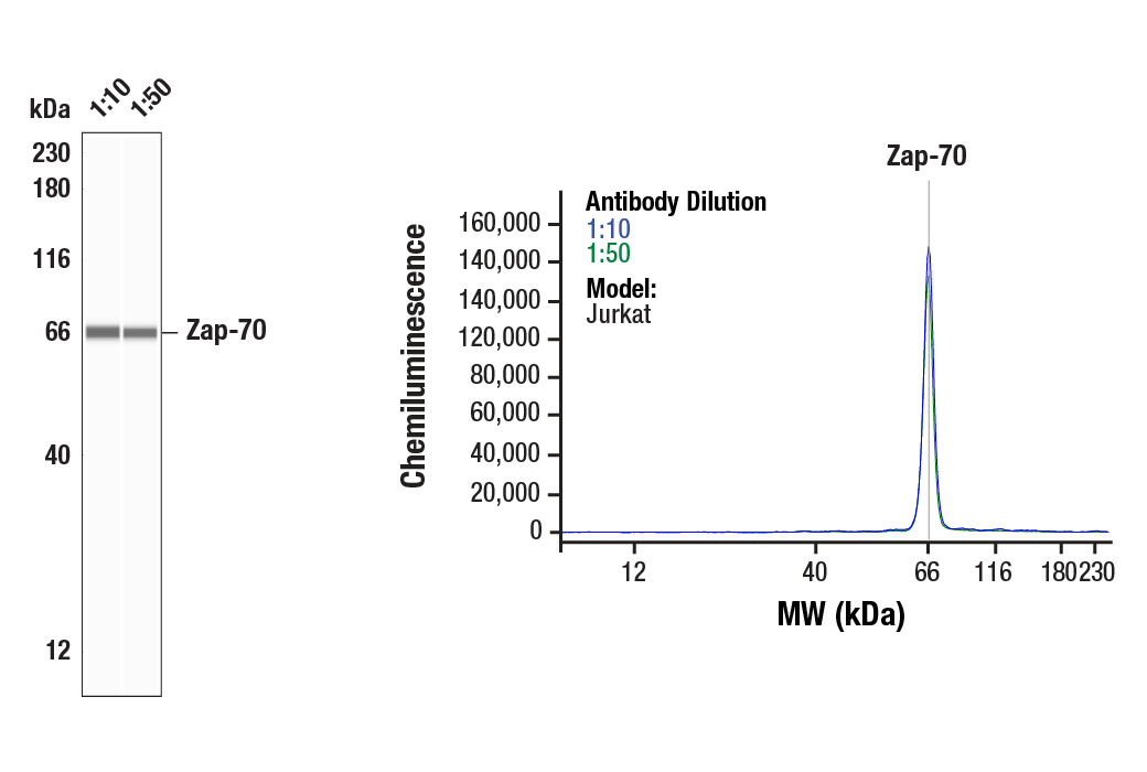
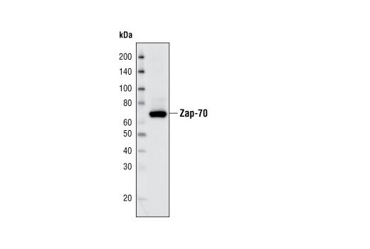
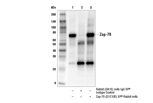
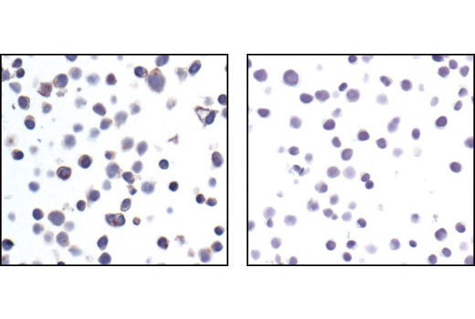
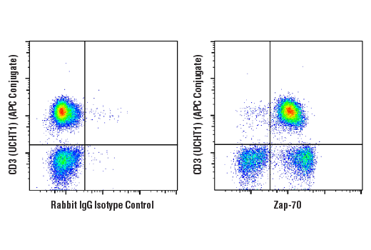
| Zap-70 (D1C10E) XP® Rabbit mAb | 3165 | 40 µl | 70 kDa | Rabbit IgG |
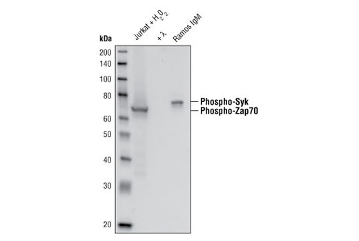
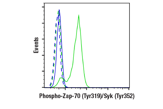
| Phospho-Zap-70 (Tyr319)/Syk (Tyr352) (65E4) Rabbit mAb | 2717 | 40 µl | 70, 72 kDa | Rabbit IgG |
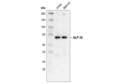
| SLP-76 Antibody | 4958 | 40 µl | 76 kDa | Rabbit |
| Phospho-LAT (Tyr220) Antibody | 3584 | 40 µl | 36, 38 kDa | Rabbit |
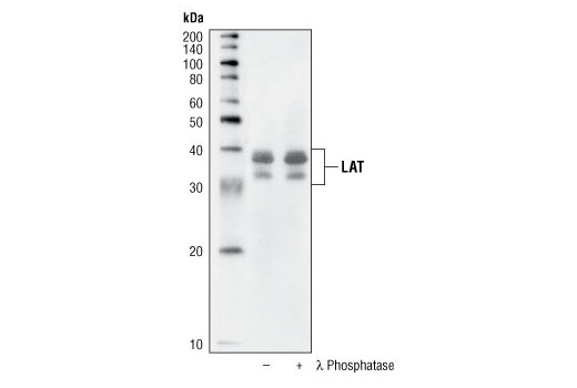
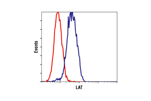
| LAT Antibody | 9166 | 40 µl | 36, 38 kDa | Rabbit |
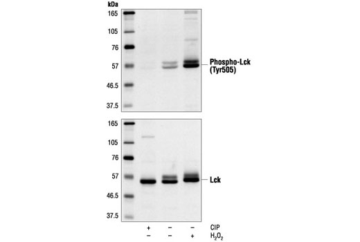
| Phospho-Lck (Tyr505) Antibody | 2751 | 40 µl | 56 kDa | Rabbit |
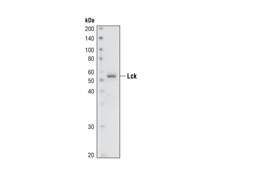
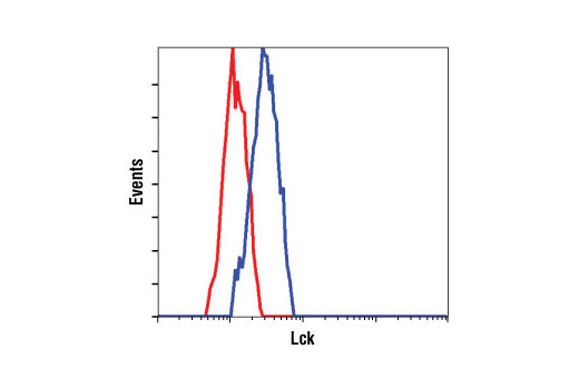
| Lck (73A5) Rabbit mAb | 2787 | 40 µl | 56 kDa | Rabbit IgG |
| Anti-rabbit IgG, HRP-linked Antibody | 7074 | 100 µl | Goat |
Please visit cellsignal.com for individual component applications, species cross-reactivity, dilutions, protocols, and additional product information.
Description
The T Cell Signaling Antibody Sampler Kit contains primary and secondary antibodies to perform four Western blots with each antibody.
Storage
Background
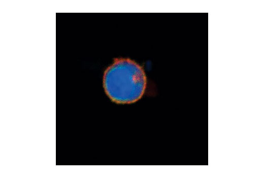
The Syk family protein tyrosine kinase Zap-70 is expressed in T and NK cells and plays a critical role in mediating T cell activation in response to T cell receptor (TCR) engagement (1). Following TCR engagement, Zap-70 is rapidly phosphorylated on several tyrosine residues through autophosphorylation and transphosphorylation by the Src family tyrosine kinase Lck (2-6). Tyrosine phosphorylation correlates with increased Zap-70 kinase activity and downstream signaling events. Expression of Zap-70 is correlated with disease progression and survival in patients with chronic lymphocytic leukemia (7,8).
LAT, a transmembrane adaptor protein expressed in T, NK and mast cells, is an important mediator for T cell receptor (TCR) signaling (9). Upon TCR engagement, activated Zap-70 phosphorylates LAT at multiple conserved tyrosine residues within SH2 binding motifs, exposing these motifs as the docking sites for downstream signaling targets (10,11). The phosphorylation of LAT at Tyr171 and 191 enables the binding of Grb2, Gads/SLP-76, PLCgamma1 and PI3 kinase through their SH2 domain and translocates them to the membrane. This process eventually leads to activation of the corresponding signaling pathways (11-12).
- Chu, D.H. et al. (1998) Immunol Rev 165, 167-80.
- Iwashima, M. et al. (1994) Science 263, 1136-9.
- Neumeister, E.N. et al. (1995) Mol Cell Biol 15, 3171-8.
- Chan, A.C. et al. (1995) EMBO J 14, 2499-508.
- Williams, B.L. et al. (1999) EMBO J 18, 1832-44.
- Di Bartolo, V. et al. (1999) J Biol Chem 274, 6285-94.
- Wiestner, A. et al. (2003) Blood 101, 4944-51.
- Crespo, M. et al. (2003) N Engl J Med 348, 1764-75.
- Wonerow, P. and Watson, S.P. (2001) Oncogene 20, 6273-83.
- Zhang, W. et al. (1998) Cell 92, 83-92.
- Paz, P.E. et al. (2001) Biochem J 356, 461-71.
- Zhang, W. et al. (2000) J Biol Chem 275, 23355-61.
Background References
Trademarks and Patents
Limited Uses
Except as otherwise expressly agreed in a writing signed by a legally authorized representative of CST, the following terms apply to Products provided by CST, its affiliates or its distributors. Any Customer's terms and conditions that are in addition to, or different from, those contained herein, unless separately accepted in writing by a legally authorized representative of CST, are rejected and are of no force or effect.
Products are labeled with For Research Use Only or a similar labeling statement and have not been approved, cleared, or licensed by the FDA or other regulatory foreign or domestic entity, for any purpose. Customer shall not use any Product for any diagnostic or therapeutic purpose, or otherwise in any manner that conflicts with its labeling statement. Products sold or licensed by CST are provided for Customer as the end-user and solely for research and development uses. Any use of Product for diagnostic, prophylactic or therapeutic purposes, or any purchase of Product for resale (alone or as a component) or other commercial purpose, requires a separate license from CST. Customer shall (a) not sell, license, loan, donate or otherwise transfer or make available any Product to any third party, whether alone or in combination with other materials, or use the Products to manufacture any commercial products, (b) not copy, modify, reverse engineer, decompile, disassemble or otherwise attempt to discover the underlying structure or technology of the Products, or use the Products for the purpose of developing any products or services that would compete with CST products or services, (c) not alter or remove from the Products any trademarks, trade names, logos, patent or copyright notices or markings, (d) use the Products solely in accordance with CST Product Terms of Sale and any applicable documentation, and (e) comply with any license, terms of service or similar agreement with respect to any third party products or services used by Customer in connection with the Products.