| Product Includes | Product # | Quantity | Mol. Wt | Isotype/Source |
|---|---|---|---|---|
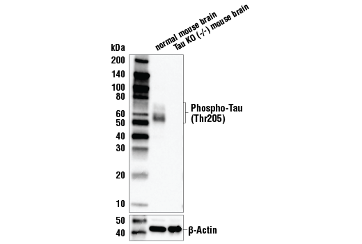
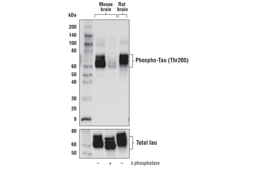
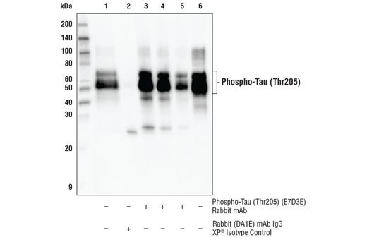
| Phospho-Tau (Thr205) (E7D3E) Rabbit mAb | 49561 | 20 µl | 50-80 kDa | Rabbit IgG |
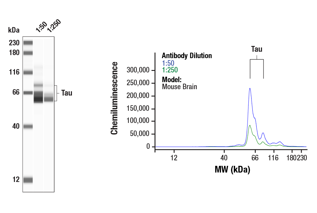
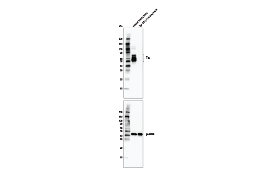
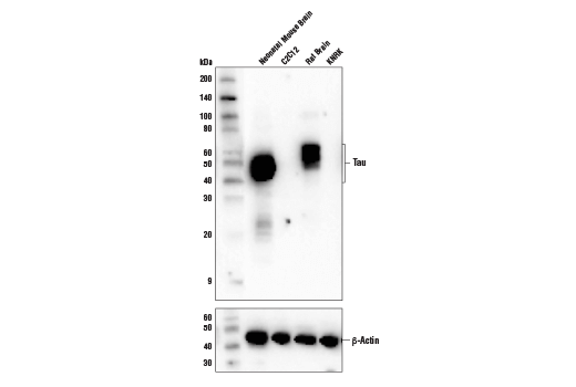
| Tau (D1M9X) XP® Rabbit mAb | 46687 | 20 µl | 50-80 kDa | Rabbit IgG |
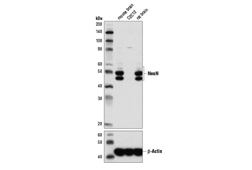
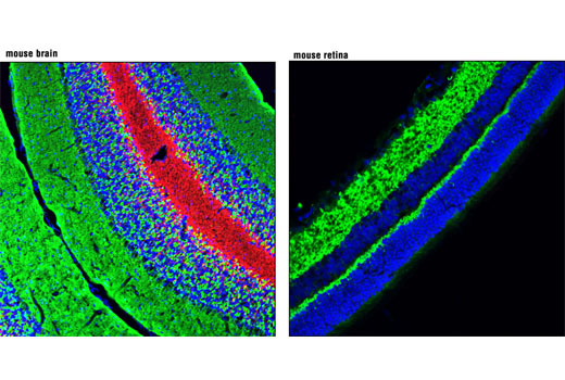
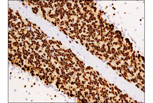
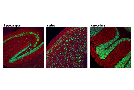
| NeuN (D4G4O) XP® Rabbit mAb | 24307 | 20 µl | 46-55 kDa | Rabbit IgG |
| Synaptophysin (7H12) Mouse mAb (IF Formulated) | 9020 | 20 µl | Mouse IgG1 | |
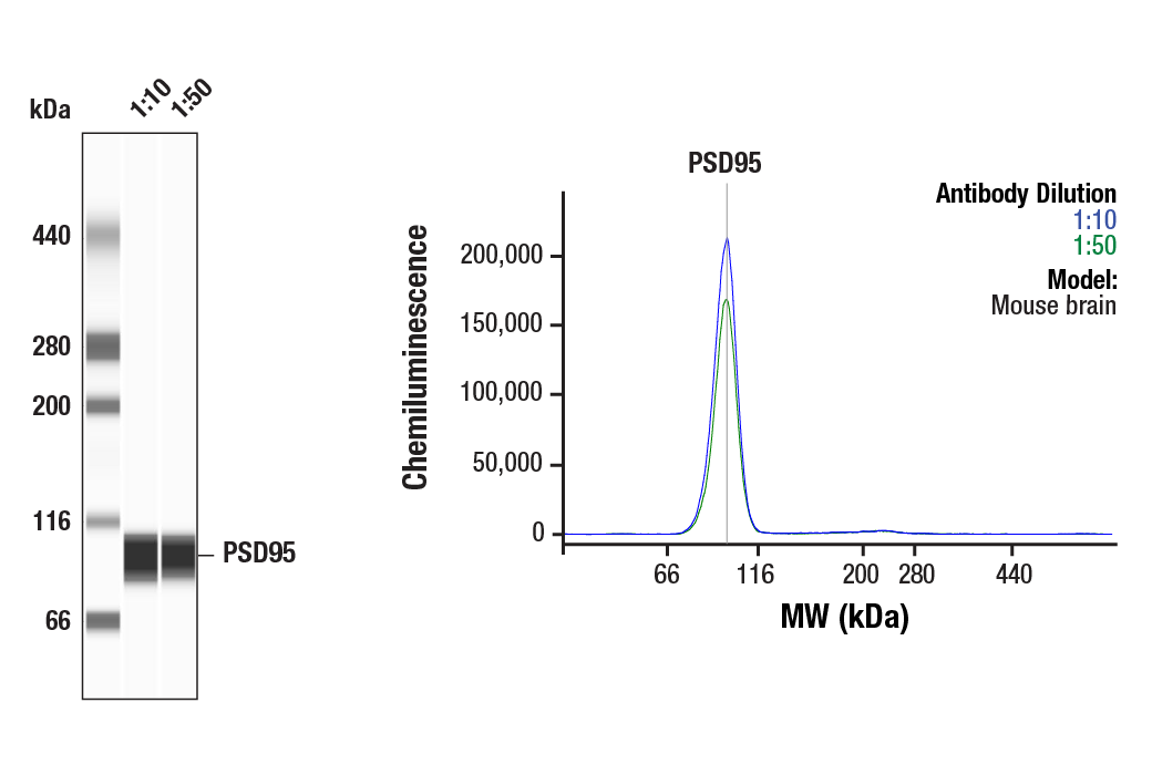
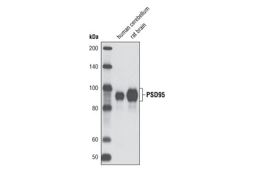
| PSD95 (D27E11) XP® Rabbit mAb | 3450 | 20 µl | 95 kDa | Rabbit IgG |
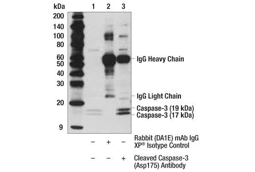
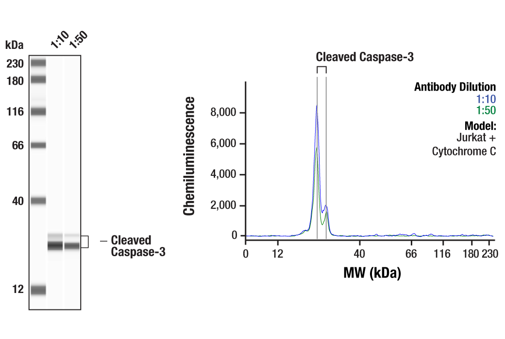
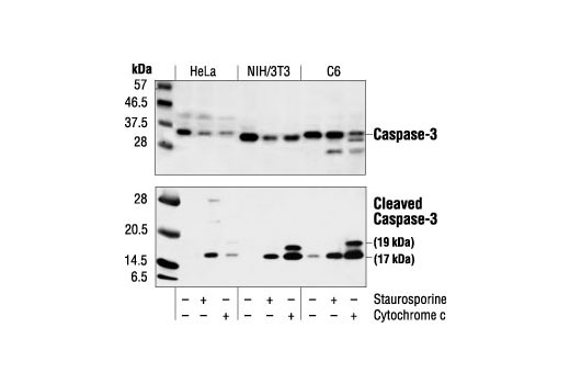
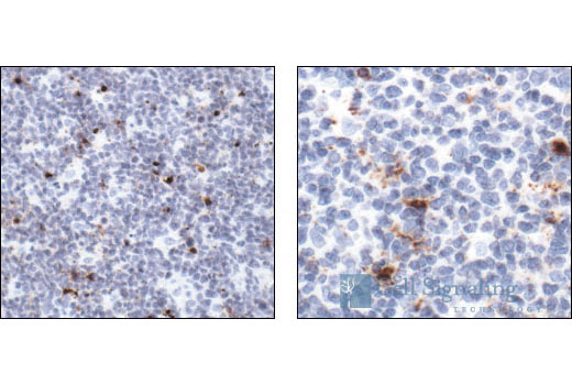
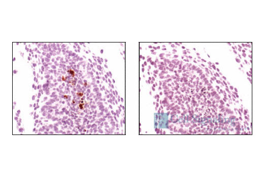
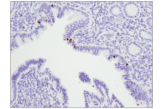
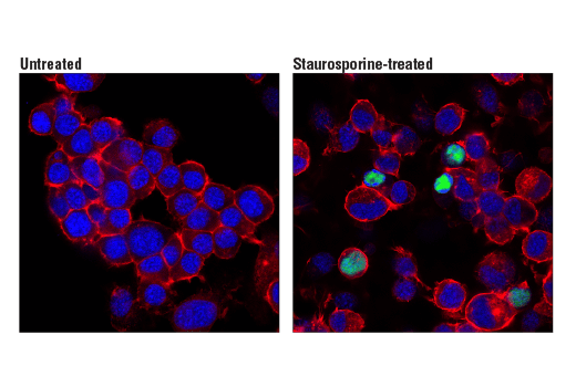
| Cleaved Caspase-3 (Asp175) Antibody | 9661 | 20 µl | 17, 19 kDa | Rabbit |
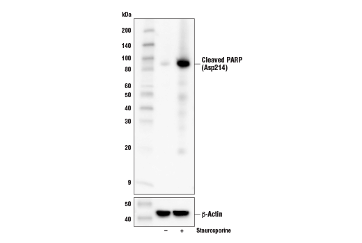
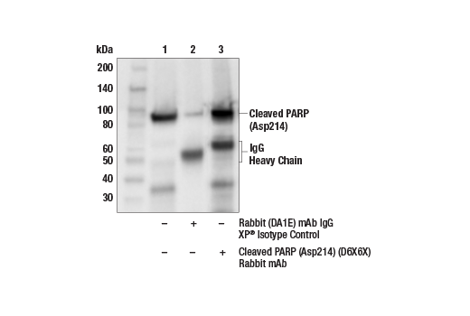
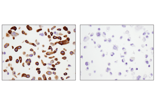
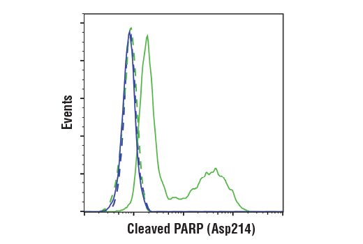
| Cleaved PARP (Asp214) (D6X6X) Rabbit mAb | 94885 | 20 µl | 89 kDa | Rabbit IgG |
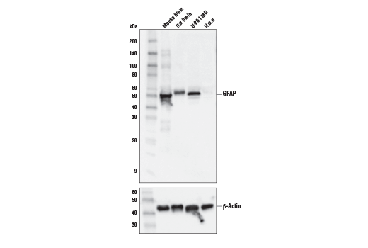
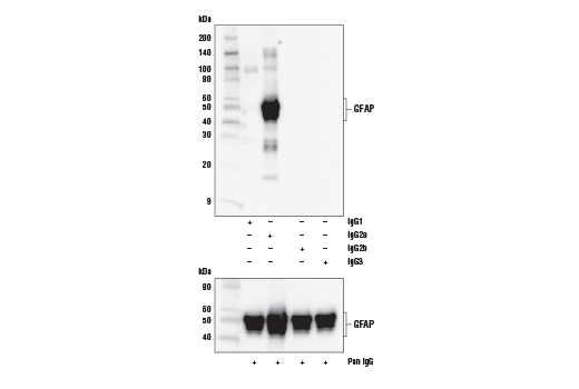
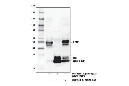
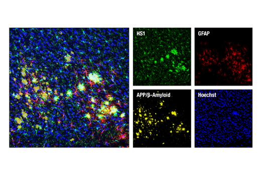
| GFAP (E6N9L) Mouse mAb | 34001 | 20 µl | 50 kDa | Mouse IgG2a |
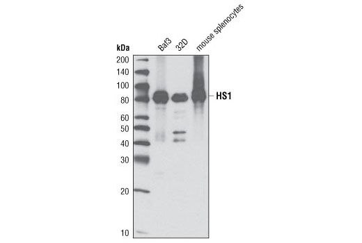
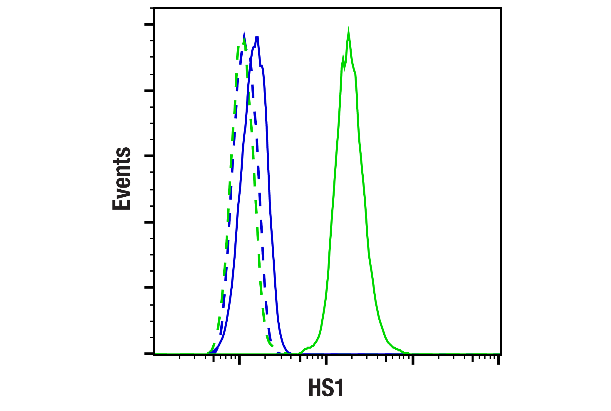
| HS1 (D5A9) XP® Rabbit mAb | 3892 | 20 µl | 80 kDa | Rabbit IgG |
Please visit cellsignal.com for individual component applications, species cross-reactivity, dilutions, protocols, and additional product information.
Description
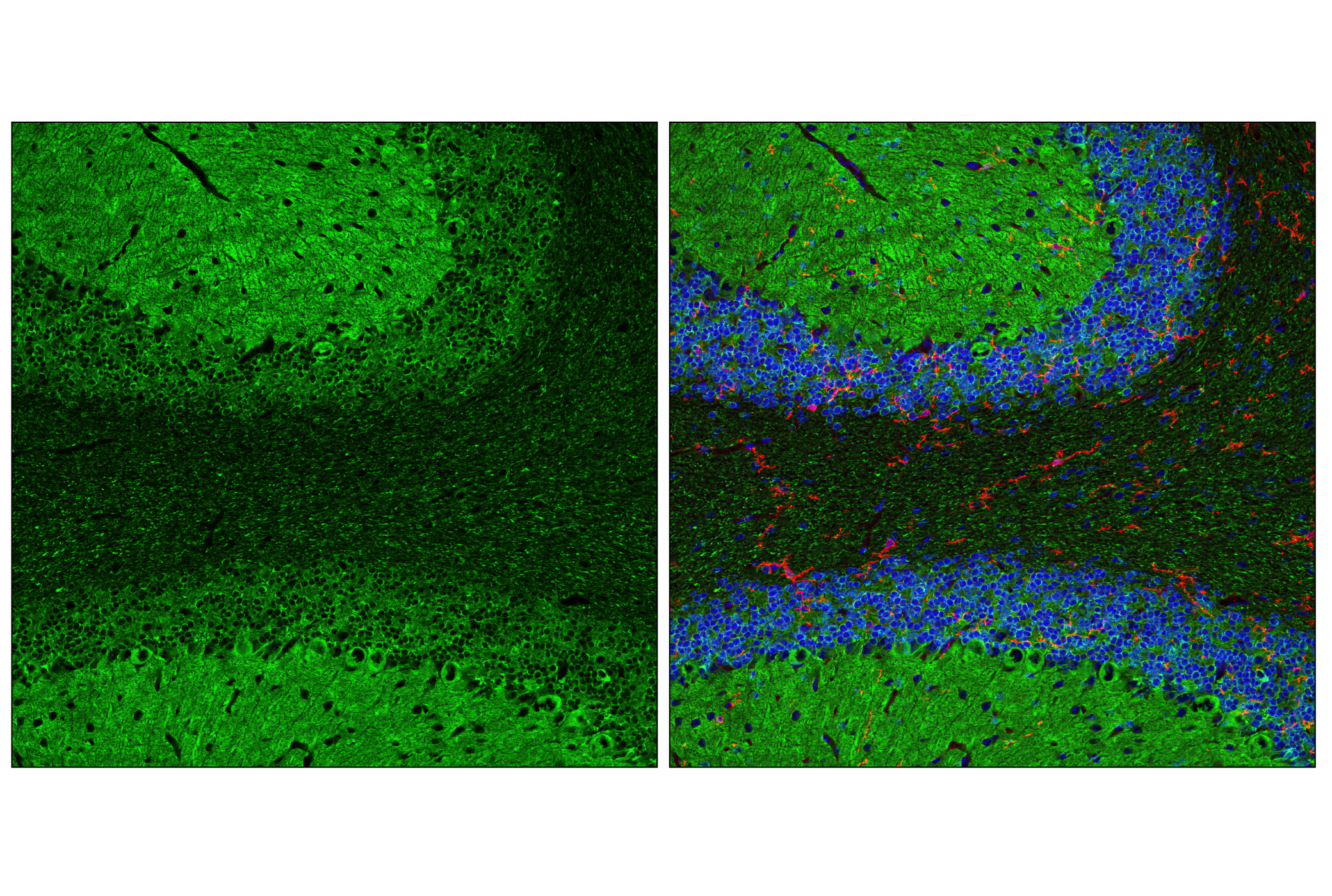
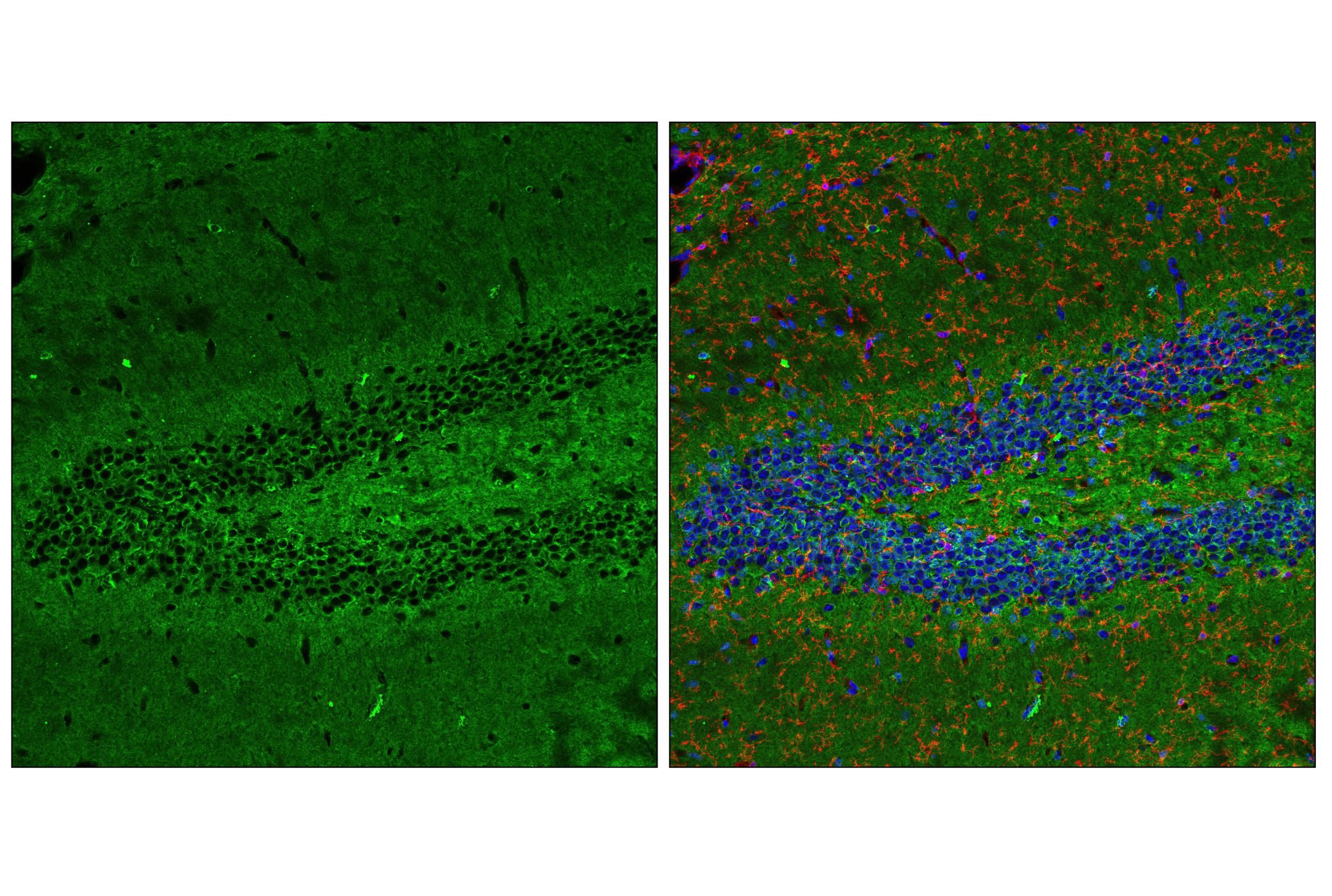
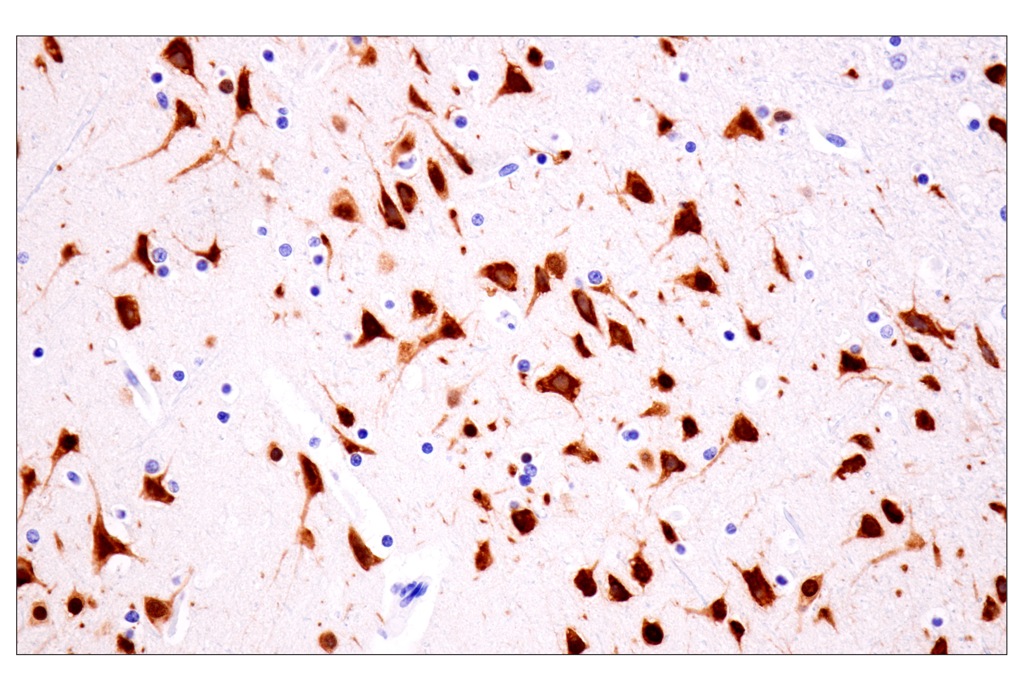
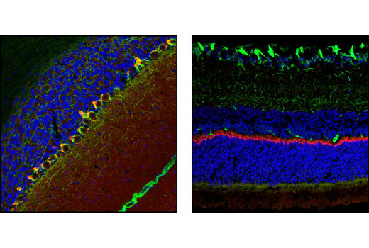
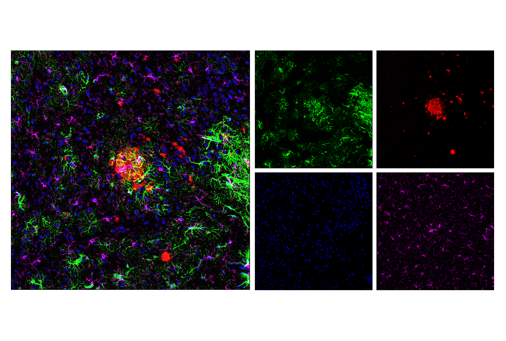
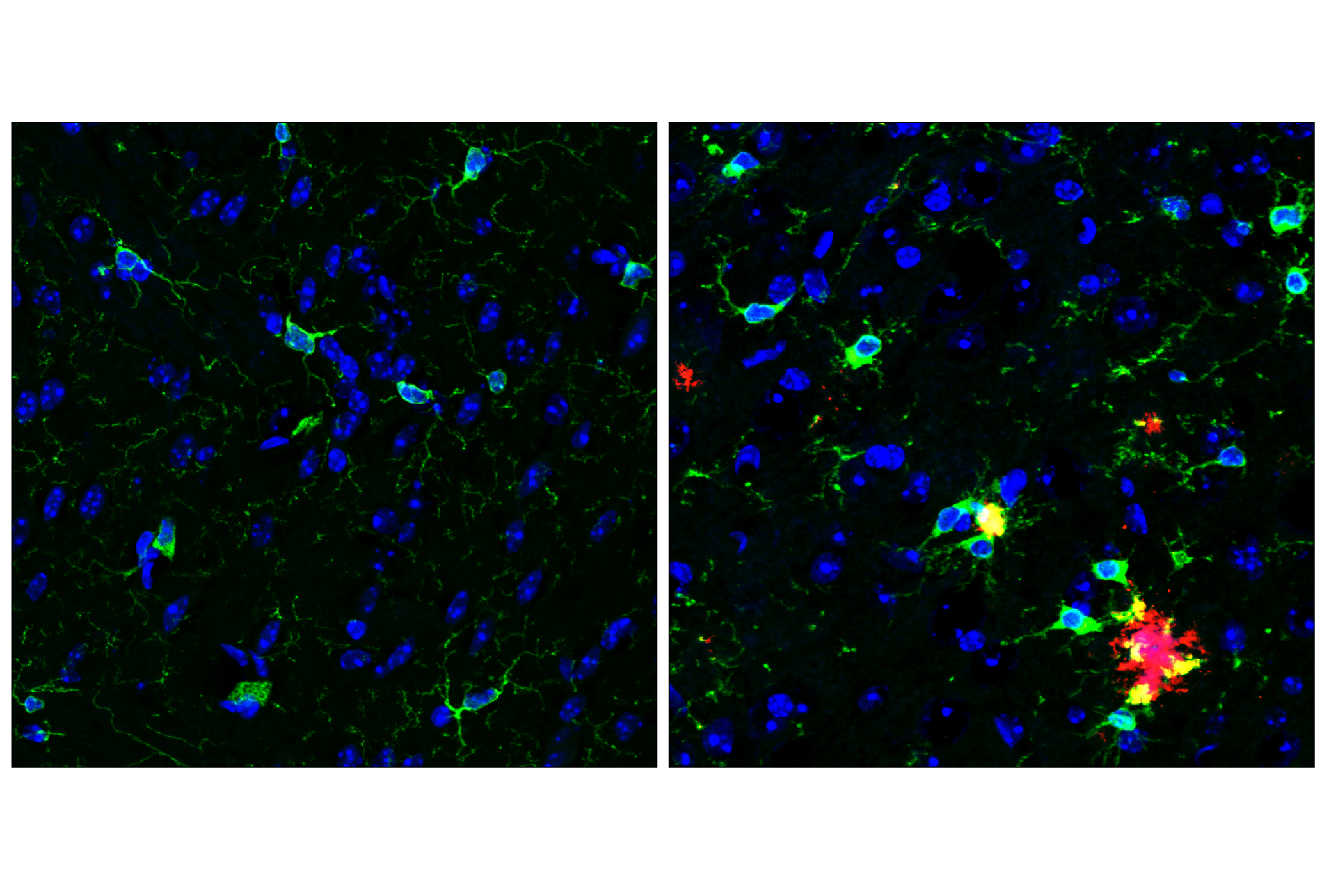
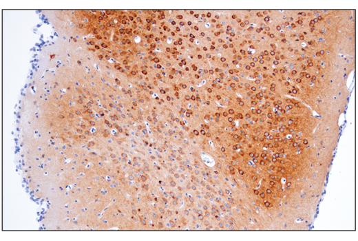
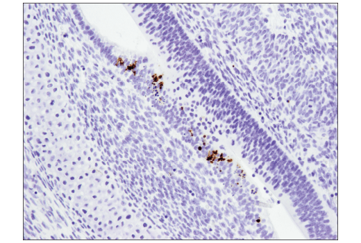
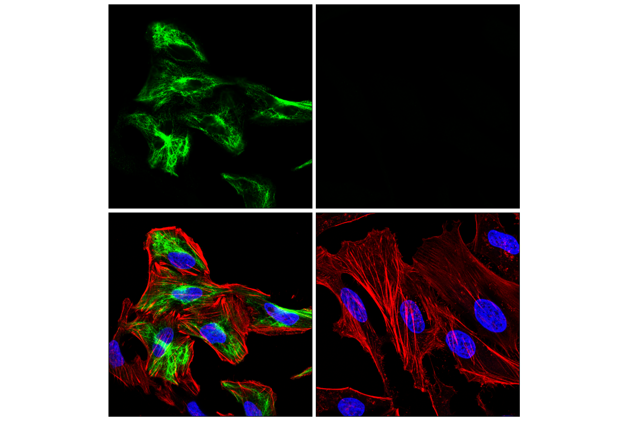
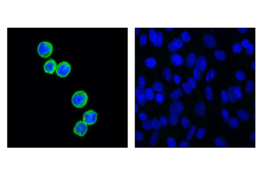
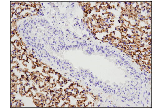
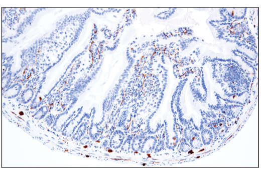
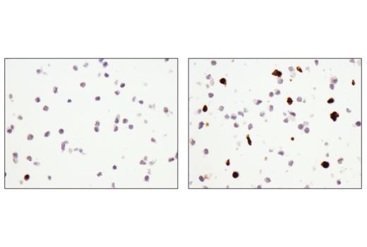
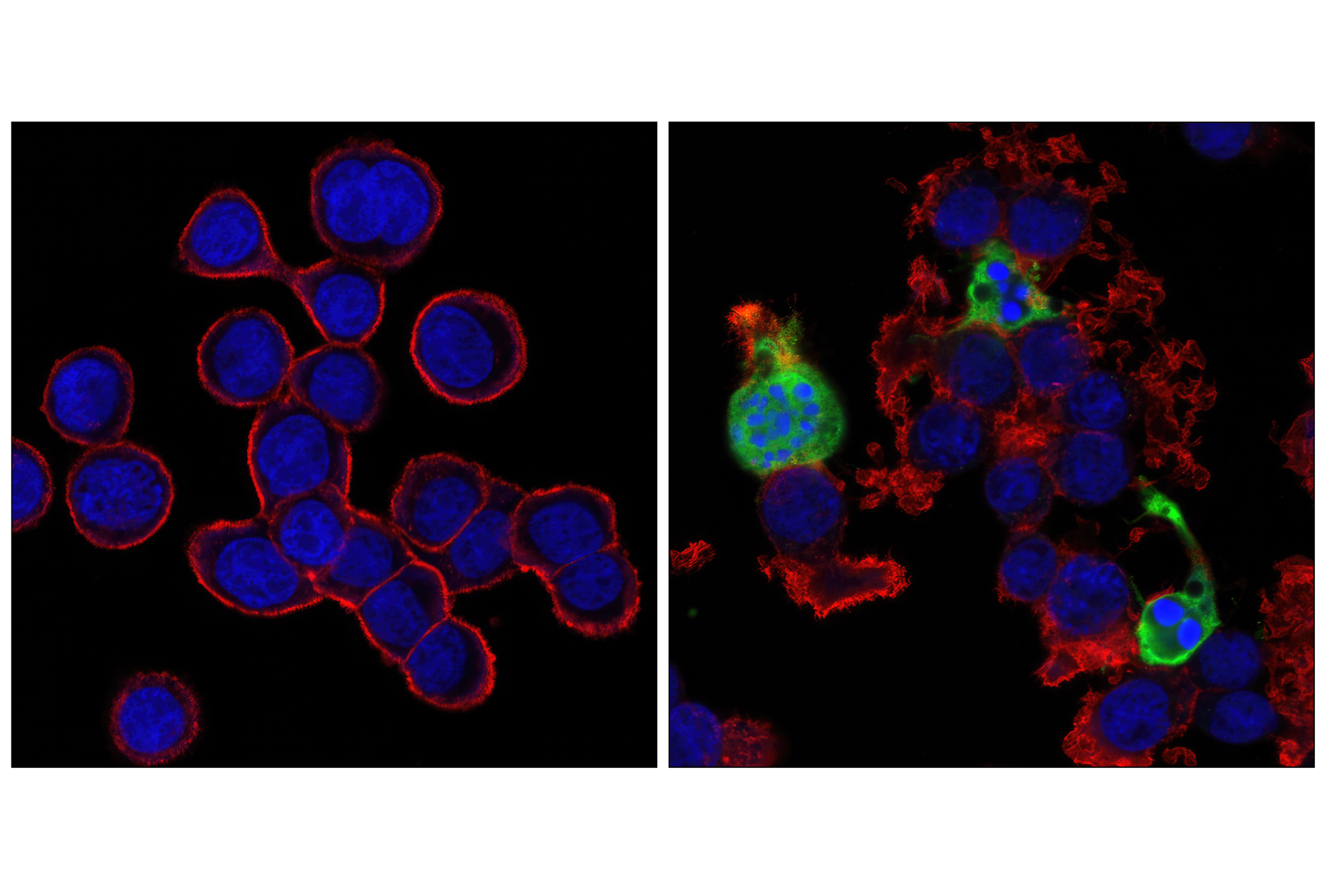
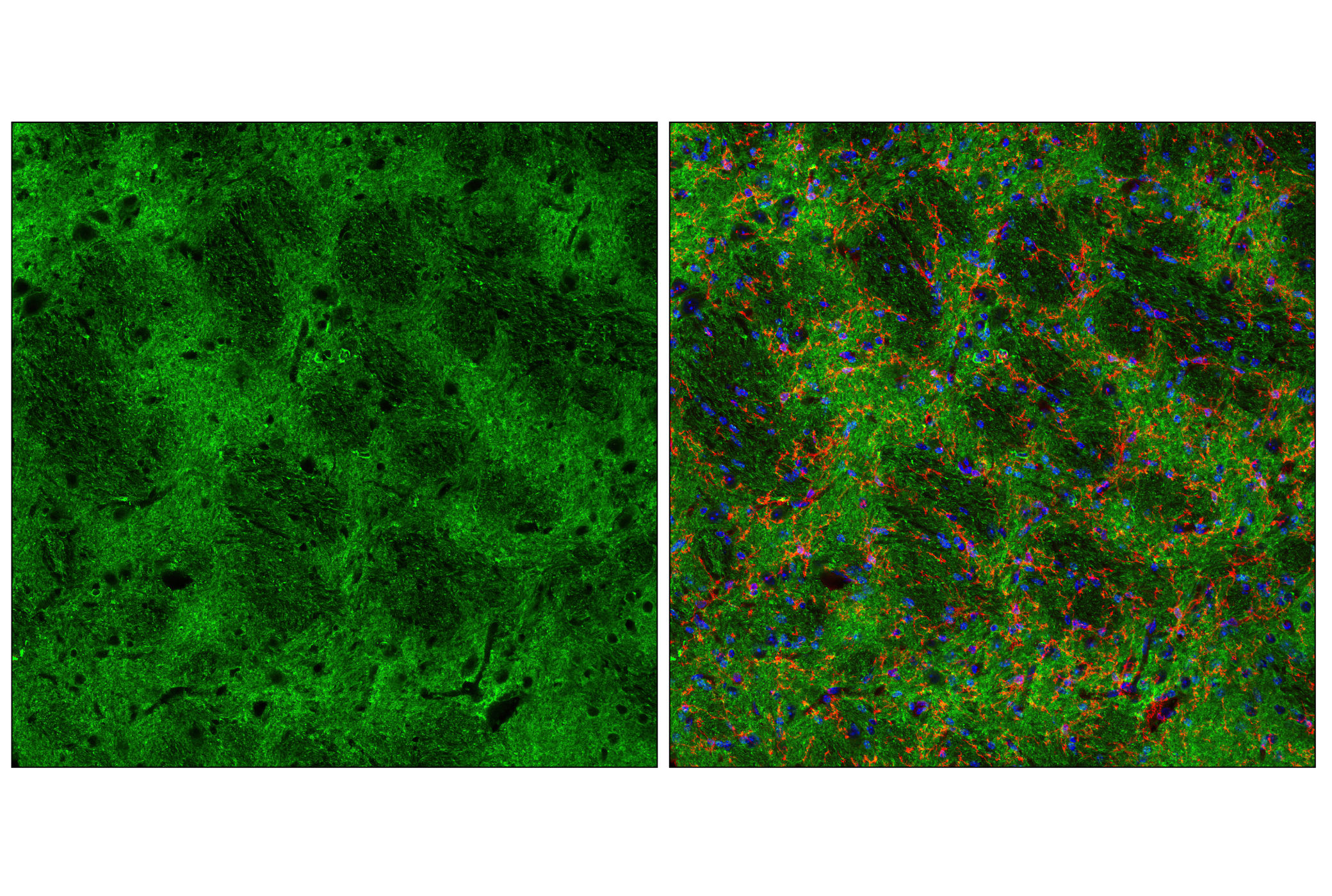
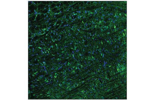
The Tau Mouse Model Neuronal Viability IF Antibody Sampler Kit provides an economical means of detecting proteins to confirm neuronal viability and surrounding astrocytes and microglia in mouse models by immunofluorescence.
Storage
Background
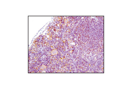
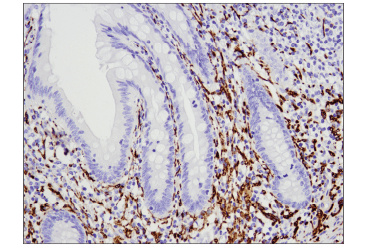
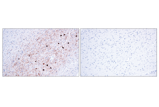
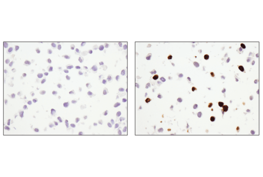
Tau is a heterogeneous microtubule-associated protein that promotes and stabilizes microtubule assembly, especially in axons. Neurofibrillary tangles are a major pathological hallmark of Alzheimer's disease; these tangles are bundles of paired helical filaments composed of hyperphosphorylated tau, including phosphorylation of tau at Thr205 (1,2). Research studies have shown that inclusions of tau are found in a number of other neurodegenerative diseases, collectively known as tauopathies (1,3). Neuronal nuclei (NeuN, Fox-3, RBFOX3) is a nuclear protein expressed in most post-mitotic neurons of the central and peripheral nervous systems. NeuN is not detected in Purkinje cells, sympathetic ganglion cells, Cajal-Retzius cells, INL retinal cells, inferior olivary, or dentate nucleus neurons (4). Glial fibrillary acidic protein (GFAP) is the main intermediate filament in mature brain astroglial and radial glial cells. GFAP plays an important role in modulating astroglial motility and shape (5). HS1 is a protein kinase substrate that is expressed only in tissues and cells of hematopoietic origin (6). Previous work identifying markers of specific brain cell types using RNA-seq has shown HS1 to be a useful and specific tool to study microglia (7). Synaptophysin (SYP) is a neuronal synaptic vesicle glycoprotein that occurs in presynaptic vesicles of neurons (8). Postsynaptic density protein 95 (PSD95) is a member of the membrane-associated guanylate kinase (MAGUK) family of proteins. PSD95 is a scaffolding protein involved in the assembly and function of the postsynaptic density complex (9,10). Caspase-3 (CPP-32, Apoptain, Yama, SCA-1) is a critical executioner of apoptosis, as it is either partially or totally responsible for the proteolytic cleavage of many key proteins, including nuclear enzyme poly (ADP-ribose) polymerase (PARP) (11). PARP, a 116 kDa nuclear poly (ADP-ribose) polymerase, appears to be involved in DNA repair in response to environmental stress (12). PARP helps cells to maintain their viability; cleavage of PARP facilitates cellular disassembly and serves as a marker of cells undergoing apoptosis (13).
- Johnson, G.V. and Stoothoff, W.H. (2004) J Cell Sci 117, 5721-9.
- Wang, J. et al. (2000) Zhongguo Yi Xue Ke Xue Yuan Xue Bao 22, 120-3.
- Bramblett, G.T. et al. (1993) Neuron 10, 1089-99.
- Mullen, R.J. et al. (1992) Development 116, 201-11.
- Eng, L.F. et al. (2000) Neurochem Res 25, 1439-51.
- Kitamura, D. et al. (1995) Biochem Biophys Res Commun 208, 1137-46.
- Zhang, Y. et al. (2014) J Neurosci 34, 11929-47.
- Wiedenmann, B. et al. (1986) Proc Natl Acad Sci U S A 83, 3500-4.
- Cao, J. et al. (2005) J Cell Biol 168, 117-26.
- Chetkovich, D.M. et al. (2002) J Neurosci 22, 6415-25.
- Fernandes-Alnemri, T. et al. (1994) J Biol Chem 269, 30761-4.
- Satoh, M.S. and Lindahl, T. (1992) Nature 356, 356-8.
- Oliver, F.J. et al. (1998) J Biol Chem 273, 33533-9.
Background References
Trademarks and Patents
Limited Uses
Except as otherwise expressly agreed in a writing signed by a legally authorized representative of CST, the following terms apply to Products provided by CST, its affiliates or its distributors. Any Customer's terms and conditions that are in addition to, or different from, those contained herein, unless separately accepted in writing by a legally authorized representative of CST, are rejected and are of no force or effect.
Products are labeled with For Research Use Only or a similar labeling statement and have not been approved, cleared, or licensed by the FDA or other regulatory foreign or domestic entity, for any purpose. Customer shall not use any Product for any diagnostic or therapeutic purpose, or otherwise in any manner that conflicts with its labeling statement. Products sold or licensed by CST are provided for Customer as the end-user and solely for research and development uses. Any use of Product for diagnostic, prophylactic or therapeutic purposes, or any purchase of Product for resale (alone or as a component) or other commercial purpose, requires a separate license from CST. Customer shall (a) not sell, license, loan, donate or otherwise transfer or make available any Product to any third party, whether alone or in combination with other materials, or use the Products to manufacture any commercial products, (b) not copy, modify, reverse engineer, decompile, disassemble or otherwise attempt to discover the underlying structure or technology of the Products, or use the Products for the purpose of developing any products or services that would compete with CST products or services, (c) not alter or remove from the Products any trademarks, trade names, logos, patent or copyright notices or markings, (d) use the Products solely in accordance with CST Product Terms of Sale and any applicable documentation, and (e) comply with any license, terms of service or similar agreement with respect to any third party products or services used by Customer in connection with the Products.