| Product Includes | Product # | Quantity | Mol. Wt | Isotype/Source |
|---|---|---|---|---|
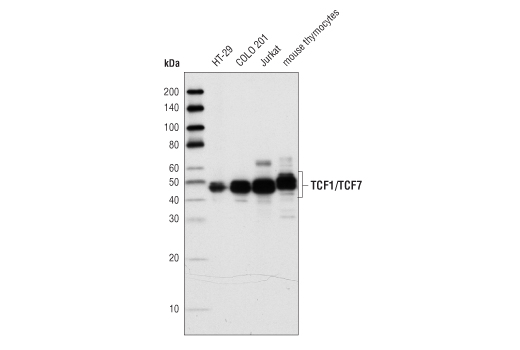
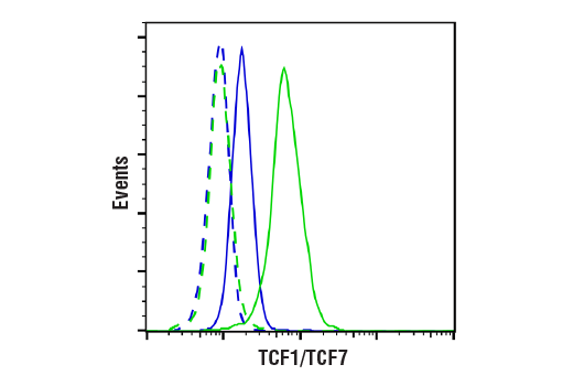
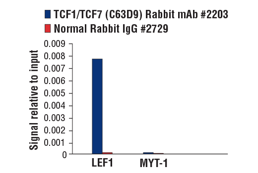
| TCF1/TCF7 (C63D9) Rabbit mAb | 2203 | 20 µl | 48, 50 kDa | Rabbit IgG |
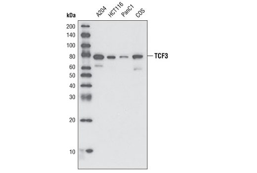
| TCF3/TCF7L1 (D15G11) Rabbit mAb | 2883 | 20 µl | 78 kDa | Rabbit |
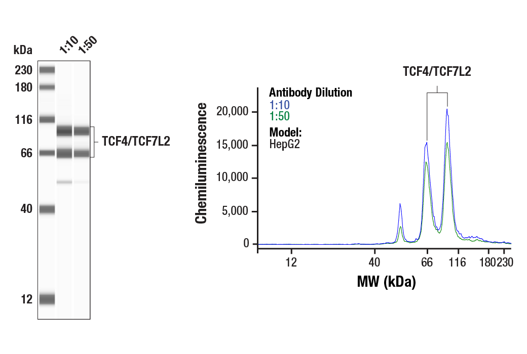
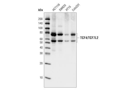
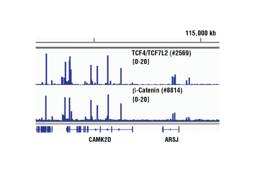
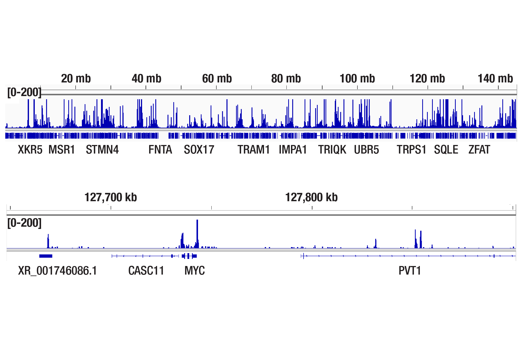
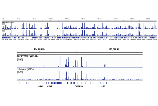
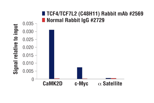
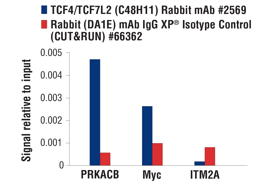
| TCF4/TCF7L2 (C48H11) Rabbit mAb | 2569 | 20 µl | 58, 79 kDa | Rabbit IgG |
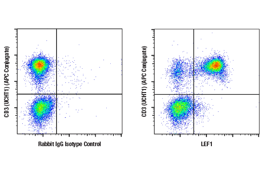
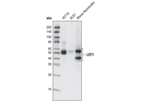
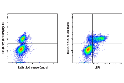
| LEF1 (C12A5) Rabbit mAb | 2230 | 20 µl | 25-58 kDa | Rabbit IgG |
| Anti-rabbit IgG, HRP-linked Antibody | 7074 | 100 µl | Goat |
Please visit cellsignal.com for individual component applications, species cross-reactivity, dilutions, protocols, and additional product information.
Description
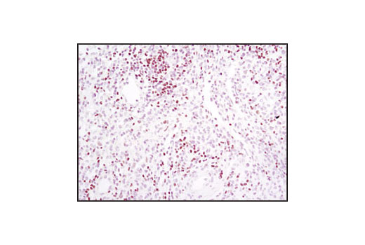
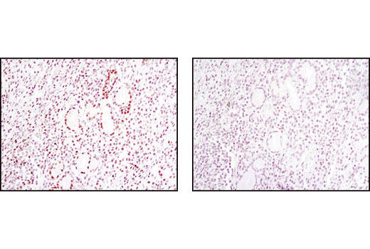
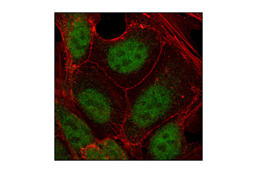
The TCF/LEF Family Antibody Sampler Kit provides an economical means of detecting total protein from the TCF/LEF1 family members. The kit contains enough primary and secondary antibodies to perform two Western blots with each antibody.
Storage
Background
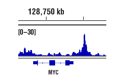
LEF1 and TCF are members of the high mobility group (HMG) DNA binding protein family of transcription factors which consists of the following: Lymphoid Enhancer Factor 1 (LEF1), T Cell Factor 1 (TCF1, also known as TCF7), TCF3 (also known as TCF7L1) and TCF4 (also known as TCF7L2) (1). LEF1 and TCF1/TCF7 were originally identified as important factors regulating early lymphoid development (2) that act downstream in Wnt signaling. LEF1 and TCF bind to Wnt response elements to provide docking sites for β-catenin, which translocates to the nucleus to promote the transcription of target genes upon activation of Wnt signaling (3). LEF1 and TCF proteins are dynamically expressed during development and aberrant activation of the Wnt signaling pathway is involved in many types of cancers including colon cancer (4,5).
Background References
Trademarks and Patents
Limited Uses
Except as otherwise expressly agreed in a writing signed by a legally authorized representative of CST, the following terms apply to Products provided by CST, its affiliates or its distributors. Any Customer's terms and conditions that are in addition to, or different from, those contained herein, unless separately accepted in writing by a legally authorized representative of CST, are rejected and are of no force or effect.
Products are labeled with For Research Use Only or a similar labeling statement and have not been approved, cleared, or licensed by the FDA or other regulatory foreign or domestic entity, for any purpose. Customer shall not use any Product for any diagnostic or therapeutic purpose, or otherwise in any manner that conflicts with its labeling statement. Products sold or licensed by CST are provided for Customer as the end-user and solely for research and development uses. Any use of Product for diagnostic, prophylactic or therapeutic purposes, or any purchase of Product for resale (alone or as a component) or other commercial purpose, requires a separate license from CST. Customer shall (a) not sell, license, loan, donate or otherwise transfer or make available any Product to any third party, whether alone or in combination with other materials, or use the Products to manufacture any commercial products, (b) not copy, modify, reverse engineer, decompile, disassemble or otherwise attempt to discover the underlying structure or technology of the Products, or use the Products for the purpose of developing any products or services that would compete with CST products or services, (c) not alter or remove from the Products any trademarks, trade names, logos, patent or copyright notices or markings, (d) use the Products solely in accordance with CST Product Terms of Sale and any applicable documentation, and (e) comply with any license, terms of service or similar agreement with respect to any third party products or services used by Customer in connection with the Products.