Revision 3

#16551Store at -20C

Cell Signaling Technology

Orders: 877-616-CELL (2355) [email protected]

Support: 877-678-TECH (8324)

Web: [email protected] cellsignal.com

3 Trask LaneDanversMassachusetts01923USA
For Research Use Only. Not for Use in Diagnostic Procedures.
Applications:

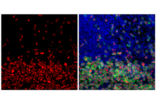
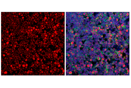
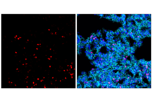
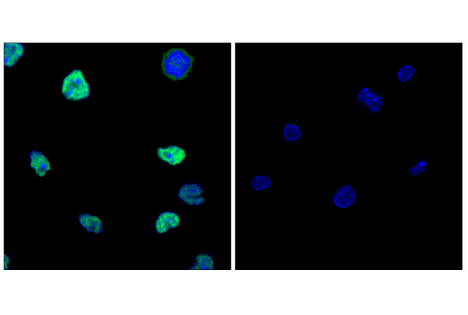
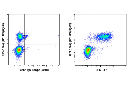
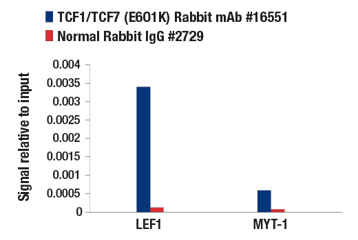
WB, IF-F, IF-IC, FC-FP, ChIP, C&R

REACTIVITY:

H M R

SENSITIVITY:

Endogenous

MW (kDa):

28-50

Source/Isotype:

Rabbit IgG

UniProt ID:

#Q00417

Entrez-Gene Id:

21414

Product Information

Product Usage Information

For optimal ChIP results, use 10 μl of antibody and 10 μg of chromatin (approximately 4 x 10⁶ cells) per IP. This antibody has been validated using SimpleChIP® Enzymatic Chromatin IP Kits.

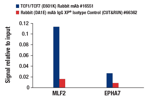
The CUT&RUN dilution was determined using CUT&RUN Assay Kit #86652.
Application Dilution
Western Blotting 1:1000
Immunofluorescence (Frozen) 1:400 - 1:800
Immunofluorescence (Immunocytochemistry) 1:1600 - 1:3200
Flow Cytometry (Fixed/Permeabilized) 1:100 - 1:400
Chromatin IP 1:50
CUT&RUN 1:50

Storage

Supplied in 10 mM sodium HEPES (pH 7.5), 150 mM NaCl, 100 µg/mL BSA, 50% glycerol, and less than 0.02% sodium azide. Store at –20°C. Do not aliquot the antibody.

Specificity / Sensitivity

TCF1/TCF7 (E6O1K) Rabbit mAb detects endogenous levels of total TCF1/TCF7 proteins. This antibody should detect all TCF1/TCF7 isoforms, including those lacking the amino-terminal β-catenin binding domain. This antibody does not cross-react with LEF1, TCF3/TCF7L1, or TCF4/TCF7L2 protein.

Species Reactivity:

Human, Mouse, Rat

Source / Purification

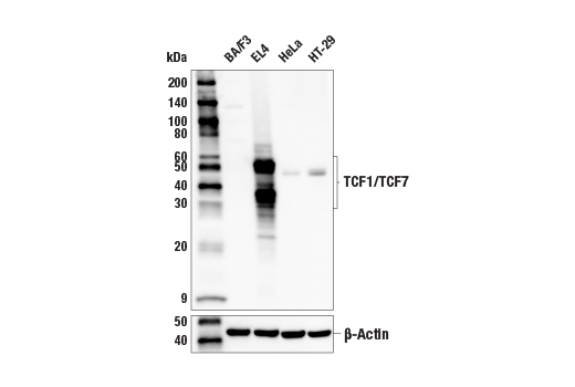
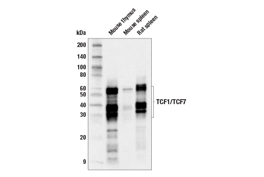
Monoclonal antibody is produced by immunizing animals with recombinant protein specific to mouse TCF1/TCF7 protein.

Background

LEF1 and TCF are members of the high mobility group (HMG) DNA-binding protein family of transcription factors that consists of the following: Lymphoid Enhancer Factor 1 (LEF1), T Cell Factor 1 (TCF1/TCF7), TCF3/TCF7L1, and TCF4/TCF7L2 (1). LEF1 and TCF1/TCF7 were originally identified as important factors that regulate early lymphoid development (2) and act downstream in Wnt signaling. LEF1 and TCF bind to Wnt response elements to provide docking sites for β-catenin, which translocates to the nucleus to promote the transcription of target genes upon activation of Wnt signaling (3). LEF1 and TCF are dynamically expressed during development and aberrant activation of the Wnt signaling pathway is involved in many types of cancers, including colon cancer (4,5).

TCF1/TCF7 has several isoforms due to alternative splicing and transcription from an alternative promoter. The isoforms generated by the alternative promoter do not contain the amino-terminal β-catenin binding domain and therefore may function in a dominant negative manner (6). TCF1/TCF7 displays dynamic expression both in the total amount and the type of isoforms expressed in T cells during development and differentiation (7).

  1. Waterman, M.L. (2004) Cancer Metastasis Rev 23, 41-52.
  2. Schilham, M.W. and Clevers, H. (1998) Semin Immunol 10, 127-32.
  3. Brantjes, H. et al. (2002) Biol Chem 383, 255-61.
  4. Reya, T. and Clevers, H. (2005) Nature 434, 843-50.
  5. Logan, C.Y. and Nusse, R. (2004) Annu Rev Cell Dev Biol 20, 781-810.
  6. Waterman, M.L. (2004) Cancer Metastasis Rev 23, 41-52.
  7. Willinger, T. et al. (2006) J Immunol 176, 1439-46.

Species Reactivity

Species reactivity is determined by testing in at least one approved application (e.g., western blot).

Western Blot Buffer

IMPORTANT: For western blots, incubate membrane with diluted primary antibody in 5% w/v nonfat dry milk, 1X TBS, 0.1% Tween® 20 at 4°C with gentle shaking, overnight.

Applications Key

WB: Western Blotting IF-F: Immunofluorescence (Frozen) IF-IC: Immunofluorescence (Immunocytochemistry) FC-FP: Flow Cytometry (Fixed/Permeabilized) ChIP: Chromatin IP C&R: CUT&RUN

Cross-Reactivity Key

H: human M: mouse R: rat Hm: hamster Mk: monkey Vir: virus Mi: mink C: chicken Dm: D. melanogaster X: Xenopus Z: zebrafish B: bovine Dg: dog Pg: pig Sc: S. cerevisiae Ce: C. elegans Hr: horse GP: Guinea Pig Rab: rabbit All: all species expected

Trademarks and Patents

Cell Signaling Technology is a trademark of Cell Signaling Technology, Inc.
SimpleChIP is a registered trademark of Cell Signaling Technology, Inc.
XP is a registered trademark of Cell Signaling Technology, Inc.
All other trademarks are the property of their respective owners. Visit cellsignal.com/trademarks for more information.

Limited Uses

Except as otherwise expressly agreed in a writing signed by a legally authorized representative of CST, the following terms apply to Products provided by CST, its affiliates or its distributors. Any Customer's terms and conditions that are in addition to, or different from, those contained herein, unless separately accepted in writing by a legally authorized representative of CST, are rejected and are of no force or effect.

Products are labeled with For Research Use Only or a similar labeling statement and have not been approved, cleared, or licensed by the FDA or other regulatory foreign or domestic entity, for any purpose. Customer shall not use any Product for any diagnostic or therapeutic purpose, or otherwise in any manner that conflicts with its labeling statement. Products sold or licensed by CST are provided for Customer as the end-user and solely for research and development uses. Any use of Product for diagnostic, prophylactic or therapeutic purposes, or any purchase of Product for resale (alone or as a component) or other commercial purpose, requires a separate license from CST. Customer shall (a) not sell, license, loan, donate or otherwise transfer or make available any Product to any third party, whether alone or in combination with other materials, or use the Products to manufacture any commercial products, (b) not copy, modify, reverse engineer, decompile, disassemble or otherwise attempt to discover the underlying structure or technology of the Products, or use the Products for the purpose of developing any products or services that would compete with CST products or services, (c) not alter or remove from the Products any trademarks, trade names, logos, patent or copyright notices or markings, (d) use the Products solely in accordance with CST Product Terms of Sale and any applicable documentation, and (e) comply with any license, terms of service or similar agreement with respect to any third party products or services used by Customer in connection with the Products.

Revision 3
#16551

TCF1/TCF7 (E6O1K) Rabbit mAb

Western Blotting Image 1: TCF1/TCF7 (E6O1K) Rabbit mAb Expand Image
Western blot analysis of extracts from various mouse and human cell lines using TCF1/TCF7 (E6O1K) Rabbit mAb (upper) or β-Actin (D6A8) Rabbit mAb #8457 (lower). Negative expression of TCF1/TCF7 protein in BA/F3 cells is consistent with the predicted expression pattern.
Western Blotting Image 2: TCF1/TCF7 (E6O1K) Rabbit mAb Expand Image
Western blot analysis of extracts from mouse thymus, mouse spleen, and rat spleen tissue using TCF1/TCF7 (E6O1K) Rabbit mAb.
Immunofluorescence Image 1: TCF1/TCF7 (E6O1K) Rabbit mAb Expand Image
Confocal immunofluorescent analysis of fixed frozen mouse spleen, labeled with TCF1/TCF7 (E6O1K) Rabbit mAb (left, red) and CD3 (17A2) Rat mAb (FITC Conjugate) #86603 (right, green). Samples were mounted in ProLong Gold Antifade Reagent with DAPI #8961 (right, blue).
Immunofluorescence Image 2: TCF1/TCF7 (E6O1K) Rabbit mAb Expand Image
Confocal immunofluorescent analysis of fixed frozen mouse thymus, labeled with TCF1/TCF7 (E6O1K) Rabbit mAb (left, red) and CD3 (17A2) Rat mAb (FITC Conjugate) #86603 (right, green). Samples were mounted in ProLong Gold Antifade Reagent with DAPI #8961 (right, blue).
Immunofluorescence Image 3: TCF1/TCF7 (E6O1K) Rabbit mAb Expand Image
Confocal immunofluorescent analysis of fixed frozen mouse lung, labeled with TCF1/TCF7 (E6O1K) Rabbit mAb (left, red) and DyLight 650 Phalloidin #12956 (right, cyan pseudocolor). Samples were mounted in ProLong Gold Antifade Reagent with DAPI #8961 (right, blue).
Immunofluorescence Image 1: TCF1/TCF7 (E6O1K) Rabbit mAb Expand Image
Confocal immunofluorescent analysis of EL4 cells (left, positive) and BA/F3 cells (right, negative) using TCF1/TCF7 (E6O1K) Rabbit mAb (green) and DAPI #4083 (blue).
Flow Cytometry Image 1: TCF1/TCF7 (E6O1K) Rabbit mAb Expand Image
Flow cytometric analysis of mouse splenocytes using TCF1/TCF7 (E6O1K) Rabbit mAb (right) or concentration-matched Rabbit (DA1E) mAb IgG XP® Isotype Control #3900 (left), co-stained with CD3 (17A2) Rat mAb (APC Conjugate) #24265. Anti-rabbit IgG (H+L), F(ab')2 Fragment (Alexa Fluor® 488 Conjugate) #4412 was used as a secondary antibody.
Chromatin Immunoprecipitation Image 1: TCF1/TCF7 (E6O1K) Rabbit mAb Expand Image
Chromatin immunoprecipitations were performed with cross-linked chromatin from EL4 cells and either TCF1/TCF7 (E6O1K) Rabbit mAb or Normal Rabbit IgG #2729, using SimpleChIP® Plus Enzymatic Chromatin IP Kit (Magnetic Beads) #9005. The enriched DNA was quantified by real-time PCR using mouse LEF1 upstream primers and SimpleChIP® Mouse MYT-1 Promoter Primers #8985. The amount of immunoprecipitated DNA in each sample is represented as signal relative to the total amount of input chromatin, which is equivalent to one.
CUT and RUN Image 1: TCF1/TCF7 (E6O1K) Rabbit mAb Expand Image
CUT&RUN was performed with EL4 cells and TCF1/TCF7 (E6O1K) Rabbit mAb, using CUT&RUN Assay Kit #86652. DNA library was prepared using DNA Library Prep Kit for Illumina Systems (ChIP-seq, CUT&RUN) #56795. The figures show binding across chromosome 6 (upper), including Mlf2 (lower), a known target gene of TCF1/TCF7 (see additional figure containing qPCR data).
CUT and RUN Image 2: TCF1/TCF7 (E6O1K) Rabbit mAb Expand Image
CUT&RUN was performed with EL4 cells and TCF1/TCF7 (E6O1K) Rabbit mAb, using CUT&RUN Assay Kit #86652. DNA library was prepared using DNA Library Prep Kit for Illumina Systems (ChIP-seq, CUT&RUN) #56795. The figure shows binding across Mlf2, a known target gene of TCF1/TCF7 (see additional figure containing qPCR data).
CUT and RUN Image 3: TCF1/TCF7 (E6O1K) Rabbit mAb Expand Image
CUT&RUN was performed with EL4 cells and TCF1/TCF7 (E6O1K) Rabbit mAb or Rabbit (DA1E) mAb IgG XP® Isotype Control (CUT&RUN) #66362, using CUT&RUN Assay Kit #86652. The enriched DNA was quantified by real-time PCR using mouse MLF2 promoter primers and mouse EPHA7 upstream primers. The amount of immunoprecipitated DNA in each sample is represented as signal relative to the total amount of input chromatin, which is equivalent to one.