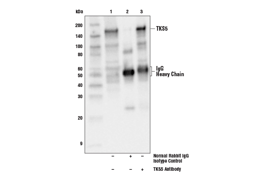
WB, IP
H
Endogenous
150
Rabbit
#Q5TCZ1
9644
Product Information
Product Usage Information
| Application | Dilution |
|---|---|
| Western Blotting | 1:1000 |
| Immunoprecipitation | 1:50 |
Storage
Specificity / Sensitivity
Species Reactivity:
Human
Source / Purification
Polyclonal antibodies are produced by immunizing animals with a synthetic peptide corresponding to residues surrounding Asp910 of human TKS5 protein. Antibodies are purified by protein A and peptide affinity chromatography.
Background
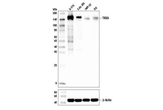
TKS5 (SH3PXD2A, FISH) is a scaffold protein expressed on invadosomes of both normal and transformed cell lines. Research studies suggest that TKS5 is functionally required for both the formation and invasive behavior of invadosomes (1, 2). TKS5 has an N-terminal PX domain that mediates invadosome initiation and localization of MMP-rich vesicles to the invadosome (3). TKS5 also has five SH3 domains, which recruit ADAM family proteinases to the invadosome to degrade extracellular matrix. These SH3 domains interact with adaptor proteins to facilitate F-actin polymerization during invadosome formation (4-6). Src tyrosine kinase has been shown to phosphorylate TKS5 at Tyr557 and Tyr619, which was shown to be necessary and sufficient for TKS5-mediated invadopia formation and invasion (7). Elevated TKS5 expression is positively associated with invasive behavior of cancer cells, suggesting TKS5 may have prognostic potential in cancer (8, 9).
- Paterson, E.K. and Courtneidge, S.A. (2017) FEBS J doi:10.1111/febs.14123.
- Courtneidge, S.A. (2012) Biochem Soc Trans 40, 129-32.
- Jacob, A. et al. (2016) J Cell Sci 129, 4341-4353.
- Abram, C.L. et al. (2003) J Biol Chem 278, 16844-51.
- Crimaldi, L. et al. (2009) Exp Cell Res 315, 2581-92.
- Stylli, S.S. et al. (2009) J Cell Sci 122, 2727-40.
- Burger, K.L. et al. (2014) Prostate 74, 134-48.
- Stylli, S.S. et al. (2014) Oncol Rep 32, 989-1002.
- Blouw, B. et al. (2015) PLoS One 10, e0121003.
Species Reactivity
Species reactivity is determined by testing in at least one approved application (e.g., western blot).
Western Blot Buffer
IMPORTANT: For western blots, incubate membrane with diluted primary antibody in 5% w/v BSA, 1X TBS, 0.1% Tween® 20 at 4°C with gentle shaking, overnight.
Applications Key
WB: Western Blotting IP: Immunoprecipitation
Cross-Reactivity Key
H: human M: mouse R: rat Hm: hamster Mk: monkey Vir: virus Mi: mink C: chicken Dm: D. melanogaster X: Xenopus Z: zebrafish B: bovine Dg: dog Pg: pig Sc: S. cerevisiae Ce: C. elegans Hr: horse GP: Guinea Pig Rab: rabbit All: all species expected
Trademarks and Patents
Limited Uses
Except as otherwise expressly agreed in a writing signed by a legally authorized representative of CST, the following terms apply to Products provided by CST, its affiliates or its distributors. Any Customer's terms and conditions that are in addition to, or different from, those contained herein, unless separately accepted in writing by a legally authorized representative of CST, are rejected and are of no force or effect.
Products are labeled with For Research Use Only or a similar labeling statement and have not been approved, cleared, or licensed by the FDA or other regulatory foreign or domestic entity, for any purpose. Customer shall not use any Product for any diagnostic or therapeutic purpose, or otherwise in any manner that conflicts with its labeling statement. Products sold or licensed by CST are provided for Customer as the end-user and solely for research and development uses. Any use of Product for diagnostic, prophylactic or therapeutic purposes, or any purchase of Product for resale (alone or as a component) or other commercial purpose, requires a separate license from CST. Customer shall (a) not sell, license, loan, donate or otherwise transfer or make available any Product to any third party, whether alone or in combination with other materials, or use the Products to manufacture any commercial products, (b) not copy, modify, reverse engineer, decompile, disassemble or otherwise attempt to discover the underlying structure or technology of the Products, or use the Products for the purpose of developing any products or services that would compete with CST products or services, (c) not alter or remove from the Products any trademarks, trade names, logos, patent or copyright notices or markings, (d) use the Products solely in accordance with CST Product Terms of Sale and any applicable documentation, and (e) comply with any license, terms of service or similar agreement with respect to any third party products or services used by Customer in connection with the Products.