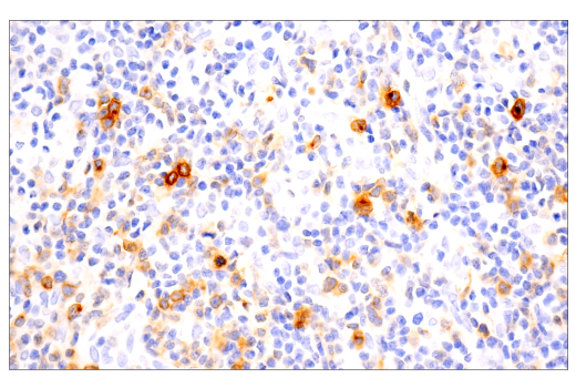
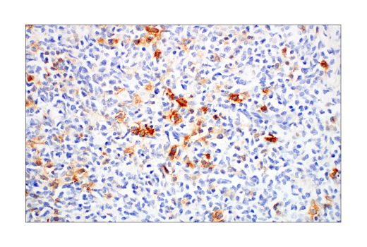
WB, IHC-Bond, IHC-P, IF-IC, FC-L
H Mk
Endogenous
90, 120
Rabbit IgG
#P28908
943
Product Information
Product Usage Information
| Application | Dilution |
|---|---|
| Western Blotting | 1:1000 |
| IHC Leica Bond | 1:50 |
| Immunohistochemistry (Paraffin) | 1:50 |
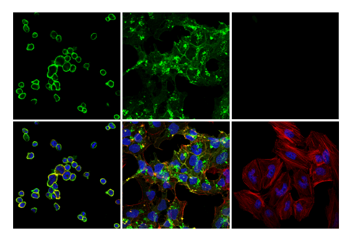
| Immunofluorescence (Immunocytochemistry) | 1:400 |
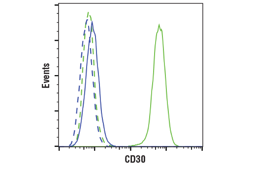
| Flow Cytometry (Live) | 1:200 - 1:800 |
Storage
For a carrier free (BSA and azide free) version of this product see product #18445.
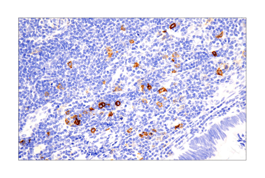
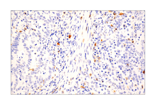
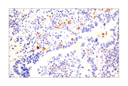
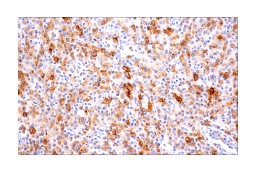
Specificity / Sensitivity
Species Reactivity:
Human, Monkey
Source / Purification
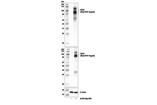
Monoclonal antibody is produced by immunizing animals with recombinant protein specific to the extracellular domain of human TNFRSF8/CD30 protein.
Background
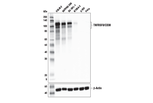
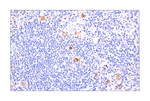
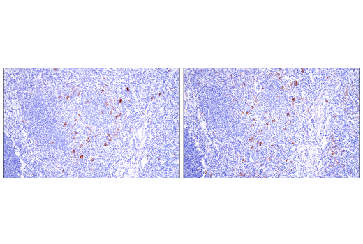
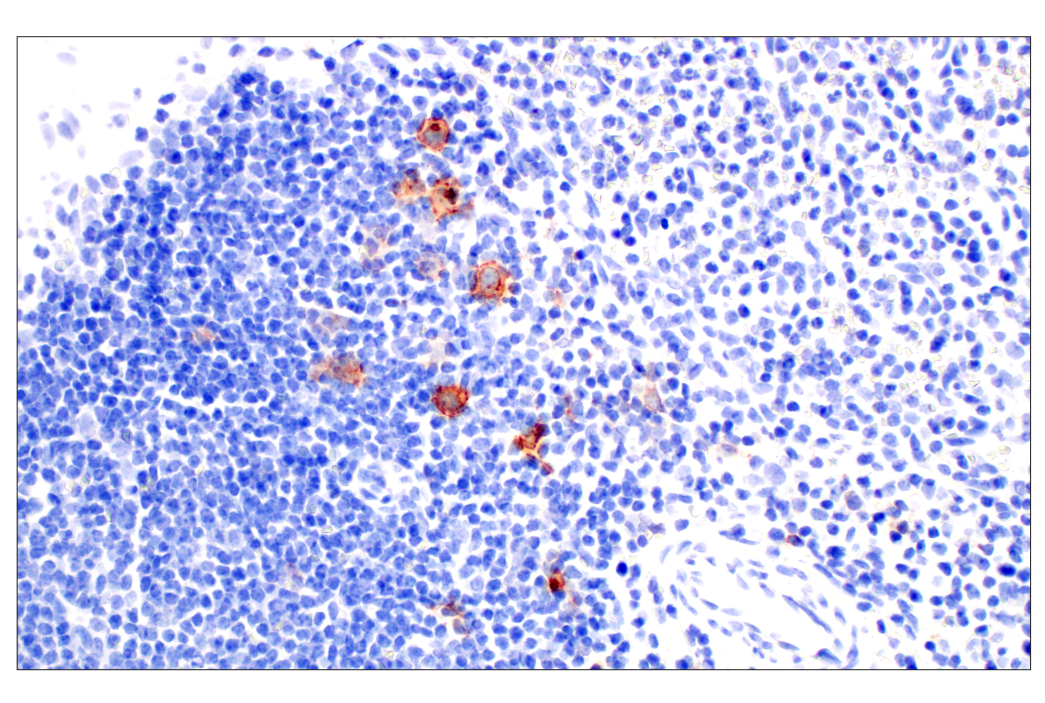
TNFRSF8/CD30 is a type-I transmembrane glycoprotein that is a member of the TNFR superfamily. CD30 is synthesized as a precursor protein that undergoes extensive post-translational modification before becoming embedded in the plasma membrane as a 120-kDa transmembrane protein (1,2). The expression of CD30 is upregulated in activated T cells and may trigger costimulatory signaling pathways upon its engagement (3,4). While its expression is normally restricted to subsets of activated T cells and B cells, CD30 expression is robustly upregulated in hematologic malignancies, such as Hodgkin lymphoma (HL), anaplastic large cell lymphoma (ALCL), and adult T-cell leukemia, thus making it an attractive target for therapeutic intervention (5,6). Research studies have suggested that in certain disease contexts, CD30 recruits TRAF2 and TRAF5 adaptor proteins to drive NF-kappa B activation, aberrant cell growth, and cytokine production (7-9). CD30 signaling is also regulated by TACE-dependent proteolytic cleavage of its ectodomain, which results in reduced CD30L-dependent activation of CD30+ cells (10,11).
- Froese, P. et al. (1987) J Immunol 139, 2081-7.
- Nawrocki, J.F. et al. (1988) J Immunol 141, 672-80.
- Del Prete, G. et al. (1995) J Exp Med 182, 1655-61.
- Gilfillan, M.C. et al. (1998) J Immunol 160, 2180-7.
- Stein, H. et al. (1985) Blood 66, 848-58.
- Chiarle, R. et al. (1999) Clin Immunol 90, 157-64.
- Horie, R. et al. (2002) Am J Pathol 160, 1647-54.
- Horie, R. et al. (2002) Oncogene 21, 2493-503.
- Horie, R. et al. (2004) Cancer Cell 5, 353-64.
- Hansen, H.P. et al. (2000) J Immunol 165, 6703-9.
- Gruss, H.J. et al. (1997) Immunol Today 18, 156-63.
Species Reactivity
Species reactivity is determined by testing in at least one approved application (e.g., western blot).
Western Blot Buffer
IMPORTANT: For western blots, incubate membrane with diluted primary antibody in 5% w/v BSA, 1X TBS, 0.1% Tween® 20 at 4°C with gentle shaking, overnight.
Applications Key
WB: Western Blotting IHC-Bond: IHC Leica Bond IHC-P: Immunohistochemistry (Paraffin) IF-IC: Immunofluorescence (Immunocytochemistry) FC-L: Flow Cytometry (Live)
Cross-Reactivity Key
H: human M: mouse R: rat Hm: hamster Mk: monkey Vir: virus Mi: mink C: chicken Dm: D. melanogaster X: Xenopus Z: zebrafish B: bovine Dg: dog Pg: pig Sc: S. cerevisiae Ce: C. elegans Hr: horse GP: Guinea Pig Rab: rabbit All: all species expected
Trademarks and Patents
Limited Uses
Except as otherwise expressly agreed in a writing signed by a legally authorized representative of CST, the following terms apply to Products provided by CST, its affiliates or its distributors. Any Customer's terms and conditions that are in addition to, or different from, those contained herein, unless separately accepted in writing by a legally authorized representative of CST, are rejected and are of no force or effect.
Products are labeled with For Research Use Only or a similar labeling statement and have not been approved, cleared, or licensed by the FDA or other regulatory foreign or domestic entity, for any purpose. Customer shall not use any Product for any diagnostic or therapeutic purpose, or otherwise in any manner that conflicts with its labeling statement. Products sold or licensed by CST are provided for Customer as the end-user and solely for research and development uses. Any use of Product for diagnostic, prophylactic or therapeutic purposes, or any purchase of Product for resale (alone or as a component) or other commercial purpose, requires a separate license from CST. Customer shall (a) not sell, license, loan, donate or otherwise transfer or make available any Product to any third party, whether alone or in combination with other materials, or use the Products to manufacture any commercial products, (b) not copy, modify, reverse engineer, decompile, disassemble or otherwise attempt to discover the underlying structure or technology of the Products, or use the Products for the purpose of developing any products or services that would compete with CST products or services, (c) not alter or remove from the Products any trademarks, trade names, logos, patent or copyright notices or markings, (d) use the Products solely in accordance with CST Product Terms of Sale and any applicable documentation, and (e) comply with any license, terms of service or similar agreement with respect to any third party products or services used by Customer in connection with the Products.