| Product Includes | Product # | Quantity | Mol. Wt | Isotype/Source |
|---|---|---|---|---|
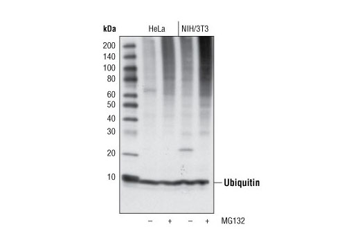
| Ubiquitin Antibody | 3933 | 40 µl | Rabbit | |
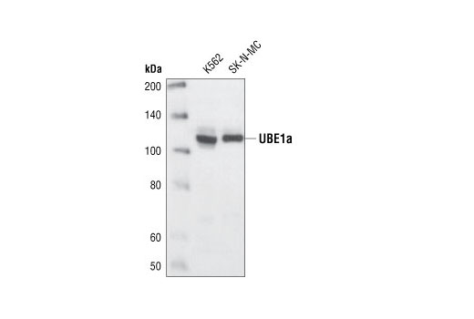
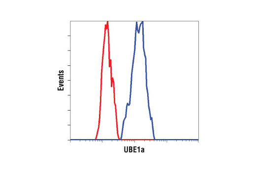
| UBE1a Antibody | 4890 | 40 µl | 117 kDa | Rabbit |
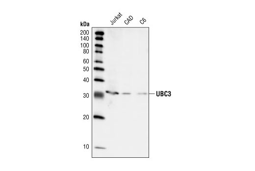
| UBC3 Antibody | 4997 | 40 µl | 32 kDa | Rabbit |
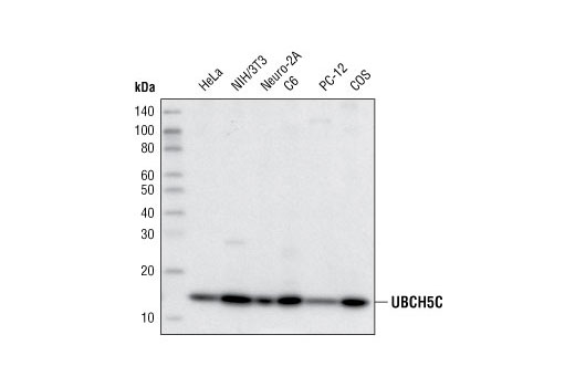
| UbcH5C (D60E2) Rabbit mAb | 4330 | 40 µl | 14 kDa | Rabbit IgG |
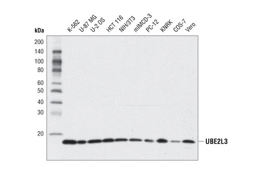
| UBE2L3 (D5G1) Rabbit mAb | 8721 | 40 µl | 18 kDa | Rabbit IgG |
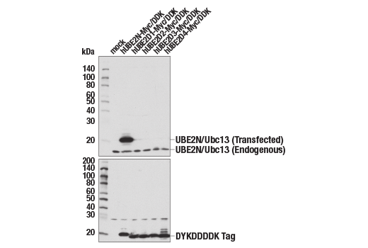
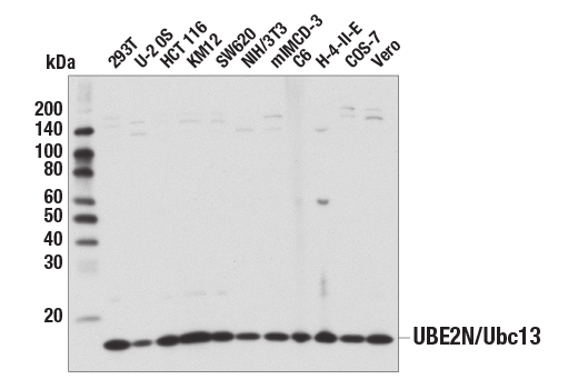
| UBE2N/Ubc13 (D2A1) Rabbit mAb | 6999 | 40 µl | 17 kDa | Rabbit IgG |
| Anti-rabbit IgG, HRP-linked Antibody | 7074 | 100 µl | Goat |
Please visit cellsignal.com for individual component applications, species cross-reactivity, dilutions, protocols, and additional product information.
Description
The Ubiquitin Activation (E1, E2 Enzymes) Antibody Sampler Kit provides an economical means to study ubiquitin activation and conjugation. This kit contains enough primary antibody to perform four western blots per primary.
Storage
Background
Ubiquitin is a conserved polypeptide unit that plays an important role in the ubiquitin-proteasome pathway. Ubiquitin can be covalently linked to many cellular proteins for degradation by the 26S proteasome. Three components are involved in the target protein-ubiquitin conjugation process. Ubiquitin is first activated by forming a thioester complex with the ubiquitin-activating enzyme (UBE1 or E1). The activated ubiquitin is subsequently transferred to the ubiqutin-carrier protein (conjugating enzyme) E2, and then from E2 to ubiqutin ligase E3 for final delivery to the epsilon-NH2 of the target protein lysine residue (1-3). The ubiquitin-proteasome pathway has been implicated in a wide range of normal biological processes and in disease-related abnormalities. Several proteins such as IκB, p53, cdc25a, and Bcl-2 have been shown to be targets for the ubiquitin-proteasome process as part of the regulation of cell cycle progression, differentiation, cell stress response, and apoptosis (4-7). UBC3, the mammalian ortholog of yeast cdc34, and UBC3B, a UBC3 family member, are E2 ubiquitin-carrier proteins. UBC3, in concert with SCF-Skp2 (Skp1, Cullin, F-box protein/Skp2) complex, mediates cell cycle progression from G1 to S phase by targeting the CDK inhibitor p27 for proteolysis (8). UBC3B, in concert with SCFb-Trcp (Skp1, Cullin and F-box protein/b-Trcp) complex, mediates degradation of β-catenin (9). UbcH5C is a universally expressed E2 ubiquitin conjugating enzyme and member of the UbcH5 family that also includes UbcH5A and UbcH5B (10). Evidence suggests that UbcH5 plays an important role in regulating a number of signaling pathways by catalyzing the ubiquitination of key target proteins, including p53, PCNA, the IκB kinase proein NEMO, and the apoptosis inhibitor BRUCE (11-14). UBE2L3, also commonly referred to as UBCH7, is a ubiquitin-conjugating enzyme that has been linked to the ubiquitination of numerous substrates via its interaction with protein-ubiquitin E3 ligases, such as NEDD4 (15), E6AP (16), Parkin (17), c-Cbl (18), and Triad1 (19,20). UBE2N/Ubc13 is a ubiquitin-E2-conjugating enzyme that catalyzes K63-linked polyubiquitin chain formation (21,22). UBE2N forms a heterodimer with MMS2 or Uev1A to exert its E2 ligase function. The UBE2N/MMS2 and UBE2N/Uev1A heterodimers catalyze different modes of target protein ubiquitination to mediate various signaling pathways (23-25) including DNA damage and recombination, p53 and check point control, cell cycle (26-30), immunoreceptor signaling (31,32), and endocytosis (33).
- Ciechanover, A. (1998) EMBO J 17, 7151-60.
- Hochstrasser, M. (2000) Nat Cell Biol 2, E153-7.
- Hochstrasser, M. (2000) Science 289, 563-4.
- Bernardi, R. et al. (2000) Oncogene 19, 2447-54.
- Aberle, H. et al. (1997) EMBO J 16, 3797-804.
- Salomoni, P. and Pandolfi, P.P. (2002) Nat Cell Biol 4, E152-3.
- Jesenberger, V. and Jentsch, S. (2002) Nat Rev Mol Cell Biol 3, 112-21.
- Pagano, M. et al. (1995) Science 269, 682-5.
- Semplici, F. et al. (2002) Oncogene 21, 3978-87.
- Jensen, J.P. et al. (1995) J Biol Chem 270, 30408-14.
- Saville, M.K. et al. (2004) J Biol Chem 279, 42169-81.
- Zhang, S. et al. (2008) Cell Cycle 7, 3399-404.
- Tang, E.D. et al. (2003) J Biol Chem 278, 37297-305.
- Qiu, X.B. et al. (2004) EMBO J 23, 800-10.
- Anan, T. et al. (1998) Genes Cells 3, 751-63.
- Huang, L. et al. (1999) Science 286, 1321-6.
- Shimura, H. et al. (2001) Science 293, 263-9.
- Yokouchi, M. et al. (1999) J Biol Chem 274, 31707-12.
- Marteijn, J.A. et al. (2009) Leukemia 23, 1480-9.
- Marteijn, J.A. et al. (2005) Blood 106, 4114-23.
- Herrmann, J. et al. (2007) Circ Res 100, 1276-91.
- Wilkinson, K.D. et al. (2005) EMBO Rep 6, 815-20.
- Hofmann, R.M. and Pickart, C.M. (1999) Cell 96, 645-53.
- Deng, L. et al. (2000) Cell 103, 351-61.
- Andersen, P.L. et al. (2005) J Cell Biol 170, 745-55.
- Zhao, G.Y. et al. (2007) Mol Cell 25, 663-75.
- Kolas, N.K. et al. (2007) Science 318, 1637-40.
- Laine, A. et al. (2006) Mol Cell Biol 26, 8901-13.
- Huen, M.S. et al. (2008) Mol Cell Biol 28, 6104-12.
- Loring, G.L. et al. (2008) Cell Cycle 7, 96-105.
- Yamamoto, M. et al. (2006) Nat Immunol 7, 962-70.
- Yamamoto, M. et al. (2006) J Immunol 177, 7520-4.
- Duncan, L.M. et al. (2006) EMBO J 25, 1635-45.
Background References
Trademarks and Patents
Limited Uses
Except as otherwise expressly agreed in a writing signed by a legally authorized representative of CST, the following terms apply to Products provided by CST, its affiliates or its distributors. Any Customer's terms and conditions that are in addition to, or different from, those contained herein, unless separately accepted in writing by a legally authorized representative of CST, are rejected and are of no force or effect.
Products are labeled with For Research Use Only or a similar labeling statement and have not been approved, cleared, or licensed by the FDA or other regulatory foreign or domestic entity, for any purpose. Customer shall not use any Product for any diagnostic or therapeutic purpose, or otherwise in any manner that conflicts with its labeling statement. Products sold or licensed by CST are provided for Customer as the end-user and solely for research and development uses. Any use of Product for diagnostic, prophylactic or therapeutic purposes, or any purchase of Product for resale (alone or as a component) or other commercial purpose, requires a separate license from CST. Customer shall (a) not sell, license, loan, donate or otherwise transfer or make available any Product to any third party, whether alone or in combination with other materials, or use the Products to manufacture any commercial products, (b) not copy, modify, reverse engineer, decompile, disassemble or otherwise attempt to discover the underlying structure or technology of the Products, or use the Products for the purpose of developing any products or services that would compete with CST products or services, (c) not alter or remove from the Products any trademarks, trade names, logos, patent or copyright notices or markings, (d) use the Products solely in accordance with CST Product Terms of Sale and any applicable documentation, and (e) comply with any license, terms of service or similar agreement with respect to any third party products or services used by Customer in connection with the Products.