WB, IF-IC
H Mk
Endogenous
62
Rabbit IgG
#Q9BZI7
65109
Product Information
Product Usage Information
| Application | Dilution |
|---|---|
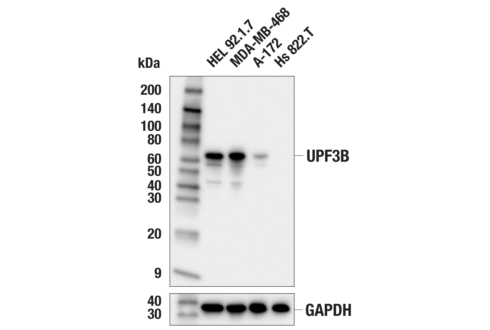
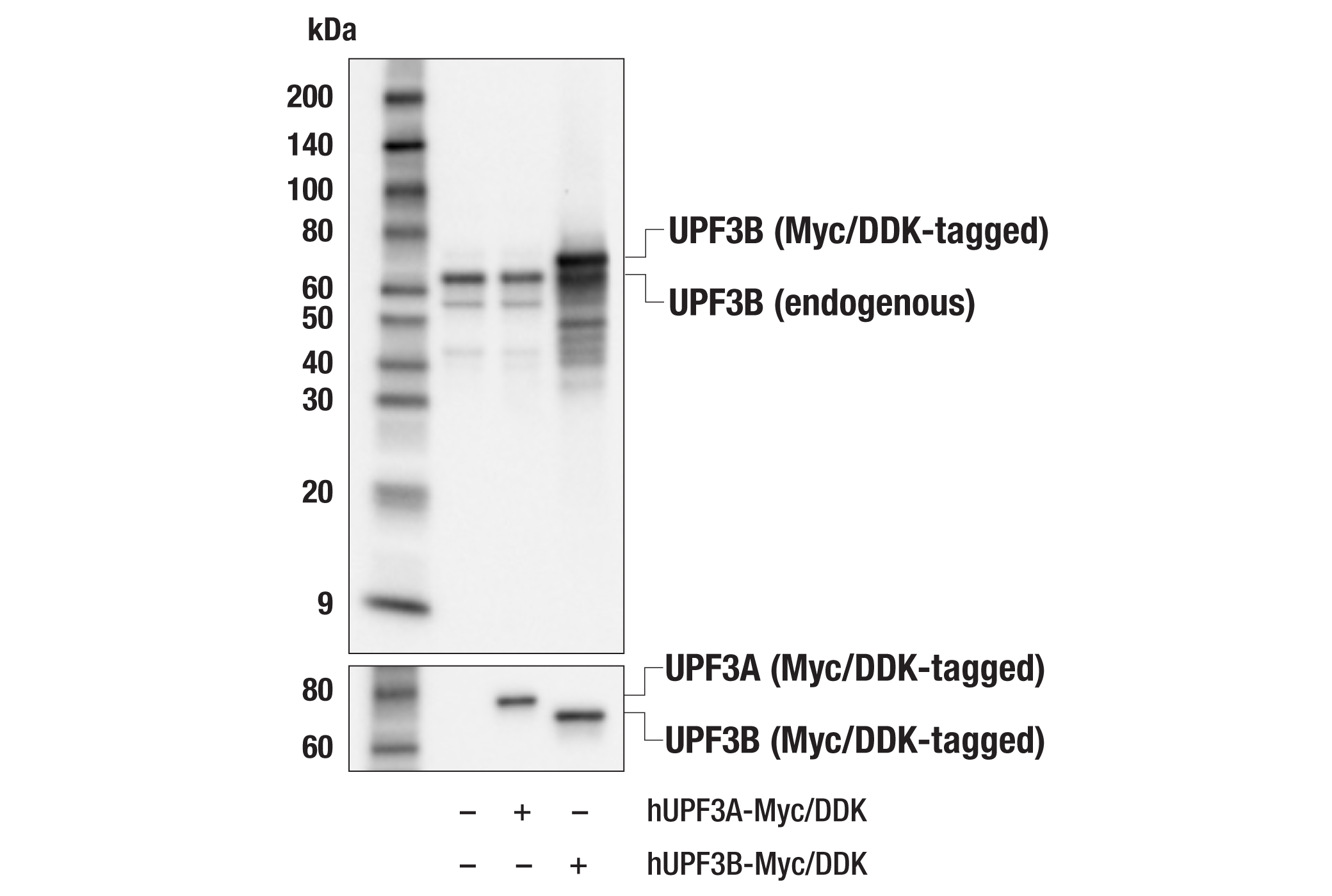
| Western Blotting | 1:1000 |
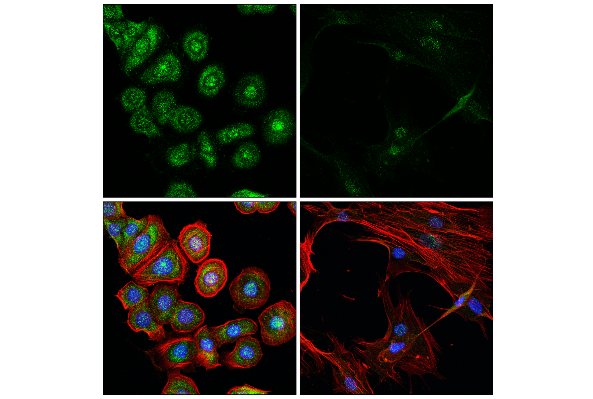
| Immunofluorescence (Immunocytochemistry) | 1:100 - 1:400 |
Storage
Specificity / Sensitivity
Species Reactivity:
Human, Monkey
Source / Purification
Monoclonal antibody is produced by immunizing animals with a synthetic peptide corresponding to residues surrounding Leu262 of human UPF3B protein.
Background
UPF3A and UPF3B are paralogs derived from a gene duplication and are involved in nonsense-mediated decay (NMD), a process that eliminates aberrantly formed mRNAs (1,2). While UPF3A seems to be an NMD repressor, UPF3B serves as an NMD activator, by binding to the exon junction complex (EJC) and shuttling to the cytoplasm after mRNA export (2-4). Under normal conditions, UPF3B will outcompete UPF3A binding to the EJC and key NMD factor UPF2, but upon UPF3B loss, UPF3A is upregulated and can become an NMD activator (4,5). UPF3 activation of NMD, however, can be independent of EJC binding (6). Though UPF3B is dispensable for global NMD, certain transcripts rely on it, suggesting there is both a UPF3B-dependent and independent pathway (7,8). UPF3B also plays a role in translation termination by causing dissociation of ribosomal complexes bound to mRNAs with premature termination codons (9). UPF3B can regulate differentiation of neural stem cells, and mutations and aberrant expression has been described in various neurological disorders, including autism and ALS (10-13).
- Gehring, N.H. et al. (2003) Mol Cell 11, 939-49.
- Kim, V.N. et al. (2001) Science 293, 1832-6.
- Lu, S. and Cullen, B.R. (2004) RNA Biol 1, 42-7.
- Shum, E.Y. et al. (2016) Cell 165, 382-95.
- Chan, W.K. et al. (2009) Nat Struct Mol Biol 16, 747-53.
- Yi, Z. et al. (2022) EMBO J 41, e109202.
- Chan, W.K. et al. (2007) EMBO J 26, 1820-30.
- Huang, L. et al. (2011) Mol Cell 43, 950-61.
- Neu-Yilik, G. et al. (2017) EMBO J 36, 2968-2986.
- Alrahbeni, T. et al. (2015) Mol Brain 8, 33.
- Huang, L. et al. (2018) Mol Psychiatry 23, 1773-1786.
- Lovrečić, L. et al. (2018) J Appl Genet 59, 179-185.
- Kamelgarn, M. et al. (2018) Proc Natl Acad Sci U S A 115, E11904-E11913.
Species Reactivity
Species reactivity is determined by testing in at least one approved application (e.g., western blot).
Western Blot Buffer
IMPORTANT: For western blots, incubate membrane with diluted primary antibody in 5% w/v BSA, 1X TBS, 0.1% Tween® 20 at 4°C with gentle shaking, overnight.
Applications Key
WB: Western Blotting IF-IC: Immunofluorescence (Immunocytochemistry)
Cross-Reactivity Key
H: human M: mouse R: rat Hm: hamster Mk: monkey Vir: virus Mi: mink C: chicken Dm: D. melanogaster X: Xenopus Z: zebrafish B: bovine Dg: dog Pg: pig Sc: S. cerevisiae Ce: C. elegans Hr: horse GP: Guinea Pig Rab: rabbit All: all species expected
Trademarks and Patents
Limited Uses
Except as otherwise expressly agreed in a writing signed by a legally authorized representative of CST, the following terms apply to Products provided by CST, its affiliates or its distributors. Any Customer's terms and conditions that are in addition to, or different from, those contained herein, unless separately accepted in writing by a legally authorized representative of CST, are rejected and are of no force or effect.
Products are labeled with For Research Use Only or a similar labeling statement and have not been approved, cleared, or licensed by the FDA or other regulatory foreign or domestic entity, for any purpose. Customer shall not use any Product for any diagnostic or therapeutic purpose, or otherwise in any manner that conflicts with its labeling statement. Products sold or licensed by CST are provided for Customer as the end-user and solely for research and development uses. Any use of Product for diagnostic, prophylactic or therapeutic purposes, or any purchase of Product for resale (alone or as a component) or other commercial purpose, requires a separate license from CST. Customer shall (a) not sell, license, loan, donate or otherwise transfer or make available any Product to any third party, whether alone or in combination with other materials, or use the Products to manufacture any commercial products, (b) not copy, modify, reverse engineer, decompile, disassemble or otherwise attempt to discover the underlying structure or technology of the Products, or use the Products for the purpose of developing any products or services that would compete with CST products or services, (c) not alter or remove from the Products any trademarks, trade names, logos, patent or copyright notices or markings, (d) use the Products solely in accordance with CST Product Terms of Sale and any applicable documentation, and (e) comply with any license, terms of service or similar agreement with respect to any third party products or services used by Customer in connection with the Products.