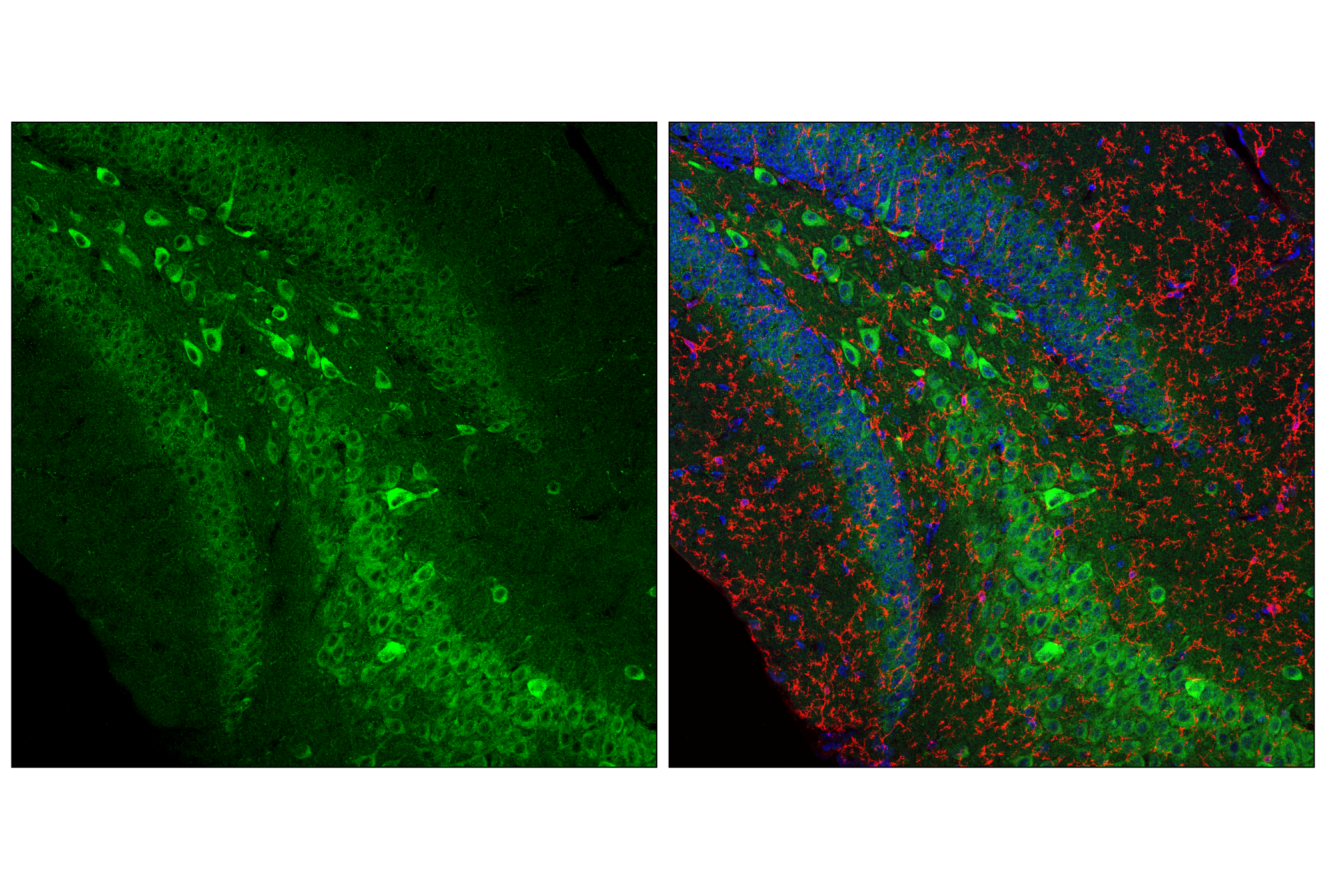
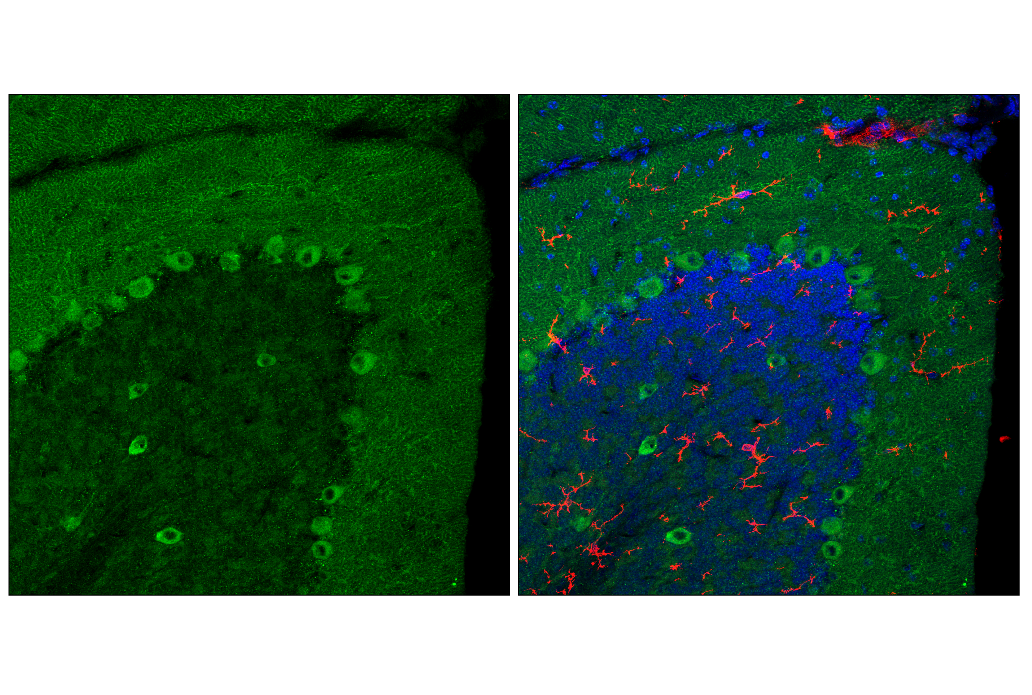
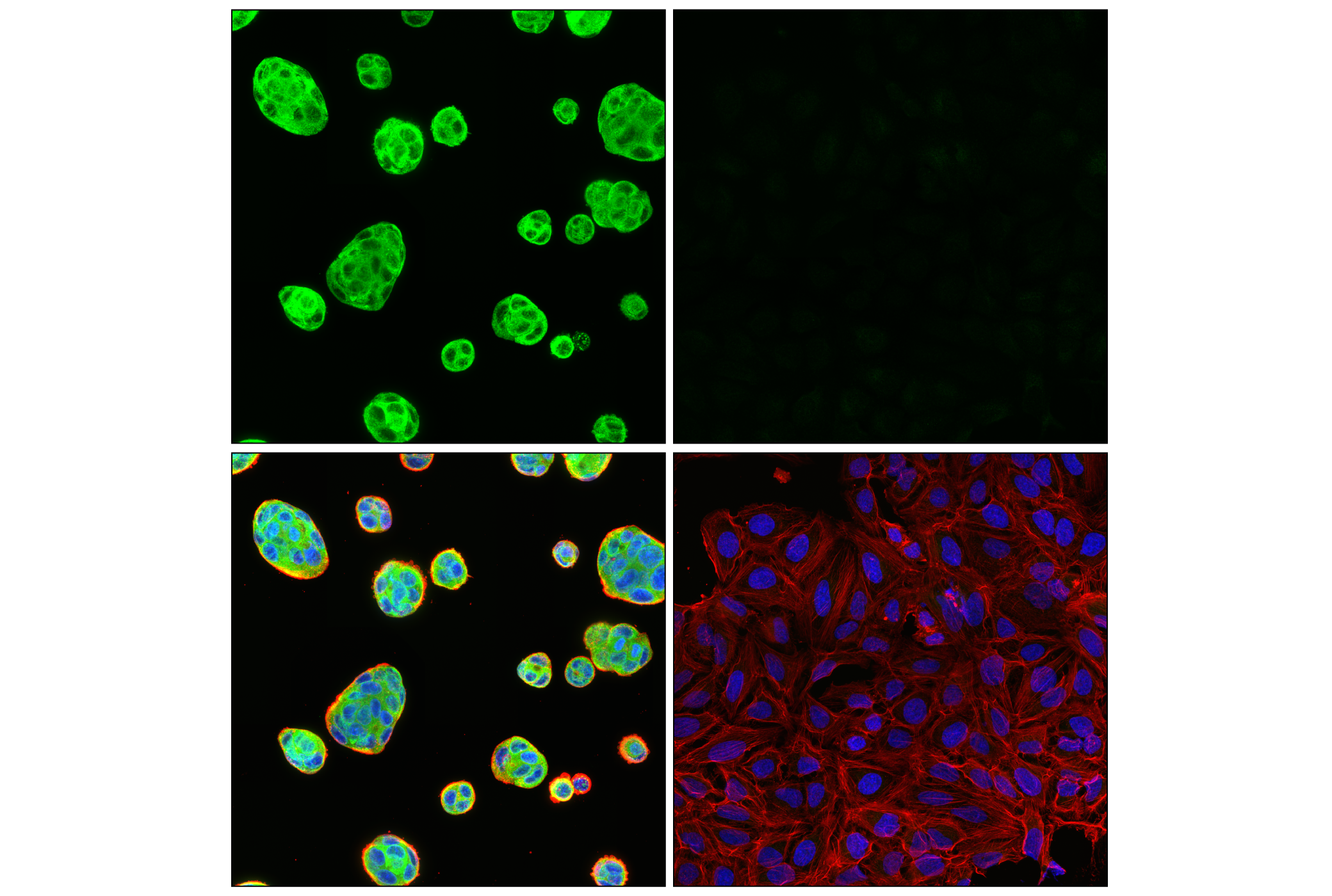
WB, IF-F, IF-IC
H M R
Endogenous
27
Rabbit IgG
#O95292
9217
Product Information
Product Usage Information
| Application | Dilution |
|---|---|
| Western Blotting | 1:1000 |
| Immunofluorescence (Frozen) | 1:50 - 1:200 |
| Immunofluorescence (Immunocytochemistry) | 1:400 - 1:1600 |
Storage
Specificity / Sensitivity
Species Reactivity:
Human, Mouse, Rat
Source / Purification
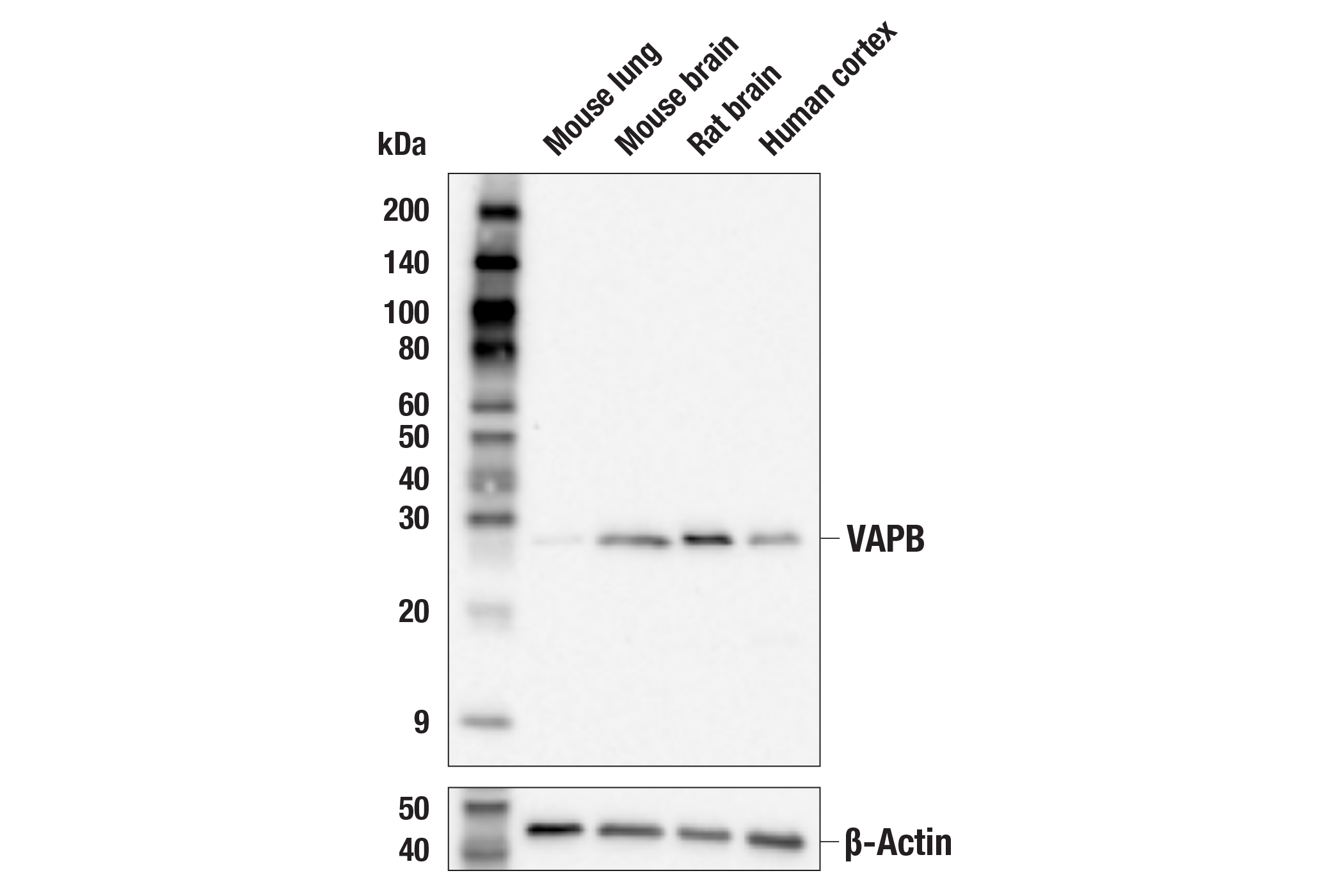
Monoclonal antibody is produced by immunizing animals with a synthetic peptide corresponding to residues near the amino terminus of human VAPB protein.
Background
The human VAP family proteins, which include VAPA (also known as VAP33), VAPB, and VAPC, were initially identified as homologues of vesicle-associated membrane protein (VAMP)-associated protein (VAP) that is involved in exocytosis of neurotransmitters (1). VAPB is an integral endoplasmic reticulum (ER) protein whose amino terminus projects into the cytosol. A proline to serine substitution at position-56 (VAPBP56S) leads to an autosomal-dominant form of amyotrophic lateral sclerosis (ALS), classified as ALS-8. (2). VAPB has been implicated in a variety of processes, including ER stress and the unfolded protein response (UPR), ER to Golgi transport, and bouton formation at the neuromuscular junction (1,2). The P56S mutation causes almost complete loss of function of VAPB to mediate UPR by inducing its misfolding and localization shift to non-ER compartments (1). A possible role of VAPB as a pathologic marker in PBMCs from ALS patients has been published (3).
Species Reactivity
Species reactivity is determined by testing in at least one approved application (e.g., western blot).
Western Blot Buffer
IMPORTANT: For western blots, incubate membrane with diluted primary antibody in 5% w/v BSA, 1X TBS, 0.1% Tween® 20 at 4°C with gentle shaking, overnight.
Applications Key
WB: Western Blotting IF-F: Immunofluorescence (Frozen) IF-IC: Immunofluorescence (Immunocytochemistry)
Cross-Reactivity Key
H: human M: mouse R: rat Hm: hamster Mk: monkey Vir: virus Mi: mink C: chicken Dm: D. melanogaster X: Xenopus Z: zebrafish B: bovine Dg: dog Pg: pig Sc: S. cerevisiae Ce: C. elegans Hr: horse GP: Guinea Pig Rab: rabbit All: all species expected
Trademarks and Patents
Limited Uses
Except as otherwise expressly agreed in a writing signed by a legally authorized representative of CST, the following terms apply to Products provided by CST, its affiliates or its distributors. Any Customer's terms and conditions that are in addition to, or different from, those contained herein, unless separately accepted in writing by a legally authorized representative of CST, are rejected and are of no force or effect.
Products are labeled with For Research Use Only or a similar labeling statement and have not been approved, cleared, or licensed by the FDA or other regulatory foreign or domestic entity, for any purpose. Customer shall not use any Product for any diagnostic or therapeutic purpose, or otherwise in any manner that conflicts with its labeling statement. Products sold or licensed by CST are provided for Customer as the end-user and solely for research and development uses. Any use of Product for diagnostic, prophylactic or therapeutic purposes, or any purchase of Product for resale (alone or as a component) or other commercial purpose, requires a separate license from CST. Customer shall (a) not sell, license, loan, donate or otherwise transfer or make available any Product to any third party, whether alone or in combination with other materials, or use the Products to manufacture any commercial products, (b) not copy, modify, reverse engineer, decompile, disassemble or otherwise attempt to discover the underlying structure or technology of the Products, or use the Products for the purpose of developing any products or services that would compete with CST products or services, (c) not alter or remove from the Products any trademarks, trade names, logos, patent or copyright notices or markings, (d) use the Products solely in accordance with CST Product Terms of Sale and any applicable documentation, and (e) comply with any license, terms of service or similar agreement with respect to any third party products or services used by Customer in connection with the Products.